

SimpleMed - Learning Medicine, SimplifiedSimpleMed - Learning Medicine, Simplified
Hit enter to search or ESC to close

Get in Touch

Contact Us

Use our contact form.

Gametogenesis

Share:

By Dr. Maddie Swannack

Next Lesson - Sex, Infertility and Contraception

Reproductive System


Contents

Contents

  1. The Male Gamete
    • Spermatogenesis
    • Spermiogenesis
    • Sperm Capacitation
    • Spermatogenic Wave
  2. The Female Gamete
    • Oocyte
      • Preantral Stage
      • Antral Stage
      • Preovulatory Stage
    • Ovulation
    • Ovarian Cancer
    • Mittelschmerz
    • The Corpus Luteum
    • The Corpus Albicans
  3. Quiz
Abstract

Abstract

  • Gametogenesis is the production of sex cells (gametes) through meiosis.
  • Spermatogenesis involves the following transitions: primordial germ cells -> spermatogonia -> type B spermatogonia -> primary spermatocyte -> secondary spermatocyte -> spermatids. Spermatogonia also produce type A spermatogonia which are recycled to replenish the spermatogonia supply.
  • Spermiogenesis is the transition of spermatids into mature sperm cells, with spermiation ending with mobile sperm.
  • Sperm capacitation is the final step to allow for fertilisation. It involves the removal of glycoproteins and cholesterol and activating signalling pathways.
  • The Spermatogenic cycle is the time taken for the same stage of spermatogenesis to appear in the same segment of the seminiferous tubule.
  • The Spermatogenic wave is the appearance of the different stages of spermatogenesis in the seminiferous tubules in a sequential and ‘wave like’ motion.
  • Formation of oocytes begins before birth, arresting in prophase I, becoming primary oocytes surrounded by follicular cells, called primordial follicles. Most of these undergo atresia before puberty. Around 15-20 primordial follicles are recruited into growth each month, developing cuboidal epithelium with granulosa and theca cells in the preantral stage. In the antral stage, spaces between granulosa cells coalesce to form the antrum. Only one oocyte reaches the preovulatory phase, finishing meiosis I and forming a secondary oocyte.
  • Ovulation occurs, releasing the oocyte into the fimbriae of the fallopian tubes, passing down it by peristalsis towards the uterus. Because ovulation is a traumatic action, increased ovulation may be related to increased ovarian cancer incidence.
  • The corpus luteum is formed from the theca interna and granulosa cells, secreting progesterone and oestrogen causing the uterus to enter the secretory phase. If β-hCG is released by the implanted zygote, the corpus luteum persists and releases progesterone for 4 months until the placenta takes over. If no β-hCG is released, the corpus luteum forms a mass of scar tissue called the corpus albicans, stopping progesterone release and causing menstruation.
Core

Core

Gametogenesis - the process of making sex cells (gametes) through meiosis. This occurs in the testes in males, and in the ovaries in females.


The Male Gamete

Spermatogenesis

Spermatogenesis is the process that creates sperm from primordial germ cells in the testicles. It has a number of steps, which are listed below and included in the diagram:

  1. The primordial germ cell divides by mitosis to produce spermatogonia.
  2. The spermatogonia divide by mitosis to produce two types of cells:
    • Type A Spermatogonium - replace the spermatogonia in a loop, will divide into type A and type B spermatogonia in the future.
    • Type B Spermatogonium - continue along the spermatogenesis pathway to produce sperm.
  3. Type B spermatogonium divides by mitosis to form primary spermatocytes.
  4. Primary spermatocytes divide by meiosis to form secondary spermatocytes.
  5. Secondary spermatocytes go through meiosis to turn into spermatids.
  6. Spermatids continue through spermiogenesis to produce sperm cells.


Spermiogenesis

Spermiogenesis is the final stage in the production of sperm cells and involves the transition of spermatids into motile sperm cells.

Spermiation is the outcome of spermiogenesis. Spermiation occurs when the spermatids are released into the lumen of the seminiferous tubules. They then pass through the rete testis and ductuli efferentes and into the epididymis. Spermatids are not mobile until they pass through the epididymis. Before this, they are transported by secretions from Sertoli cells and peristalsis, and after transit through the epididymis, they acquire motility and become mature sperm cells.
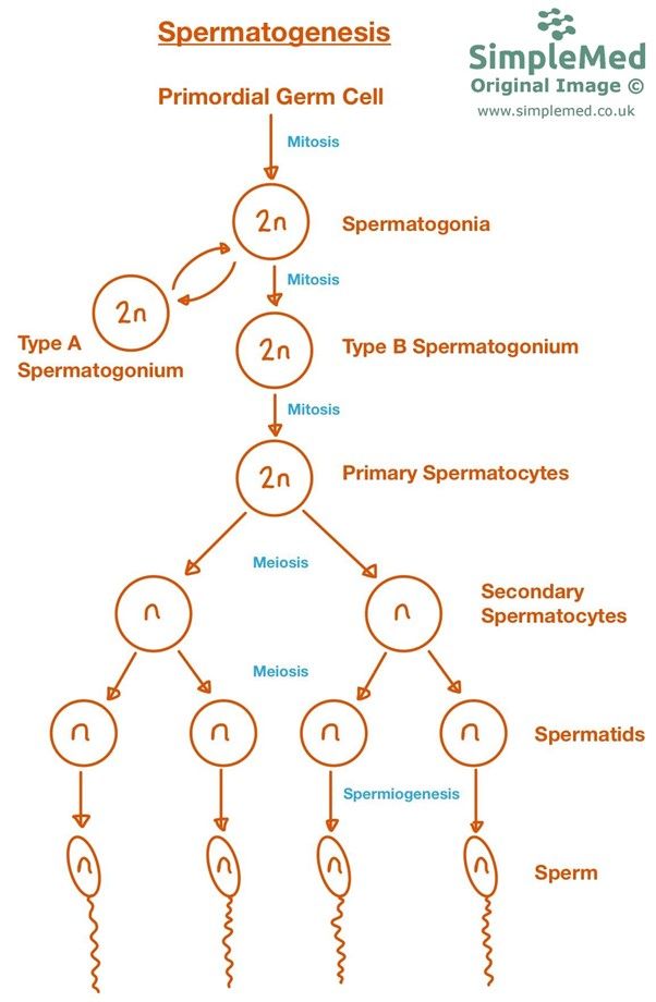
 

Spermatogenesis and Spermiogenesis SimpleMed

Diagram - Spermatogenesis and spermiogenesis

SimpleMed original by Dr. Maddie Swannack


Sperm Capacitation

Capacitation of sperm is the final step before sperm becomes able to fertilise an oocyte. Capacitation occurs in the female reproductive tract following ejaculation. It involves the removal of glycoproteins and cholesterol from the sperm membrane and activation of signalling pathways inside the sperm cells. Removal of the glycoproteins allows the sperm to bind to the zona pellucida of the oocyte and initiate the acrosome reaction. This is the process that allows the sperm to penetrate the egg cell and begin fertilisation.

Capacitation is an important consideration in in vitro fertilisation (IVF). As the sperm never encounters the female reproductive tract before meeting the egg, the sperm must be treated in a capacitation medium to trigger the process artificially.


Spermatogenic Wave

Not all the stages of spermatogenesis are visible histologically in a single cross-section of a seminiferous tubule. This observation confirms that sperm is produced continuously rather than batch produced.

The spermatogenic cycle is the time taken for the same stage of spermatogenesis to appear in the same segment of the seminiferous tubule. In humans this takes around 16 days. The total time to produce one fully mature sperm cell from a spermatogonium is much longer.

The spermatogenic wave is the appearance of the different stages of spermatogenesis in the seminiferous tubules in a sequential and ‘wave like’ motion. This wave-like movement is the spermatogenic wave.


The Female Gamete

Oocyte

The oocyte (pronounced o-o-cyte not oo-cyte) is the female gamete and is also known as an egg cell. A woman is born with all her oocytes formed but not fully developed in her ovaries. This stock declines after birth with age due to atresia (cell death).

Formation of oocytes begins before birth. Germ cells differentiate into oogonia and proliferate by mitosis. The oogonia then begin meiosis, but arrest in prophase I (meaning they have replicated their chromosomes but not commenced division). At this point, the oogonia become primary oocytes. These primary oocytes number approximately 2 million, and each one is surrounded by a flat layer of epithelial cells called follicular cells. The primary oocytes surrounded by follicular cells are named primordial follicles.

Most of these primordial follicles undergo atresia during childhood, leaving approximately 40,000 in the ovaries by the start of puberty. From puberty onwards, 15-20 oocytes begin to mature each month, but usually only one completes maturation and is ovulated, with the remainder undergoing atresia.


Preantral Stage 

As primordial follicles grow, the epithelium changes from flat to cuboidal. A number of new cell types develop: granulosa cells secrete a glycoprotein layer named zona pellucida, and the theca interna cells become paracrine tissue, going on to produce testosterone under the influence of LH so granulosa cells can turn it into oestrogen.


Antral Stage

As development continues, fluid filled spaces appear between granulosa cells, which coalesce to form the antrum (the name for the largest fluid filled sac). Several follicles (15-20) begin development every menstrual cycle but only one progresses to the preovulatory stage.


Preovulatory Stage

An increase in FSH and LH induce growth. The primary oocyte then enters meiosis I. However, in division the cytoplasm is not evenly distributed. The majority is given to the secondary oocyte and the minority to the first polar body. The polar bodies never become an egg and in effect are dustbins for unneeded genetic material. The secondary oocyte then enters meiosis II and halts until it is fertilised. 


Ovulation

Before ovulation occurs, the fimbriae of the fallopian tube sweep the surface of the ovary, and the peristaltic action of the fallopian tube begins. This means that as soon as ovulation occurs, the fallopian tubes are doing everything they can to make sure the oocyte progresses into the uterus.

Ovulation occurs when the graafian/ovarian follicle (the follicle containing the secondary oocyte) becomes approximately 2.5cm in size. The LH surge increases collagenase activity (an enzyme that breaks down collagen), and prostaglandin release causes ovarian wall contractions. This causes the oocyte to be extruded from the ovary in a traumatic release.

The fallopian tube then acts to move the oocyte into the uterus through peristaltic contractions and fimbriae movements. The fallopian tubes are also lined with cilia which help to waft the oocyte towards the uterus.


Ovarian Cancer 

As the oocyte leaves the ovary through a traumatic rupture, repeated repair is needed. This means that there is an increase in cellular proliferation needed at these sites, which can increase the risk of some types of ovarian cancer. This means that increased ovulation can lead to an increased risk of ovarian cancer.

Preventing ovulation can therefore be a protective factor for ovarian cancer, meaning that multiparous women (women who have had more than one child, ovulation pauses during pregnancy) and women who use birth control methods that prevent ovulation (such as the combined oral contraceptive pill) are at a lower risk of ovarian cancer.


Mittelschmerz

Mittelschmerz or ovulation pain which occurs on ovulation. It is unknown what specifically causes the pain. One theory is that fluid or blood from the follicle irritate the abdomen. Alternatively it is thought that if the epithelium of the ovary is too well innervated when the oocyte traumatically ruptures from the ovary it can cause pain. The term gets its name from the fact that the painful ‘schmerz’ ovulation occurs on the 14th day of the cycle, in the middle ‘mittel’ of the month. It is important, however, that abdominal pain in a woman is not dismissed to be Mittelschmerz pain.
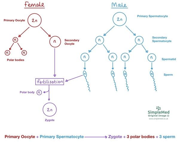
 

Meiosis to Fertilisation SimpleMed

Diagram - Processes from meiosis to fertilisation

SimpleMed original by Dr. Maddie Swannack


The Corpus Luteum

The corpus luteum (lit. ‘yellow body’) is formed from the remains of the ovarian follicle (graafian) including the remaining granulosa and theca interna cells. As the cells become vascularised they change into yellow coloured lutein cells. The corpus luteum secretes oestrogen and progesterone, which stimulates the uterine mucosa to enter the secretory stage to prepare for implantation of the fertilised oocyte.

If fertilisation occurs, β-hCG release prompts the corpus luteum to grow and form the corpus luteum graviditatis, which continues to secrete progesterone until around 10 to 12 weeks of pregnancy, when the placenta takes over progesterone production. The progesterone released maintains the thick lining of the endometrium.

If the corpus luteum does not receive β-hCG signals from the implanted embryo within around 14 days, it dies and turns into the corpus albicans. 


The Corpus Albicans

The corpus albicans (lit. ‘white body’) is essentially scar tissue formed from the regressed corpus luteum and does not release progesterone. As a result the endometrial lining breaks down which is observed as menstruation.
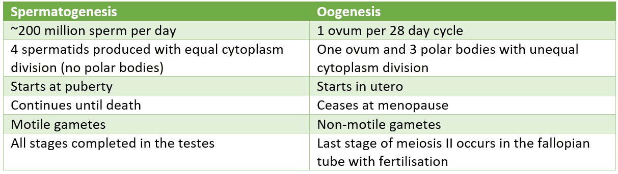


Differences Between Spermatogenesis and Oogenesis SimpleMed

Table - Differences between spermatogenesis and oogenesis

SimpleMed original by Dr. Maddie Swannack


Edited by: Dr. Ben Appleby

Reviewed by: Dr. Thomas Burnell

Quiz

Quiz

Preview the Gametogenesis quiz

In this article

Gametogenesis is the production of sex cells (gametes) through meiosis. Spermatogenesis involves the following transitions: primordial germ cells ->…

Free account

Keep reading for free

Create a free account to continue this article and save your revision progress.

Your account includes:

  • Thousands of free practice questions
  • Tools to track your revision progress
Sign up free Already have an account? Log in

No payment. No spam. Just free medical revision.

SimpleMed+

Want More?

Access more on SimpleMed+

Lock it in with practice questions, the full question bank and revision tools that track what you know.

Practice now See what's in SimpleMed+
9270
simplemedlonglogo.png
  • Home
  • Terms & Conditions
  • Privacy Policy
  • Contact

The medical information on this site is provided as an information resource only, and is not to be used or relied on for any diagnostic or treatment purposes. This information is intended for medical education, specifically to biological/medical learning inclined individuals, and does not create any doctor-patient relationship, and should not be used as a substitute for professional diagnosis and treatment.

©2026 Lili's Ark Limited, UK Company #15424953
S+

Search

  • Pre-Clinical
    • Cardiovascular System
      • 1. Anatomy of the Cardiovascular System
      • 2. The Cardiac Cycle
      • 3. Pulses and Pressures
      • 4. Control of Cardiac Output
      • 5. Cardiac Conduction and Contraction
      • 6. Embryology of the CVS
      • 7. Congenital Heart Defects
      • 8. Hypertension
      • 9. Electrocardiograms
      • 10. Electrocardiogram Pathology
      • 11. Cardiovascular Drugs
      • 12. Murmurs
      • 13. Special Circulations
      • 14. Heart Failure
      • 15. Artery and Vein Disease
      • 16. Acute Coronary Syndromes
      • 17. Haemodynamic Shock
    • Musculoskeletal System
      • The Human Body
        • 1. Anatomical Terms
        • 2. Structure of the Muscles
        • 3. Muscles in Action
        • 4. Connective Tissue
        • Cartilage: Structure, Growth and Repair
        • 5. Structure of Bones
        • 6. Basics of Bone Anatomy
        • 7. Joints
        • 8. The Spine
        • 9. Dermatomes and Myotomes
        • 10. The Brachial Plexus
        • 11. The Lumbar Plexus
        • 12. The Sacral Plexus
      • Clinical Conditions
        • 1. Conditions of the Foot
        • 2. Conditions of the Ankle
        • 3. Conditions of the Lower Leg
        • 4. Conditions of the Knee
        • 5. Conditions of the Thigh
        • 6. Conditions of the Hip
        • 7. Conditions of the Lower Back
        • 8. Conditions of the Back and Neck
        • 9. Conditions of the Shoulder and Clavicle
        • 10. Conditions of the Arm
        • 11. Conditions of the Elbow
        • 12. Conditions of the Forearm and Wrist
        • 13. Conditions of the Hand
      • Musculoskeletal Anatomy
        • 1. Muscles of the Gluteal Region
        • 2. Muscles of the Thigh
        • 3. Key Areas of the Lower Limb
        • 4. Bones of the Lower Limb
        • 5. Bones of the Foot
        • 6. Muscles of the Lower Leg
        • 7. Bones of the Upper Limb
        • 8. Muscles of the Shoulder
        • 9. Muscles of the Upper Arm
        • 10. Bones of the Hand
        • 11. Muscles of the Forearm
        • 12. Key Areas of the Upper Limb
    • Cell Physiology & Biology
      • 1. An Introduction to the Cell
      • 2. Introduction to DNA
      • 3. The Cell Cycle, Checkpoints and Apoptosis
      • 3. Structure of Proteins
      • 4. Protein Function in Oxygen Transport
      • 5. Enzymes and Regulation of Protein Function
      • 6. Molecular Techniques for Medics
      • 7. Post-translational Modification and Collagen Biosynthesis
      • 8. Protein Targeting
      • 9. Biological Membranes
      • 10. G-Protein Coupled Receptor
      • 12. Enzyme-linked and Intracellular Receptor Signalling
      • 11. Membrane Transport and Intracellular Calcium Regulation
      • 12. Membrane Potentials
      • 13. Action Potentials
      • 14. Autonomic Nervous System Introduction
    • Metabolism
      • 1. Nutrition, Diet and Body Weight
      • 2. Energy Reactions in Cells
      • 3. Dietary Energy Production
      • 4. Vitamins, Cofactors, Minerals and Trace Elements
      • 4. Glycolysis
      • 5. Oxygen-dependent Pathway
      • 6. Lipid Metabolism and Transport
      • 7. Protein and amino acid metabolism
      • 9. Purine and Pyrimidine Metabolism
      • 10. Inborn Errors of Metabolism and Newborn Screening
      • 8. Alcohol Metabolism
      • 9. Oxidative Stress
      • 10. Energy Storage
      • 11. Temperature Regulation
      • 12. Metabolic and Endocrine Regulation
    • Endocrinology
      • 1. An Introduction to Endocrinology
      • 2. Control of Appetite
      • 3. The Adrenal Glands and Adrenal Disorders
      • 4. HPA and Growth Hormone
      • 5. Pituitary Disorders
      • 6. The Thyroid Gland
      • 7. The Endocrine Pancreas
      • 8. Diabetes Mellitus
      • 9. Calcium Metabolism
    • Haematology
      • 1. Haematopoiesis and the Spleen
      • 2. Full Blood Count
      • 3. Blood Groups, Crossmatching and Transfusion Reactions
      • 3. Anaemia
      • 4. Vitamin B12 and Folate
      • 5. Iron Metabolism
      • 6. Haemoglobinopathies
      • 7. When Haematopoiesis goes wrong
      • 8. Haematology in Systemic Disease
    • Genetics
      • 1. Mitosis and Meiosis
      • 2. Genotype, Phenotype and Inheritance
      • 3. Gene Expression
      • 4. Epigenetics, Genomic Imprinting and Mitochondrial Inheritance
      • 4. Introduction to Gene Mutation
      • 5. DNA Repair and Cancer
      • 7. Genetic Testing and Counselling
    • Infection
      • 1. An Introduction To Infection
      • 2. The Infection Model
      • 3. Bacterial Structure, Classification and Growth
      • 3. Biofilms
      • 4. Innate Immunity
      • 5. Adaptive Immunity
      • 6. Gram Staining and Infection Investigations
      • 8. Staphylococci and Other Gram-positive Bacteria
      • 9. Streptococci
      • 10. Gram-negative Bacteria: Structure, Entry Routes and Endotoxin
      • 11. The Organisms Gram Staining Misses
      • 7. Sepsis
      • 8. Antibiotics
      • 14. Principles of Medical Virology
      • 10. Hepatitis
      • 11. Human Immunodeficiency Virus
      • 17. Fungi and Parasites: The Eukaryotic Pathogens
      • 12. The Immunocompromised Host
      • 13. Travel-related Infections
    • Pathology
      • 1. An Introduction to Pathology
      • 2. How to Read a Histology Slide
      • 2. The Basics of Pathology
      • 3. Epithelium, Skin and Exocrine Glands
      • 4. Acute Inflammation
      • 5. Chronic Inflammation
      • 6. Regeneration and Repair of Tissues
      • 7. The Lymphatic System
      • 8. Haemostasis and Clotting
      • 9. Thrombosis and Embolism
      • 10. Atherosclerosis
      • 11. Cellular Adaptations
      • 12. Introduction to Neoplasia
      • 13. Pathogenesis of Neoplasms
      • 14. Behaviour of Neoplasms
    • Population & Social Science
      • 1. Introduction to Population Science
      • 2. Describing Data and Testing Hypotheses
      • 2. Epidemiological Studies
      • 3. Models of Health
      • 4. Beliefs of the Public
      • 5. Health Economics
      • 6. Evidence-Based Practice
      • 7. Patient Safety and Quality Control
      • 8. Summary and Glossary
    • Patient-Centred Care
      • 1. Lifestyle Medicine
      • 2. Childhood Immunisations
      • 3. Sepsis in Paediatrics
      • 4. Child Safeguarding
      • 5. Ageing
      • 6. Falls
      • 7. Men's Health
      • 8. Palliative Care
    • Reproductive System
      • 1. Anatomy of the Man
      • 2. Anatomy of the Woman
      • 3. The Pelvic Floor
      • 4. Development of the Reproductive System
      • 5. Puberty
      • 6. The Menstrual Cycle and Menstrual Abnormalities
      • 7. Gametogenesis
      • 8. Sex, Infertility and Contraception
      • 9. Physiology of the Mother
      • 10. Diseases of the Mother in Pregnancy
      • 11. Physiology of the Fetus
      • 12. The Placenta
      • 13. Labour and Delivery
      • 14. The Postpartum Period
      • 15. Infections of the Reproductive Tract
      • 16. Pelvic Inflammatory Disease
      • 17. The Menopause
      • 18. Malignancies of the Reproductive Tract
      • 19. Introduction to the Breast
      • 20. Benign Conditions of the Breast
      • 21. Malignant Breast Disease
    • Respiratory System
      • 1. Anatomy of the Respiratory System
      • 2. Development of the Respiratory System
      • 3. Ventilation and Lung Mechanics
      • 4. Properties of Gas Exchange
      • 5. Carbon Dioxide Transport
      • 6. Chemical Control of Breathing and Plasma pH
      • 7. Lung Function Testing
      • 8. Pulmonary Embolism
      • 9. Asthma
      • 10. Chronic Obstructive Pulmonary Disease
      • 11. Pneumonia and other Lower Respiratory Tract Infections
      • 12. Bronchiectasis and Cystic Fibrosis
      • 13. Tuberculosis
      • 14. Pneumothorax and Pleural Effusion
      • 15. Lung Cancer
      • 16. Respiratory Failure
    • Urinary System
      • 1. Anatomy of the Kidneys, Bladder and Urethra
      • 2. Development of the Urinary System
      • 3. The Nephron
      • 4. The Glomerulus – Tubular Reabsorption and Secretion
      • 5. Control of Plasma Volume
      • 6. Control of Plasma Osmolarity
      • 7. Control of Potassium
      • 8. Micturition
      • 9. Diuretics
      • 10. Glomerular Filtration Rate and the Measurement of Kidney Function
      • 11. Hyponatraemia
      • 12. Urinary Incontinence
      • 13. Glomerular Disease
      • 14. Diabetic Nephropathy
      • 15. Acute Kidney Injury
      • 16. Chronic Kidney Disease
      • 17. Urinary Tract Obstruction
      • 18. Urinary Tract Infections
      • 19. Prostate Pathology
      • 20. Male Urological Conditions
      • 21. Urological Malignancy
    • Gastrointestinal System
      • 1. Development of the Gastrointestinal Tract
      • 2. Abdominal Wall and Inguinal Canal
      • 2. Anatomy and Physiology of the Foregut
      • 3. Anatomy and Physiology of the Midgut
      • 4. Anatomy and Physiology of the Hindgut
      • 6. Gastrointestinal Motility and the Enteric Nervous System
      • 7. Liver, Biliary and Exocrine Pancreatic Physiology
      • 8. Gastrointestinal Secretion, Digestion and Absorption
      • 5. Upper Gastrointestinal Pathology
      • 6. Hepatic, Biliary and Pancreatic Pathology
      • 7. Intestinal Pathology
      • 8. Distal Gastrointestinal Tract Pathology
      • 9. Gastrointestinal Malignancy
      • 10. Gastroenteritis and Enterocolitis
      • 11. Gastrointestinal Emergencies
    • Head & Neck
      • 1. Introduction to Head & Neck Anatomy
      • 2. Blood Supply of the Head and Neck
      • 3. Lymph Nodes and Neck Lumps
      • 4. The Skull
      • 5. The Temporomandibular Joint
      • 6. The Meninges
      • 7. Cranial Nerves I-VI
      • 8. Cranial Nerves VII-XII
      • 9. Development of the Head and Neck
      • 10. Autonomics in the Head and Neck
      • 11. Anatomy of the Ear
      • 12. Ear Pathology
      • 13. The Nose
      • 14. The Oral Cavity and Pharynx
      • 15. The Eye
      • 16. The Larynx
      • 17. Head and Neck Cancers
      • 18. Imaging of the Head and Neck
      • 19. Fascial Planes, Muscles and Triangles
    • Immunology
      • 1. An Introduction to Hypersensitivity
      • 2. Type 1 Hypersensitivity
      • 3. Food Allergy
      • 4. Introduction to Autoimmunity
      • 5. Rheumatoid Arthritis
      • 6. Systemic Lupus Erythematosus (SLE)
      • 7. Immunological Therapies | Monoclonal Antibodies
    • Neuroanatomy & Psychiatry
      • 1. Basics of Neuroanatomy
      • 2. Cells and Components of the Nervous System
      • 3. Arterial Supply to the Brain
      • 4. Development of the Nervous System
      • 5. Production and Circulation of CSF and Related Disorders
      • 6. Somatosensory System
      • 7. Motor System
      • 8. The Cerebellum: Circuits, Coordination and Clinical Signs
      • 8. Visual System
      • 10. Auditory and Vestibular Systems
      • 11. The Hypothalamus and Limbic System
      • 9. Higher Cortical Function
      • 10. Reticular Formation
      • 11. Movement Disorders
      • 12. Infections of the Central Nervous System
      • 13. Dementia
      • 14. Delirium
      • 15. Headaches
      • 16. Stroke
      • 17. Raised Intracranial Pressure (ICP)
      • 18. Subarachnoid Haemorrhage
      • 19. Anxiety
      • 20. Mood Disorders
      • 21. Psychosis
    • Embryology
      • 1. The Basics of Embryology
      • 2. Development of the Limbs and Body Wall
      • 3. Teratology and Congenital Malformations
      • 2. Development of the Gastrointestinal Tract
      • 3. Development of the Reproductive Systems
      • 5. Development of the Respiratory System
      • 6. Development of the Head and Neck
      • 8. Embryology of the Cardiovascular System
    • Pharmacology
      • 1. Research Methods in Pharmacology
      • 2. Pharmacokinetics
      • 3. Pharmacodynamics
      • 4. Pharmacovigilance and Pharmacogenetics
      • 5. Safe Prescribing
      • 6. Cholinergic Pharmacology
      • 7. Adrenergic Pharmacology
      • 6. Hypertension and Heart Failure
      • 7. Cardiac Arrhythmias
      • 8. Antiplatelets, Anticoagulants and Thrombolysis
      • 9. Hyperlipidaemias
      • 10. Diuretics and Renal Pharmacology
      • 11. Pharmacology of Insulin
      • 12. Reproductive and Post-reproductive Health
      • 13. Antibiotics
      • 14. Antivirals
      • 15. Cancer Chemotherapy
      • 16. Anaesthetics
      • 17. Opioids
      • 20. Introduction to Psychopharmacology
      • 18. Epilepsy and Anti-epileptic Drugs
      • 19. Parkinson's Disease and Myasthenia Gravis
      • 20. NSAIDs
      • 21. Immunosuppression and Disease-modifying Therapy
      • 22. Pharmacology of Airway Control
      • 23. Drugs Affecting Acid Secretion
      • 24. Antiemetics and Laxatives
      • 25. Poisoning and STOPP/START
  • OSCE
    • Index Conditions for OSCE exams
    • Cardiovascular System Examination
    • Shoulder Examination
    • Hand Examination
    • Venepuncture
    • Blood Pressure
    • Respiratory System OSCE Examination
    • Peripheral Vascular OSCE Examination
    • Varicose Vein OSCE Examination
    • Deep Vein Thrombosis (DVT) OSCE Examination
    • Abdominal OSCE Examination
    • Rectal (PR) OSCE Examination
    • Stoma OSCE Examination
    • Hernia OSCE Examination
    • Upper Limb Neurological OSCE Examination
    • Lower Limb Neurological OSCE Examination
    • Cranial Nerve OSCE Examination
    • Cerebellar OSCE Examination
    • GALS Screening OSCE Examination
    • Elbow OSCE Examination
    • Hip OSCE Examination
    • Knee OSCE Examination
    • Ankle and Foot OSCE Examination
    • Spine OSCE Examination
    • Thyroid Status OSCE Examination
    • Diabetic Foot OSCE Examination
    • Cushing's Syndrome OSCE Examination
    • Acromegaly OSCE Examination
    • Ear, Otoscopy and Hearing OSCE Examination
    • Nasal OSCE Examination
    • Eye and Vision OSCE Examination
    • Fundoscopy (Ophthalmoscopy) OSCE Examination
    • Visual Acuity OSCE Assessment
    • Anterior Segment Eye OSCE Examination
    • Pigmented Skin Lesion OSCE Examination
    • Rash and Non-Pigmented Skin Lesion OSCE Examination
    • Breast OSCE Examination
    • Testicular OSCE Examination
    • Neck Lump OSCE Examination
    • Lymphoreticular OSCE Examination
  • Question Bank
  • About
  • How to Use SimpleMed
  • SimpleMed Lecture Series
  • DON'T PANIC - Guide to the Wards
  • SimpleMed Plus
Your privacy Allow anonymous analytics to help us improve SimpleMed. Privacy policy
