

SimpleMed - Learning Medicine, SimplifiedSimpleMed - Learning Medicine, Simplified
Hit enter to search or ESC to close

Get in Touch

Contact Us

Use our contact form.

Anatomy of the Respiratory System

Share:

By Dr. Peter Parkinson

Next Lesson - Development of the Respiratory System

Respiratory System


Contents

Contents

  1. The Nasal Cavity
  2. The Pharynx and Larynx 
  3. The Upper Airways
  4. Chronic Obstructive Pulmonary Disease
  5. Cystic Fibrosis
  6. The Thoracic Cavity
  7. The Intercostal Muscles
  8. The Intercostal Arteries, Veins and Nerves
  9. The Diaphragm
  10. The Movements of the Thoracic Wall and Diaphragm During Respiration
  11. The Pleura and Pleural Cavity
  12. The Trachea and the Bronchial Tree
  13. The Lungs
  14. The Neurovascular Supply of the Lungs
  15. Visualising the Lobes of the Lungs and The Pleural Cavity
  16. Quiz
Abstract

Abstract

  • The nasal cavity is divided into the left and the right, providing an entrance for air. This air is warmed & moistened as it enters the nasal cavity, whilst expired air is cooled to recover the water.
  • The pharynx is split into three regions: nasopharynx, oropharynx and laryngopharynx. The pharynx and larynx work together to ensure food enters the oesophagus and air enters the trachea.
  • The airways are divided into the conducting and respiratory portions. The conducting portion extends from the nasal cavity to the terminal bronchioles and transports air to the gas exchange surfaces. The respiratory portion extends from the respiratory bronchioles to the alveoli and is the site of gas exchange.
  • Patients suffering from chronic obstructive pulmonary disease have inflammation of the airways, meaning they have an increased risk of developing infections.
  • Patients suffering from cystic fibrosis have thickened mucus in their airways due to less water being able to enter the mucus from the cells lining the respiratory tract. This leads to patients having an increased risk of developing respiratory infections.
  • There are three muscles present in the intercostal space, and from superficial to deep they are: external, internal and innermost intercostal muscles.
  • The neurovascular bundle of each rib runs along the inferior border of the rib above (superiorly in the intercostal space). Clinically, procedures such as chest drain insertion are performed just above the upper border of the lower rib to reduce the risk of neurovascular injury.
  • The diaphragm is the main muscle involved in inspiration and when it contracts, it flattens to increase thoracic space. This causes the lungs to inflate.
  • The pleura is made up of two serous membranes - parietal membrane, which lines the inside of each hemi-thorax, and the visceral pleura lines the outside of the lungs and extends into the interlobar fissures.
  • The trachea commences at the lower border of the cricoid cartilage and terminates by dividing into the right and left main bronchi at the level of the sternal angle.
  • The right lung has three lobes and the left lung has two lobes.
  • A tumour in the apex of either lung can lead to the patient presenting with neurovascular symptoms due to the close relationship of the apex of the lungs with blood vessels & nerves.
  • The lungs have a dual blood supply, the pulmonary system and the bronchial system.
Core

Core

The respiratory system allows the body to take in oxygen and expel carbon dioxide. This process oxygenates the blood and removes carbon dioxide produced by respiring cells within the body. This article will cover the anatomical structures that form the respiratory system as well as the neurovascular supply to it. 


The Nasal Cavity

The nasal cavity is divided into the left and right nasal cavities by the medial nasal septum. Air enters the nasal cavity through the nostrils and passes into the nasal vestibule, which is lined by keratinised, stratified squamous epithelium. The nostrils of the nasal vestibule are lined with nasal hair, which filter dust and any other foreign materials. Within the nasal vestibule, the epithelium changes to ciliated pseudostratified columnar epithelium (respiratory epithelium).

On the medial surface of the nasal cavity, there are three outgrowths called conchae or turbinates. They increase the surface area for inspired air to be warmed and moistened on the way in, and for expired air to be cooled on the way out.

As well as warming and moistening inspired air, the nasal cavity is also used as a resonance chamber in the production of speech and in olfaction (smell). 
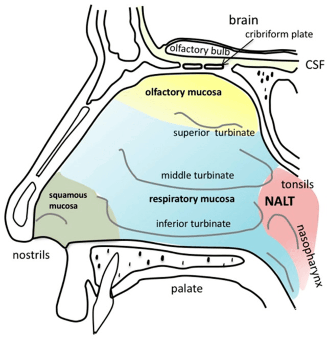
 

Nasal Cavity Anatomy SimpleMed

Diagram - The structures in the nasal cavity

Creative commons source by Stella Gänger and Katharina Schindowski [CC BY-SA 4.0 (https://creativecommons.org/licenses/by-sa/4.0)]


There are four paranasal sinuses located around the nasal cavity and they are connected to the nasal cavity via small orifices called ostia. The ostia can become blocked if inflammation is present, such as in situations like allergic reactions and infections. These can lead to the drainage of mucus from the sinuses being disrupted, causing sinusitis to develop. The four paranasal sinuses and where they are found can be seen in the diagram below.
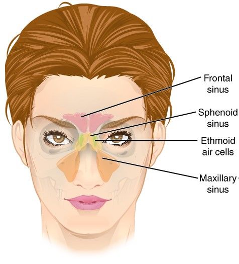
 

Paranasal Sinuses SimpleMed

Diagram - The paranasal sinuses

Creative commons source by OpenStax College [CC BY-SA 4.0 (https://creativecommons.org/licenses/by-sa/4.0)]


The Pharynx and Larynx

In the pharynx, air and food have a common passage, which connects the nasal cavity, oral cavity and trachea together. The pharynx is split into three sections: nasopharynx, oropharynx and laryngopharynx. Both the pharynx and larynx work together to ensure food enters the oesophagus and air enters the trachea.

The vocal cords are found within the larynx. During the process of swallowing, the laryngeal inlet becomes narrowed followed by the epiglottis folding downwards. Finally, the vocal cords adduct to act as a sphincter to close off the entrance to the trachea thus preventing food or any other foreign objects from entering.

If the larynx and vocal cords become dysfunctional, there is an increased risk of inhalation of food or liquid due to the inlet to the trachea not properly closing.

The coughing mechanism allows particles and objects to be expelled from the trachea. In this mechanism the vocal cords adduct, allowing intra-thoracic pressure to build up, which is then followed by quick abduction of the vocal cords that expels air and particles out of the trachea in a cough.
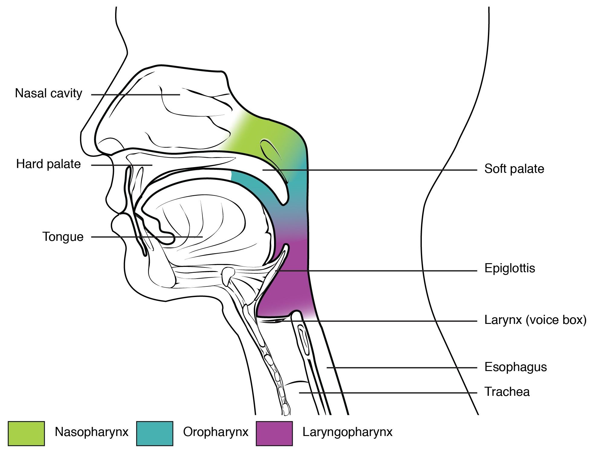
 

The Sections of the Pharynx SimpleMed

Diagram - The sections of the pharynx, with the nasopharynx in green, the oropharynx in blue and the laryngopharynx in purple

Creative commons source by OpenStax College [CC BY-SA 4.0 (https://creativecommons.org/licenses/by-sa/4.0)]


The Upper Airways

The airways are split into two regions: the conduction portion and the respiratory portion. The conducting portion is described as being from the nasal cavity to the terminal bronchioles and is responsible for the transport of air to the gaseous exchange surfaces. The respiratory portion is from the respiratory bronchioles to the alveoli and is responsible for the site of gaseous exchange.

The trachea is lined with cartilage in a C shape and the presence of this cartilage ensures the trachea remains open under the negative pressure that occurs during breathing. The cartilage changes shape in the primary bronchi to a full ring, and changes again to a crescent-shaped structure in the secondary and tertiary bronchi.

Alveoli are found at the end of respiratory bronchioles, attached to alveolar sacs (collection of alveoli). Alveoli are the sites of gaseous exchange of carbon dioxide and oxygen.
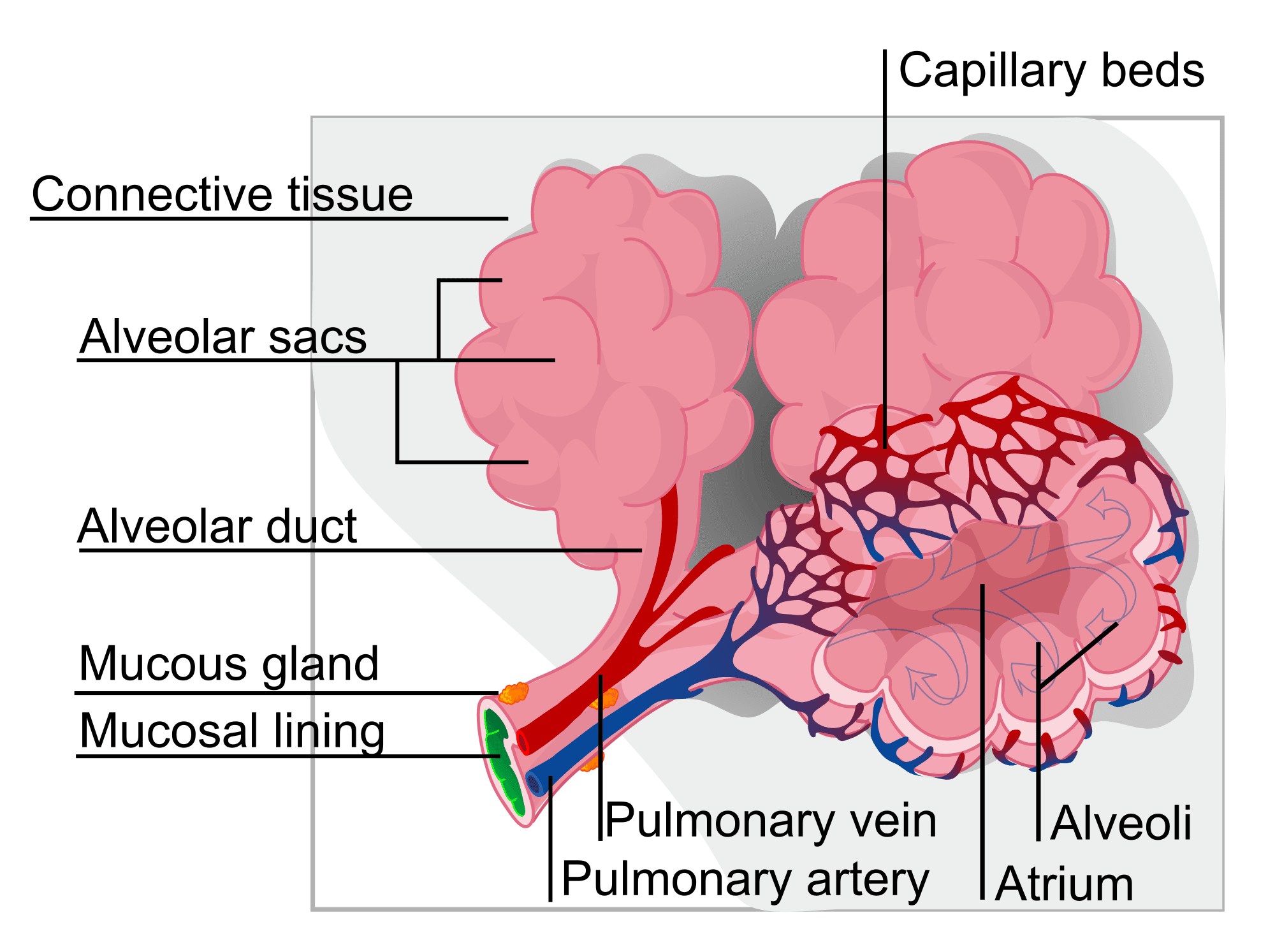
 

Structure of Alveolar Sacs SimpleMed

Diagram - The structure of alveolar sacs

Public Domain Source by LadyofHats [Public domain]


The epithelium found between the nasal cavity and the secondary bronchi is pseudostratified ciliated epithelium with goblet cells. The goblet cells produce and release mucus, which traps microorganisms and inhaled particles. The cilia then waft the mucus up to the pharynx to be swallowed and digested in the gastrointestinal tract. From the bronchioles onwards, the goblet cells are replaced by the club cells, which produce and secrete surfactant-like material that helps reduce airway collapse during expiration.
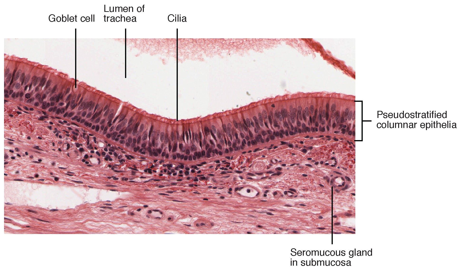
 

Histological Section of the Trachea SimpleMed

Diagram - Histological section of the trachea

Creative commons source by OpenStax College [CC BY-SA 4.0 (https://creativecommons.org/licenses/by-sa/4.0)]


Chronic Obstructive Pulmonary Disease

When a patient is suffering from Chronic Obstructive Pulmonary Disease (COPD), they will have hyperplasia of the goblet cells and hypertrophy of the submucus glands leading to an increased ratio of mucus cells to ciliated cells. This causes inflammation of the airways, and increased mucus production, meaning the patients with this disease have an increased risk of developing infections as microorganisms cannot be transported from the airways to the pharynx as efficiently anymore. For more information see our article on COPD.
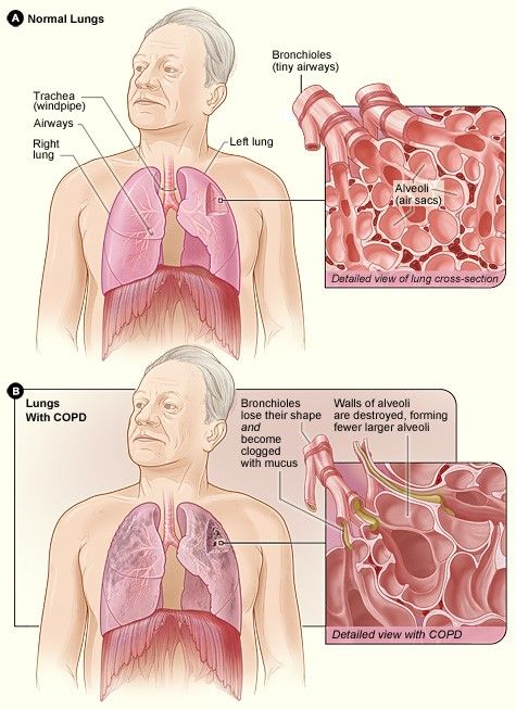
 

Lungs with COPD vs Lungs without COPD SimpleMed

Diagram - The lungs of someone with COPD versus the lungs of someone without COPD

Public Domain Source by National Heart Lung and Blood Institute [Public domain]


Cystic Fibrosis

A patient who is suffering from cystic fibrosis (CF) will have more viscous mucus due to a defect in the cystic fibrosis transmembrane conductance regulator (CFTR), which is a transporter present on the luminal side of the cells lining the airways. This defect leads to fewer chloride ions being transported from inside the cell into the mucus, meaning less water moves out of the cells causing the mucus to be more viscous. As a result of this defect, patients suffering from cystic fibrosis are more likely to suffer from repeated pulmonary infections as the mucus in the airways struggles to move up to the pharynx.


The Thoracic Cavity

The bony thorax is composed of the sternum, twelve pairs of ribs and the twelve thoracic vertebrae of the spine. The lungs are ventilated by the bony thorax, the intercostal muscles and the diaphragm functioning together.

The first seven ribs are attached to the sternum through the costal cartilage, whilst the eighth, ninth and tenth ribs are attached to the costal cartilage of the seventh rib. The eleventh and twelfth rib terminate in the abdominal musculature.


The Thoracic Cavity SimpleMed

Diagram - The thoracic cavity, showing the sternum, ribs and costal cartilages

Creative commons source by OpenStax College [CC BY-SA 4.0 (https://creativecommons.org/licenses/by-sa/4.0)]


The Intercostal Muscles

There are three muscles found in each intercostal space and they are all innervated by the intercostal nerves. These muscles are (from superficial to deep):

External Intercostal Muscles - the most superficial intercostal muscles. The fibres of these muscles project inferiorly and anteriorly. The external intercostal muscles are responsible for around 30% of chest expansion during quiet respiration.
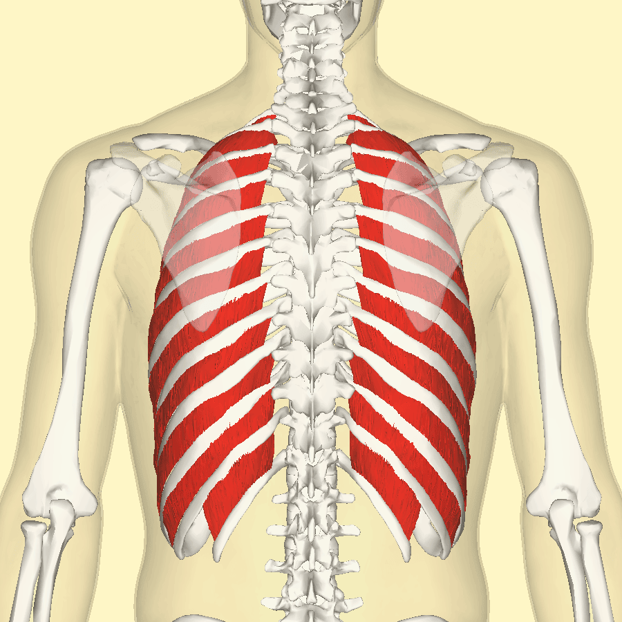
 

External Intercostal Muscles SimpleMed

Image - The external intercostal muscles

Creative commons source by Anatomography [CC BY-SA 4.0 (https://creativecommons.org/licenses/by-sa/4.0)]


Internal Intercostal Muscles - the fibres of these muscles project inferiorly and posteriorly. They pull the ribs inferiorly during forced expiration.
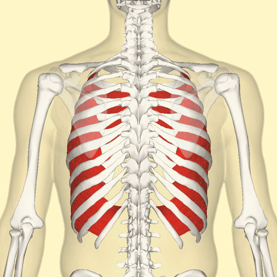
 

Internal Intercostal Muscles SimpleMed

Image - The internal intercostal muscles

Creative commons source by Anatomography [CC BY-SA 4.0 (https://creativecommons.org/licenses/by-sa/4.0)]


Innermost Intercostal Muscles - the deepest intercostal muscles. These muscles are used alongside internal intercostal muscles during forced expiration.
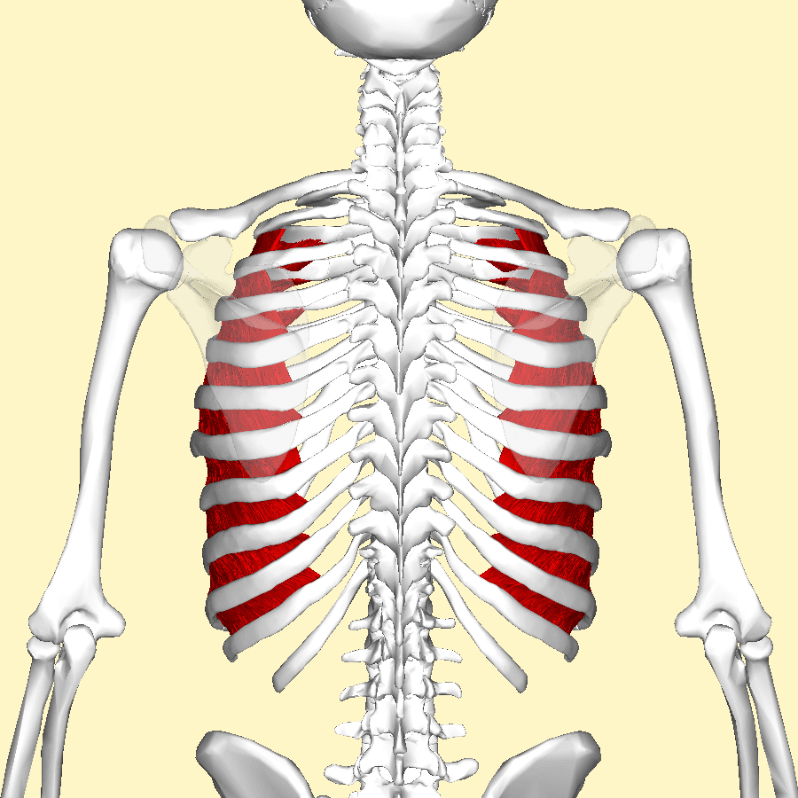
 

The Innermost Intercostal Muscles SimpleMed

Image - The innermost intercostal muscles

Creative commons source by Anatomography [CC BY-SA 4.0 (https://creativecommons.org/licenses/by-sa/4.0)]


The Intercostal Arteries, Veins and Nerves

The neurovascular bundle that supplies the intercostal muscles runs on the inferior border of each rib, and therefore superiorly in the intercostal space.

The first nine intercostal spaces are supplied by three vessels each: two anterior intercostal arteries and one posterior intercostal artery. The final two intercostal spaces (tenth and eleventh) are supplied solely by the posterior intercostal arteries.

The first two posterior intercostal arteries are branches of the superior intercostal artery whereas the final nine posterior intercostal arteries derive from the thoracic aorta.

The first six anterior intercostal arteries arise from the internal thoracic artery and the final three anterior intercostal arteries arise from the musculophrenic arteries.

Regarding the venous drainage of the intercostal spaces, there are eleven pairs of posterior intercostal veins and nine pairs of anterior intercostal veins (supplying spaces 1-9). With the anterior intercostal veins, the first six pairs drain directly into the internal thoracic vein and the final two veins drain firstly into the musculophrenic vein before draining into the internal thoracic vein. The majority of the posterior intercostal veins drain into the azygos or hemiazygous veins.
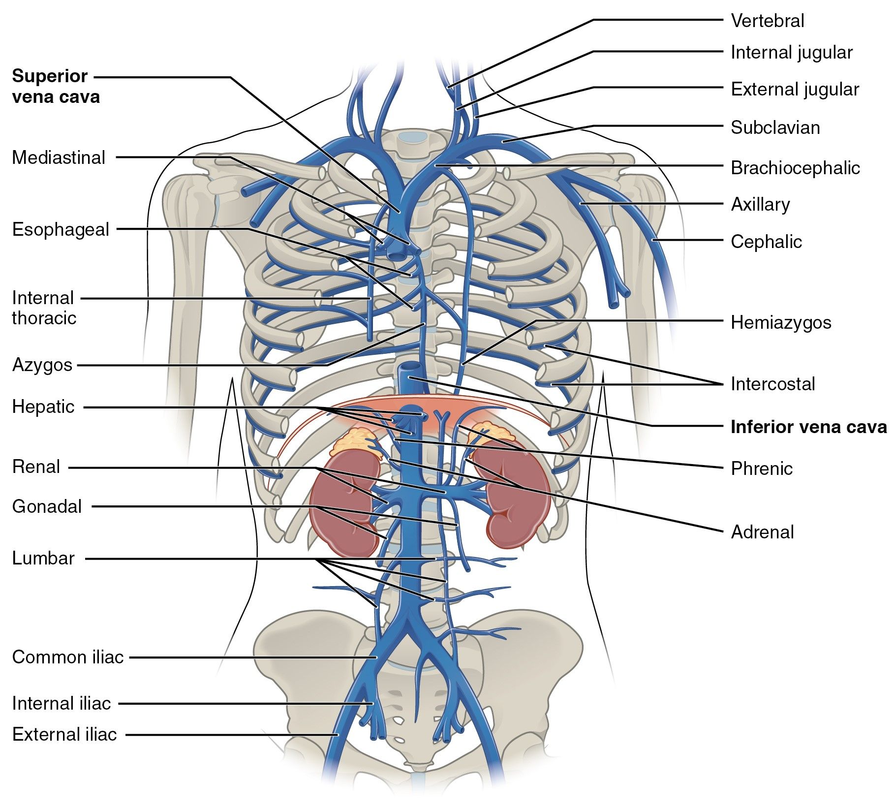
 

The Azygos and Hemiazygos Veins SimpleMed

Diagram - The azygos and hemiazygos veins alongside the other veins in the thoracic region

Creative commons source by OpenStax College [CC BY-SA 4.0 (https://creativecommons.org/licenses/by-sa/4.0)]


The intercostal nerves arise from the anterior rami of the first eleven thoracic spinal nerves and the anterior rami of the twelfth thoracic spinal nerve lies in the abdomen as the subcostal nerve.

When performing a chest drain, the tube is classically inserted into the intercostal space at the superior border of the lower rib (inferior in the space) to avoid damaging the neurovascular structures found in the intercostal space. In practice, chest drain insertion should always follow local clinical guidelines and be performed by appropriately trained clinicians.


The Diaphragm

The diaphragm is the main muscle responsible for chest expansion during inspiration. The muscle is dome-shaped at rest but during contraction it flattens with the muscle moving downwards, increasing the vertical diameter of the chest cavity.

The diaphragm is split into a right and a left dome or hemidiaphragm. The right dome of the diaphragm is found at the level of the fifth rib and the left dome is found at the level of the fifth intercostal space. The right dome is higher in comparison to the left dome due to the liver pushing the right dome superiorly.
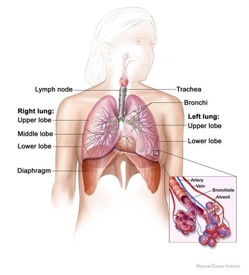
 

The Lungs and The Diaphragm SimpleMed

Diagram - The lungs and the diaphragm

Public Domain Source by National Cancer Institute [Public domain]


The diaphragm is innervated by the left and right phrenic nerves (made up of cervical nerves 3, 4 and 5) which supply the left and right hemidiaphragms respectively. This means if either nerve is damaged, there is paralysis of the affected side of the diaphragm. This can be seen on a chest x-ray, as the affected side will appear as a dome despite the x ray being taken when the patient is breathing in.

The diaphragm has three openings for three important structures. These are the inferior vena cava at the level of thoracic vertebrae 8 (T8), the oesophagus at the level of T10 and the aortic hiatus, which allows the passage of the aorta through the diaphragm, at the level of T12. This can be remembered by the vena cava having 8 letters (T8), the oesophagus having 10 letters (T10), and the aortic hiatus having 12 letters (T12)


The Movements of the Thoracic Wall and Diaphragm During Respiration

During the process of inspiration, the ribs swing superiorly and laterally, which increases the lateral diameter of the thoracic space. This movement is known as the 'bucket handle' movement, because this movement is similar to the handle of the bucket.

The sternum moves anteriorly during inspiration to increase the anterior-posterior diameter as the diaphragm descends to increase the thoracic capacity. This is called the 'pump handle' movement, because this is a similar movement to the handle of an old-fashioned water pump.


The Pleura and the Pleural Cavity

The pleura is the serous membrane that lines the lungs and the thoracic cavity. The pleura is composed of two membranes - the parietal and the visceral membranes. The parietal pleura lines the inside of each hemi-thorax, meaning it is stuck to the inside of the chest wall. The visceral pleura lines the outside of the lungs and extends into the interlobar fissures, meaning that it is stuck to the outside of the lungs. The function of the pleura is to ensure the lungs expand and inflate as the thorax expands during inspiration.

Between the parietal and visceral pleura is a small cavity, known as the pleural cavity, that contains a small volume of fluid, the pleural fluid. This fluid allows the pleura to slide effortlessly over each other during the movements of respiration. The pleural fluid also acts to keep the lung inflated against the chest wall, because of negative pressure within this cavity.

As the lungs do not completely fill the thoracic space, there are potential spaces formed between the pleura covering the diaphragm and the costal surface, these are known as the costo-diaphragmatic recesses. This is clinically important as pleural effusions can develop here through gravity, and can be seen on a chest X-ray.
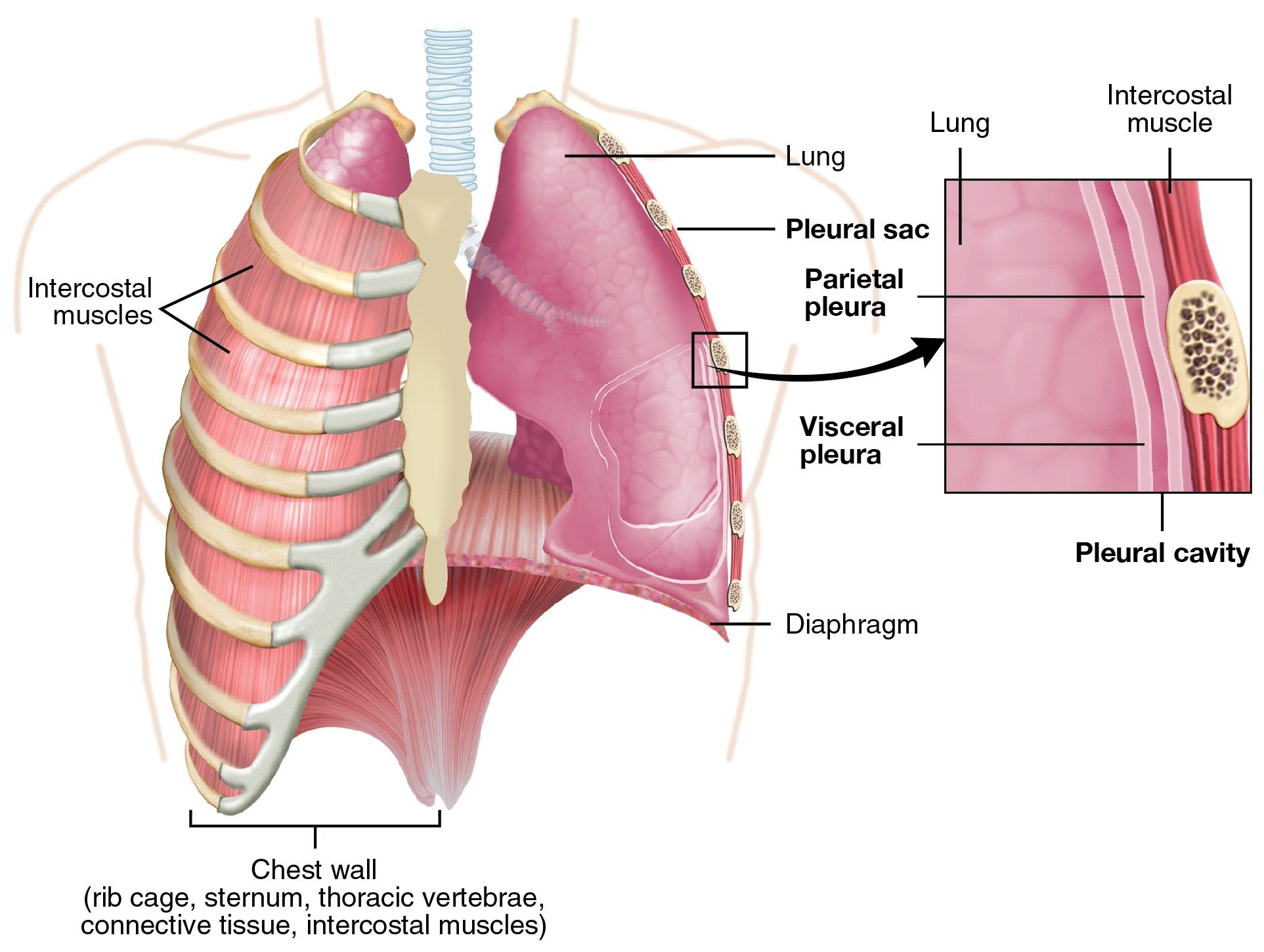
 

The Pleura SimpleMed

Diagram - The pleura and the pleural cavity

Creative commons source by OpenStax College [CC BY-SA 4.0 (https://creativecommons.org/licenses/by-sa/4.0)]


The Trachea and the Bronchial Tree

The trachea commences at the lower border of the cricoid cartilage and then terminates by dividing into the right and left main bronchi around the level of T4/5 opposite the sternal angle. The angle between the right and left bronchi is called the carina.

The right main bronchus is shorter, wider and more vertical in comparison to the left main bronchus, meaning inhaled objects are more likely to lodge in the right main bronchus.
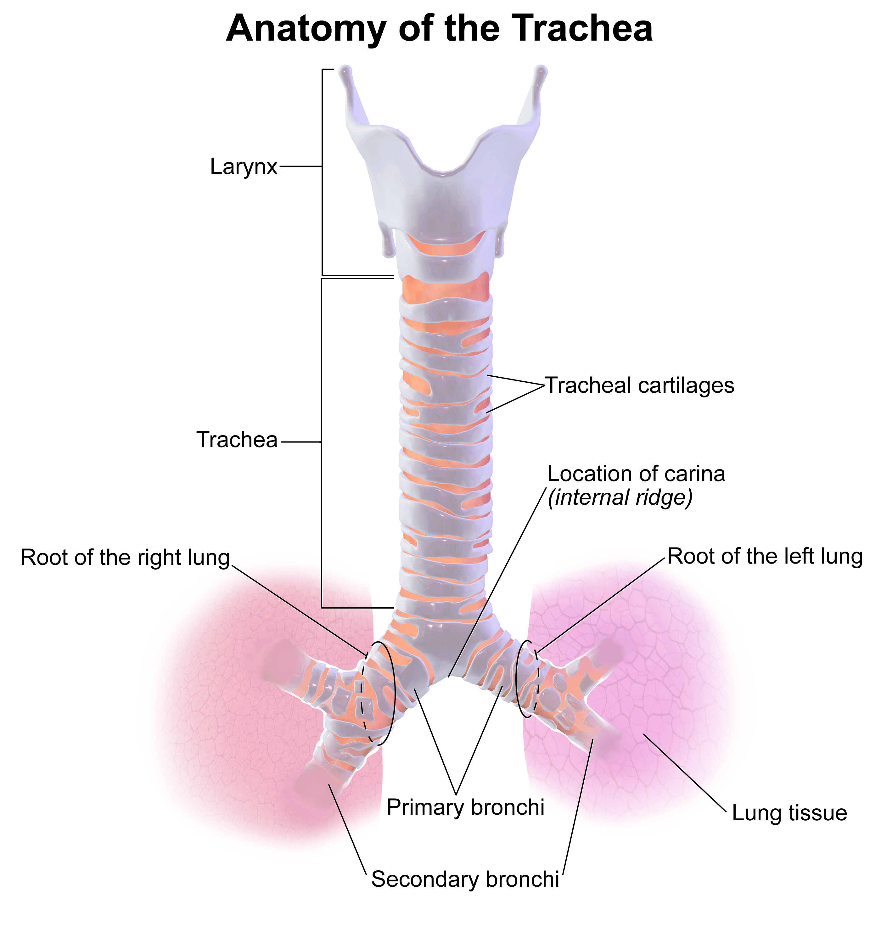
 

The Anatomy of the Trachea SimpleMed

Diagram - The anatomy of the trachea

Creative commons source by Blausen.com staff (2014). "Medical gallery of Blausen Medical 2014". WikiJournal of Medicine 1 (2). DOI:10.15347/wjm/2014.010. ISSN 2002-4436
[CC BY-SA 4.0 (https://creativecommons.org/licenses/by-sa/4.0)]


The right and left bronchi can be known as the primary bronchi and these divide into the lobar or secondary bronchi. There are three right lobar bronchi and two left lobar bronchi, relating to the number of lobes in each lung. Each lobar bronchus divides into segmental or tertiary bronchi, which each supply a bronchopulmonary segment.

A bronchopulmonary segment is an area of lung that is supplied by the segmental branch of the pulmonary artery, drained by a branch of the pulmonary vein and has a segmental bronchi. It is possible if needed to remove one bronchopulmonary segment with very little bleeding & gas leakage, because each segment is relatively separated from the next.


The Lungs

The left lung has two lobes (upper and lower) whereas the right lung has three lobes (upper, middle and lower).
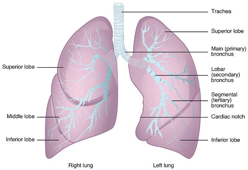
 

The Lobes and Anatomy of the Lungs SimpleMed

Diagram - The lobes and bronchi of the lungs

Creative commons source by OpenStax College [CC BY-SA 4.0 (https://creativecommons.org/licenses/by-sa/4.0)]


The apex of each lung ascends to the root of the neck, above the first rib. This area is close to many vascular and neurological structures (like the brachial plexus and subclavian arteries), meaning that a patient with a tumour in this area can present with vascular or neurological symptoms.

For example, a patient with a Pancoast tumour of the apex of the lung can present with symptoms of Horner’s syndrome (ptosis, miosis and anhydrosis of the ipsilateral side of the face including the ipsilateral eye) because the tumour impinges on the sympathetic nerves present in this area.

The base of each lung rest on a hemi-diaphragm.

The hilum of each lung is found on the mediastinal surface (closest to the heart) of each lung and contains the following structures: main bronchi, pulmonary artery and vein, lymphatics, bronchial arteries and veins.

The structures next to the mediastinal surface of the left lung include the heart, aortic arch, descending aorta, oesophagus and nerves including the phrenic, vagus and recurrent laryngeal nerves.

Structures next to the mediastinal surface of the right lung include the superior vena cava, azygos vein, right atrium, oesophagus and nerves such as the phrenic nerve, vagus nerve and the sympathetic trunk.


The Neurovascular Supply of the Lungs

The lungs have a dual blood supply via the bronchial and pulmonary arteries.

The bronchial arteries supply the bronchial tree (airways), not the alveoli, and the visceral pleura with oxygenated blood. The majority of the blood returns to the heart via the pulmonary veins with the minor portion returning to the heart via the superior vena cava. This is the blood that comes to the lungs oxygenated, and leaves deoxygenated.

The pulmonary arteries carry the entire cardiac output of the right ventricle to the lungs for the process of gaseous exchange and supply the alveoli at the same time. This is the blood that comes to the lungs deoxygenated, and leaves oxygenated.

The pulmonary veins return the oxygenated blood to the left side of the heart, which is then pumped around the body to supply oxygen to the tissues.
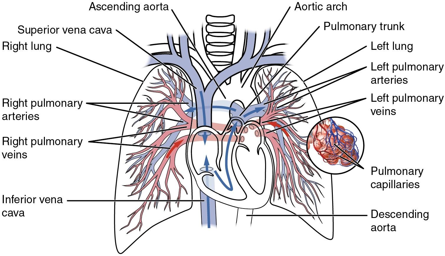
 

The Pulmonary Circulation SimpleMed

Diagram - The pulmonary circulation, showing the pulmonary arteries and veins

Creative commons source by OpenStax College [CC BY-SA 4.0 (https://creativecommons.org/licenses/by-sa/4.0)]


The lymphatics of the visceral pleura and the lungs drain into the hilar nodes (bronchopulmonary nodes) and from here, they drain into the tracheobronchial nodes.

The nerve supply to the lungs includes the right and left vagus nerves, and the sympathetic trunk.

  • The vagus nerve contains some parasympathetic efferent fibres which supply motor innervation to bronchial smooth muscle. Activation of these fibres leads to bronchoconstriction and secretomotor to the mucous glands.
  • The afferent fibres of the vagus nerve are sensory and are responsible for the coughing reflex.
  • The sympathetic efferent fibres cause bronchodilation and vasoconstriction when they are activated.


Visualising the Lobes of the Lungs and The Pleural Cavity

The upper and lower lobes of the left lung are separated by the oblique fissure. The right upper and middle lobes are separated by the horizontal fissure, whilst the oblique fissure separates the middle and lower lobes of the right lung.

The oblique fissure on both sides extends from the spinous process of T3 posteriorly to the sixth costal cartilage anteriorly.

The horizontal fissure on the right side extends from the mid-axillary line anteriorly along the fourth rib to the anterior edge of the lung and this fissure is only present on the right lung.


Edited by: Dr. Maddie Swannack and Dr. Thomas Burnell

Reviewed by: Dr. Marcus Judge

Quiz

Quiz

Preview the Anatomy of the Respiratory System quiz

In this article

The nasal cavity is divided into the left and the right, providing an entrance for air.

Free account

Keep reading for free

Create a free account to continue this article and save your revision progress.

Your account includes:

  • Thousands of free practice questions
  • Tools to track your revision progress
Sign up free Already have an account? Log in

No payment. No spam. Just free medical revision.

SimpleMed+

Want More?

Access more on SimpleMed+

Lock it in with practice questions, the full question bank and revision tools that track what you know.

Practice now See what's in SimpleMed+
12509
simplemedlonglogo.png
  • Home
  • Terms & Conditions
  • Privacy Policy
  • Contact

The medical information on this site is provided as an information resource only, and is not to be used or relied on for any diagnostic or treatment purposes. This information is intended for medical education, specifically to biological/medical learning inclined individuals, and does not create any doctor-patient relationship, and should not be used as a substitute for professional diagnosis and treatment.

©2026 Lili's Ark Limited, UK Company #15424953
S+

Search

  • Pre-Clinical
    • Cardiovascular System
      • 1. Anatomy of the Cardiovascular System
      • 2. The Cardiac Cycle
      • 3. Pulses and Pressures
      • 4. Control of Cardiac Output
      • 5. Cardiac Conduction and Contraction
      • 6. Embryology of the CVS
      • 7. Congenital Heart Defects
      • 8. Hypertension
      • 9. Electrocardiograms
      • 10. Electrocardiogram Pathology
      • 11. Cardiovascular Drugs
      • 12. Murmurs
      • 13. Special Circulations
      • 14. Heart Failure
      • 15. Artery and Vein Disease
      • 16. Acute Coronary Syndromes
      • 17. Haemodynamic Shock
    • Musculoskeletal System
      • The Human Body
        • 1. Anatomical Terms
        • 2. Structure of the Muscles
        • 3. Muscles in Action
        • 4. Connective Tissue
        • Cartilage: Structure, Growth and Repair
        • 5. Structure of Bones
        • 6. Basics of Bone Anatomy
        • 7. Joints
        • 8. The Spine
        • 9. Dermatomes and Myotomes
        • 10. The Brachial Plexus
        • 11. The Lumbar Plexus
        • 12. The Sacral Plexus
      • Clinical Conditions
        • 1. Conditions of the Foot
        • 2. Conditions of the Ankle
        • 3. Conditions of the Lower Leg
        • 4. Conditions of the Knee
        • 5. Conditions of the Thigh
        • 6. Conditions of the Hip
        • 7. Conditions of the Lower Back
        • 8. Conditions of the Back and Neck
        • 9. Conditions of the Shoulder and Clavicle
        • 10. Conditions of the Arm
        • 11. Conditions of the Elbow
        • 12. Conditions of the Forearm and Wrist
        • 13. Conditions of the Hand
      • Musculoskeletal Anatomy
        • 1. Muscles of the Gluteal Region
        • 2. Muscles of the Thigh
        • 3. Key Areas of the Lower Limb
        • 4. Bones of the Lower Limb
        • 5. Bones of the Foot
        • 6. Muscles of the Lower Leg
        • 7. Bones of the Upper Limb
        • 8. Muscles of the Shoulder
        • 9. Muscles of the Upper Arm
        • 10. Bones of the Hand
        • 11. Muscles of the Forearm
        • 12. Key Areas of the Upper Limb
    • Cell Physiology & Biology
      • 1. An Introduction to the Cell
      • 2. Introduction to DNA
      • 3. The Cell Cycle, Checkpoints and Apoptosis
      • 3. Structure of Proteins
      • 4. Protein Function in Oxygen Transport
      • 5. Enzymes and Regulation of Protein Function
      • 6. Molecular Techniques for Medics
      • 7. Post-translational Modification and Collagen Biosynthesis
      • 8. Protein Targeting
      • 9. Biological Membranes
      • 10. G-Protein Coupled Receptor
      • 12. Enzyme-linked and Intracellular Receptor Signalling
      • 11. Membrane Transport and Intracellular Calcium Regulation
      • 12. Membrane Potentials
      • 13. Action Potentials
      • 14. Autonomic Nervous System Introduction
    • Metabolism
      • 1. Nutrition, Diet and Body Weight
      • 2. Energy Reactions in Cells
      • 3. Dietary Energy Production
      • 4. Vitamins, Cofactors, Minerals and Trace Elements
      • 4. Glycolysis
      • 5. Oxygen-dependent Pathway
      • 6. Lipid Metabolism and Transport
      • 7. Protein and amino acid metabolism
      • 9. Purine and Pyrimidine Metabolism
      • 10. Inborn Errors of Metabolism and Newborn Screening
      • 8. Alcohol Metabolism
      • 9. Oxidative Stress
      • 10. Energy Storage
      • 11. Temperature Regulation
      • 12. Metabolic and Endocrine Regulation
    • Endocrinology
      • 1. An Introduction to Endocrinology
      • 2. Control of Appetite
      • 3. The Adrenal Glands and Adrenal Disorders
      • 4. HPA and Growth Hormone
      • 5. Pituitary Disorders
      • 6. The Thyroid Gland
      • 7. The Endocrine Pancreas
      • 8. Diabetes Mellitus
      • 9. Calcium Metabolism
    • Haematology
      • 1. Haematopoiesis and the Spleen
      • 2. Full Blood Count
      • 3. Blood Groups, Crossmatching and Transfusion Reactions
      • 3. Anaemia
      • 4. Vitamin B12 and Folate
      • 5. Iron Metabolism
      • 6. Haemoglobinopathies
      • 7. When Haematopoiesis goes wrong
      • 8. Haematology in Systemic Disease
    • Genetics
      • 1. Mitosis and Meiosis
      • 2. Genotype, Phenotype and Inheritance
      • 3. Gene Expression
      • 4. Epigenetics, Genomic Imprinting and Mitochondrial Inheritance
      • 4. Introduction to Gene Mutation
      • 5. DNA Repair and Cancer
      • 7. Genetic Testing and Counselling
    • Infection
      • 1. An Introduction To Infection
      • 2. The Infection Model
      • 3. Bacterial Structure, Classification and Growth
      • 3. Biofilms
      • 4. Innate Immunity
      • 5. Adaptive Immunity
      • 6. Gram Staining and Infection Investigations
      • 8. Staphylococci and Other Gram-positive Bacteria
      • 9. Streptococci
      • 10. Gram-negative Bacteria: Structure, Entry Routes and Endotoxin
      • 11. The Organisms Gram Staining Misses
      • 7. Sepsis
      • 8. Antibiotics
      • 14. Principles of Medical Virology
      • 10. Hepatitis
      • 11. Human Immunodeficiency Virus
      • 17. Fungi and Parasites: The Eukaryotic Pathogens
      • 12. The Immunocompromised Host
      • 13. Travel-related Infections
    • Pathology
      • 1. An Introduction to Pathology
      • 2. How to Read a Histology Slide
      • 2. The Basics of Pathology
      • 3. Epithelium, Skin and Exocrine Glands
      • 4. Acute Inflammation
      • 5. Chronic Inflammation
      • 6. Regeneration and Repair of Tissues
      • 7. The Lymphatic System
      • 8. Haemostasis and Clotting
      • 9. Thrombosis and Embolism
      • 10. Atherosclerosis
      • 11. Cellular Adaptations
      • 12. Introduction to Neoplasia
      • 13. Pathogenesis of Neoplasms
      • 14. Behaviour of Neoplasms
    • Population & Social Science
      • 1. Introduction to Population Science
      • 2. Describing Data and Testing Hypotheses
      • 2. Epidemiological Studies
      • 3. Models of Health
      • 4. Beliefs of the Public
      • 5. Health Economics
      • 6. Evidence-Based Practice
      • 7. Patient Safety and Quality Control
      • 8. Summary and Glossary
    • Patient-Centred Care
      • 1. Lifestyle Medicine
      • 2. Childhood Immunisations
      • 3. Sepsis in Paediatrics
      • 4. Child Safeguarding
      • 5. Ageing
      • 6. Falls
      • 7. Men's Health
      • 8. Palliative Care
    • Reproductive System
      • 1. Anatomy of the Man
      • 2. Anatomy of the Woman
      • 3. The Pelvic Floor
      • 4. Development of the Reproductive System
      • 5. Puberty
      • 6. The Menstrual Cycle and Menstrual Abnormalities
      • 7. Gametogenesis
      • 8. Sex, Infertility and Contraception
      • 9. Physiology of the Mother
      • 10. Diseases of the Mother in Pregnancy
      • 11. Physiology of the Fetus
      • 12. The Placenta
      • 13. Labour and Delivery
      • 14. The Postpartum Period
      • 15. Infections of the Reproductive Tract
      • 16. Pelvic Inflammatory Disease
      • 17. The Menopause
      • 18. Malignancies of the Reproductive Tract
      • 19. Introduction to the Breast
      • 20. Benign Conditions of the Breast
      • 21. Malignant Breast Disease
    • Respiratory System
      • 1. Anatomy of the Respiratory System
      • 2. Development of the Respiratory System
      • 3. Ventilation and Lung Mechanics
      • 4. Properties of Gas Exchange
      • 5. Carbon Dioxide Transport
      • 6. Chemical Control of Breathing and Plasma pH
      • 7. Lung Function Testing
      • 8. Pulmonary Embolism
      • 9. Asthma
      • 10. Chronic Obstructive Pulmonary Disease
      • 11. Pneumonia and other Lower Respiratory Tract Infections
      • 12. Bronchiectasis and Cystic Fibrosis
      • 13. Tuberculosis
      • 14. Pneumothorax and Pleural Effusion
      • 15. Lung Cancer
      • 16. Respiratory Failure
    • Urinary System
      • 1. Anatomy of the Kidneys, Bladder and Urethra
      • 2. Development of the Urinary System
      • 3. The Nephron
      • 4. The Glomerulus – Tubular Reabsorption and Secretion
      • 5. Control of Plasma Volume
      • 6. Control of Plasma Osmolarity
      • 7. Control of Potassium
      • 8. Micturition
      • 9. Diuretics
      • 10. Glomerular Filtration Rate and the Measurement of Kidney Function
      • 11. Hyponatraemia
      • 12. Urinary Incontinence
      • 13. Glomerular Disease
      • 14. Diabetic Nephropathy
      • 15. Acute Kidney Injury
      • 16. Chronic Kidney Disease
      • 17. Urinary Tract Obstruction
      • 18. Urinary Tract Infections
      • 19. Prostate Pathology
      • 20. Male Urological Conditions
      • 21. Urological Malignancy
    • Gastrointestinal System
      • 1. Development of the Gastrointestinal Tract
      • 2. Abdominal Wall and Inguinal Canal
      • 2. Anatomy and Physiology of the Foregut
      • 3. Anatomy and Physiology of the Midgut
      • 4. Anatomy and Physiology of the Hindgut
      • 6. Gastrointestinal Motility and the Enteric Nervous System
      • 7. Liver, Biliary and Exocrine Pancreatic Physiology
      • 8. Gastrointestinal Secretion, Digestion and Absorption
      • 5. Upper Gastrointestinal Pathology
      • 6. Hepatic, Biliary and Pancreatic Pathology
      • 7. Intestinal Pathology
      • 8. Distal Gastrointestinal Tract Pathology
      • 9. Gastrointestinal Malignancy
      • 10. Gastroenteritis and Enterocolitis
      • 11. Gastrointestinal Emergencies
    • Head & Neck
      • 1. Introduction to Head & Neck Anatomy
      • 2. Blood Supply of the Head and Neck
      • 3. Lymph Nodes and Neck Lumps
      • 4. The Skull
      • 5. The Temporomandibular Joint
      • 6. The Meninges
      • 7. Cranial Nerves I-VI
      • 8. Cranial Nerves VII-XII
      • 9. Development of the Head and Neck
      • 10. Autonomics in the Head and Neck
      • 11. Anatomy of the Ear
      • 12. Ear Pathology
      • 13. The Nose
      • 14. The Oral Cavity and Pharynx
      • 15. The Eye
      • 16. The Larynx
      • 17. Head and Neck Cancers
      • 18. Imaging of the Head and Neck
      • 19. Fascial Planes, Muscles and Triangles
    • Immunology
      • 1. An Introduction to Hypersensitivity
      • 2. Type 1 Hypersensitivity
      • 3. Food Allergy
      • 4. Introduction to Autoimmunity
      • 5. Rheumatoid Arthritis
      • 6. Systemic Lupus Erythematosus (SLE)
      • 7. Immunological Therapies | Monoclonal Antibodies
    • Neuroanatomy & Psychiatry
      • 1. Basics of Neuroanatomy
      • 2. Cells and Components of the Nervous System
      • 3. Arterial Supply to the Brain
      • 4. Development of the Nervous System
      • 5. Production and Circulation of CSF and Related Disorders
      • 6. Somatosensory System
      • 7. Motor System
      • 8. The Cerebellum: Circuits, Coordination and Clinical Signs
      • 8. Visual System
      • 10. Auditory and Vestibular Systems
      • 11. The Hypothalamus and Limbic System
      • 9. Higher Cortical Function
      • 10. Reticular Formation
      • 11. Movement Disorders
      • 12. Infections of the Central Nervous System
      • 13. Dementia
      • 14. Delirium
      • 15. Headaches
      • 16. Stroke
      • 17. Raised Intracranial Pressure (ICP)
      • 18. Subarachnoid Haemorrhage
      • 19. Anxiety
      • 20. Mood Disorders
      • 21. Psychosis
    • Embryology
      • 1. The Basics of Embryology
      • 2. Development of the Limbs and Body Wall
      • 3. Teratology and Congenital Malformations
      • 2. Development of the Gastrointestinal Tract
      • 3. Development of the Reproductive Systems
      • 5. Development of the Respiratory System
      • 6. Development of the Head and Neck
      • 8. Embryology of the Cardiovascular System
    • Pharmacology
      • 1. Research Methods in Pharmacology
      • 2. Pharmacokinetics
      • 3. Pharmacodynamics
      • 4. Pharmacovigilance and Pharmacogenetics
      • 5. Safe Prescribing
      • 6. Cholinergic Pharmacology
      • 7. Adrenergic Pharmacology
      • 6. Hypertension and Heart Failure
      • 7. Cardiac Arrhythmias
      • 8. Antiplatelets, Anticoagulants and Thrombolysis
      • 9. Hyperlipidaemias
      • 10. Diuretics and Renal Pharmacology
      • 11. Pharmacology of Insulin
      • 12. Reproductive and Post-reproductive Health
      • 13. Antibiotics
      • 14. Antivirals
      • 15. Cancer Chemotherapy
      • 16. Anaesthetics
      • 17. Opioids
      • 20. Introduction to Psychopharmacology
      • 18. Epilepsy and Anti-epileptic Drugs
      • 19. Parkinson's Disease and Myasthenia Gravis
      • 20. NSAIDs
      • 21. Immunosuppression and Disease-modifying Therapy
      • 22. Pharmacology of Airway Control
      • 23. Drugs Affecting Acid Secretion
      • 24. Antiemetics and Laxatives
      • 25. Poisoning and STOPP/START
  • OSCE
    • Index Conditions for OSCE exams
    • Cardiovascular System Examination
    • Shoulder Examination
    • Hand Examination
    • Venepuncture
    • Blood Pressure
    • Respiratory System OSCE Examination
    • Peripheral Vascular OSCE Examination
    • Varicose Vein OSCE Examination
    • Deep Vein Thrombosis (DVT) OSCE Examination
    • Abdominal OSCE Examination
    • Rectal (PR) OSCE Examination
    • Stoma OSCE Examination
    • Hernia OSCE Examination
    • Upper Limb Neurological OSCE Examination
    • Lower Limb Neurological OSCE Examination
    • Cranial Nerve OSCE Examination
    • Cerebellar OSCE Examination
    • GALS Screening OSCE Examination
    • Elbow OSCE Examination
    • Hip OSCE Examination
    • Knee OSCE Examination
    • Ankle and Foot OSCE Examination
    • Spine OSCE Examination
    • Thyroid Status OSCE Examination
    • Diabetic Foot OSCE Examination
    • Cushing's Syndrome OSCE Examination
    • Acromegaly OSCE Examination
    • Ear, Otoscopy and Hearing OSCE Examination
    • Nasal OSCE Examination
    • Eye and Vision OSCE Examination
    • Fundoscopy (Ophthalmoscopy) OSCE Examination
    • Visual Acuity OSCE Assessment
    • Anterior Segment Eye OSCE Examination
    • Pigmented Skin Lesion OSCE Examination
    • Rash and Non-Pigmented Skin Lesion OSCE Examination
    • Breast OSCE Examination
    • Testicular OSCE Examination
    • Neck Lump OSCE Examination
    • Lymphoreticular OSCE Examination
  • Question Bank
  • About
  • How to Use SimpleMed
  • SimpleMed Lecture Series
  • DON'T PANIC - Guide to the Wards
  • SimpleMed Plus
Your privacy Allow anonymous analytics to help us improve SimpleMed. Privacy policy
