

Next Lesson - Hyponatraemia
Core
There are a number of ways in which kidney function can be measured. One of these methods is through measurement of compounds filtered by the kidneys. The constituents of urine can also be analysed to check for underlying renal pathology. Imaging of the bladder, sphincter, and urethra can also be useful to check for their storage capability.
Kidney function is measured in terms of glomerular filtration rate (GFR). GFR is defined as the volume of plasma that is filtered by the glomeruli per unit time. The normal range of GFR is 90-120 mL/min/1.73m2. For a person with a body surface area of 1.73 m2, this corresponds to 129.6-172.8 L of glomerular filtrate per day (90-120 mL/min multiplied by 1,440 minutes/day). Because the quoted GFR is indexed to body surface area, the absolute volume filtered also depends on body size. GFR can be measured using the clearance of a suitable filtration marker. In routine practice, estimated GFR (eGFR) is usually calculated from serum creatinine using a validated equation; measured marker clearance and creatinine-based eGFR are different methods.
There are many factors which can cause temporary or permanent changes in GFR.
Men typically have a higher GFR than women.
In utero, nephron development is finished by the 35th-36th week of gestation, meaning premature and low birth weight infants have lower numbers of nephrons, meaning the GFR of these infants is lower. At birth, GFR is approximately 20 mL/min/1.73 m2, but normal (adult) GFR is achieved by around 18 months of age.
GFR starts to decline again after the age of 30. The rate of decline after this point is roughly 6-7ml/min per decade. This is due primarily to the loss of functioning nephrons. In order to try and offset this decline, there is some compensatory hypertrophy of the remaining nephrons, but the capacity for hypertrophy also declines with age.
Larger people tend to have larger kidneys than smaller people, and larger kidneys contain more nephrons. Therefore, GFR in larger people is higher.
In pregnancy, GFR increases by around 50% to 130-180ml/min. This is associated with an increase in kidney size of about 1cm due to hypertrophy and increased plasma volume in pregnant women. The total nephron number stays the same. Normal pre-pregnancy levels will be reached after 6 months post-partum.
Due to the above factors, there is huge variability in the GFR between individuals. If the GFR declines it may be due to the decline in the number of nephrons, or the decline in the filtration ability within individual nephrons. When kidney function declines slowly, individual nephrons may hypertrophy so overall GFR may not fall until significant kidney damage has occurred.
GFR can be measured indirectly by the clearance of a marker that is freely filtered and has no tubular reabsorption or secretion. This differs from estimating GFR using a serum-creatinine equation.
Renal clearance can be calculated with the following equation:
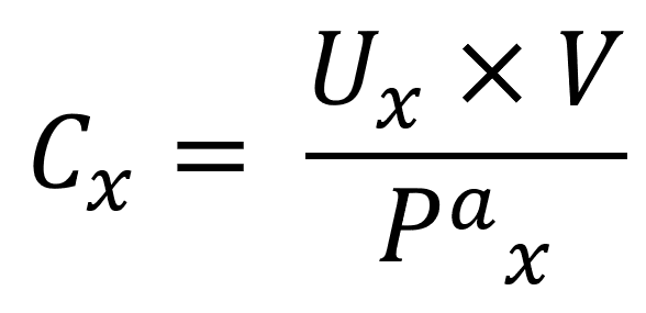
Where C = clearance, U = concentration of the marker in urine, V = urine flow rate, Pa = plasma concentration of the marker, and x = the substance whose clearance is being measured. Urinary and plasma concentrations must use consistent units.
In order to best measure GFR, the substance needs to have these four properties:
- Have an appropriately measured plasma concentration. An endogenous marker should have stable generation; an exogenous marker must be administered using a suitable measurement protocol.
- Be freely filtered across the glomerulus.
- Not be reabsorbed in the nephron.
- Not be secreted into the nephron.
If all four of these conditions are true, then the clearance rate will be equal to GFR.
Inulin is an exogenous polysaccharide that is freely filtered and neither reabsorbed nor secreted. Its urinary clearance provides a reference measurement of GFR. The conventional method uses a continuous intravenous infusion to maintain a steady plasma concentration, with timed blood sampling and urine collections. It is cumbersome and is not routinely used for everyday kidney-function assessment. Inulin should not be confused with insulin.
Creatinine is an endogenous substance produced mainly by creatine metabolism in muscle. It is freely filtered across the glomerulus and is not reabsorbed along the nephron. However, it is secreted into the nephron and the rate of production can change dependent on diet and exercise. For this reason it does not measure true GFR - but it is an estimate. Because of tubular secretion, measured creatinine clearance tends to overestimate GFR; this is different from the direction of error in a serum-creatinine eGFR equation. Creatinine clearance is measured by collecting urine over 24 hours - this is cumbersome and frequently inaccurate but it can be used in pregnancy.
51 Cr-EDTA is a radioactively labelled marker which is cleared exclusively by renal filtration. It has approximately a 10% lower clearance than inulin, meaning it slightly underestimates GFR. This can be used clinically to measure GFR in children or where a better indication of renal function is required e.g. in a kidney transplant or in a work-up to donate a kidney for transplant.
In routine clinical practice, estimated GFR (eGFR) is usually derived from measurements of serum creatinine. This is very easy to measure but can vary greatly between individuals. In order to standardise the eGFR a bit, one of two calculations can be used which take into account other patient factors.
The MDRD calculation takes into account serum creatinine, age, and sex. Historically, ethnicity adjustments were used, but current UK practice recommends race-free eGFR calculations.
This calculation is inaccurate in:
- People without kidney disease - for example transplant donors
- Children
- Pregnancy
- Old age
- Bodybuilders or other patients with unusually high muscle mass
- Amputees or other patients with significantly reduced muscle mass
- Patients with kidney function above 60 mL/min
- When true GFR changes quickly - like in AKI
These limitations do not all produce the same direction of error. During recovery from AKI, serum creatinine may remain elevated while filtration improves, so creatinine-based eGFR can underestimate current GFR. During worsening AKI, creatinine may not yet have risen to its new steady-state level, so eGFR can overestimate current GFR. Low muscle mass can also cause overestimation because creatinine generation is reduced; unusually high muscle mass can cause underestimation. Creatinine-based equations assume a steady state and should not be used to diagnose chronic kidney disease from a single result during AKI.
The CKD-EPI equation uses the same variables as MDRD, however, the calculation is slightly different. This means it is equally as accurate as MDRD when GFR is below 60ml/min but more accurate when GFR is above 60ml/min - however, it is important to note it is still not a perfect calculation.
eGFR measurement is less accurate in patients with mild kidney disease. There are three reasons for this:
- A reduction in overall nephron number leads to compensatory hyperfiltration in the remaining nephrons, which can temporarily preserve measured GFR.
- A reduced number of nephrons leads to nephron hypertrophy, which would increase filtration.
- Reduced filtration of creatinine due to reduced GFR results in increased serum creatinine which is actively excreted into the nephron in order to maintain a relatively constant plasma concentration.
These three factors mean that eGFR remains normal, despite there being some mild kidney damage.
Urinalysis is the term given to performing a dipstick test on a urine sample. Urinalysis allows practitioners to screen for common abnormalities in a patient’s urine using chemical markers.
A urinalysis dipstick will give information on the following:
- Specific gravity (density)
- pH
- Leukocytes (white blood cells)
- Blood/Haemoglobin
- Nitrites
- Ketones
- Bilirubin
- Urobilinogen
- Protein
- Glucose
Common conditions have specific findings on a urinalysis strip which make them easier to diagnose:
- Urinary Tract Infection (UTI) - blood, nitrites, or leukocytes.
- Uncontrolled Diabetes - glucose and ketones.
- Nephrotic Syndrome - protein (with potentially small amounts of blood).
- Jaundice - bilirubin or urobilinogen.
These are just some of the conditions in which urinalysis is very helpful in making a diagnosis.
There are many advantages to performing a urinalysis. It is very cheap, requires little training to be able to do, can be done at the patient’s bedside or in clinic, and all results are back within two minutes. However, a urinalysis only shows a practitioner what is present in the urine and does not give any information as to the filtration function (GFR) on the kidney.
Urodynamics is an umbrella term for a group of more invasive tests which look directly at the function of the bladder, sphincters, and urethra.
Urodynamic tests include:
- Uroflowmetry - the measurement of urine speed and volume.
- Postvoid Residual Measurement - a measurement of the amount of urine left in the bladder after urination.
- Cystometric Testing - a measurement of how much the bladder can hold, how much pressure builds up inside the bladder as it stores urine, and how full the bladder is when the urge to void is felt.
- Leak Point Pressure Measurement - a measurement of the pressure at which leakage occurs in a patient with urinary incontinence during a cystometric test.
- Pressure Flow Studies - a study which looks at the pressure required in the bladder to void and the flow rate that a given pressure generates.
- Electromyography - sensors which are used to measure the electrical stimulation to the nerves and muscles around the bladder and sphincters. This is used when a neuromuscular cause of a urological problem is suspected.
- Video Urodynamic Tests - taking images (using x-ray or ultrasound) of the bladder during filling and voiding.
These tests provide the best look at the inner workings of the bladder, sphincters, and urethra. However, they are associated with significant discomfort and in some cases a risk of developing a UTI. The results of some tests are available instantly, while others like electromyography may take some days to come back.
Edited by: Dr. Maddie Swannack
Reviewed by: Dr. Thomas Burnell
- 7500
