

Next Lesson - Ventilation and Lung Mechanics
Core
The respiratory system can be divided based on anatomical location into the upper and lower systems:
- The upper airway is made up of the nasal cavity, oral cavity, pharynx and larynx.
- The lower airway is made up of the trachea, bronchi, bronchioles and alveoli.
Opinion differs on where the upper airway ends, and the lower airway begins with some sources placing the larynx and trachea in different portions as listed here.
More helpfully for understanding physiology, the airway can also be divided based on function into the conducting portion and the respiratory portion:
- The conducting portion includes the trachea, bronchi and bronchioles up to and including the terminal bronchioles. It warms, moistens and conducts air to the respiratory portion. Respiratory bronchioles belong to the respiratory portion.
- The respiratory portion is made up of the respiratory bronchioles, alveolar ducts and alveoli, and is the site of gas exchange.
The respiratory system begins development from an outpouching of the primitive gut tube known as the respiratory diverticulum. This structure is derived from the endoderm of the primitive gut tube, which is created by folding of the embryo and will go on to develop into the gastrointestinal tract.
The outpouching occurs from the foregut (the cranial end of the gut tube) and outer surface is covered with mesoderm.
As it is an outpouching, the respiratory diverticulum is continuous with the gut tube at the start of the development. To allow the formation of two separate systems, a tracheoesophageal septum forms.
By the end of week 4, the respiratory diverticulum splits into two bronchial buds. These give rise to the branching airways. The airway and alveolar epithelial lining develops from endoderm; the surrounding mesoderm contributes supporting tissues, including connective tissue, smooth muscle and pulmonary vessels.
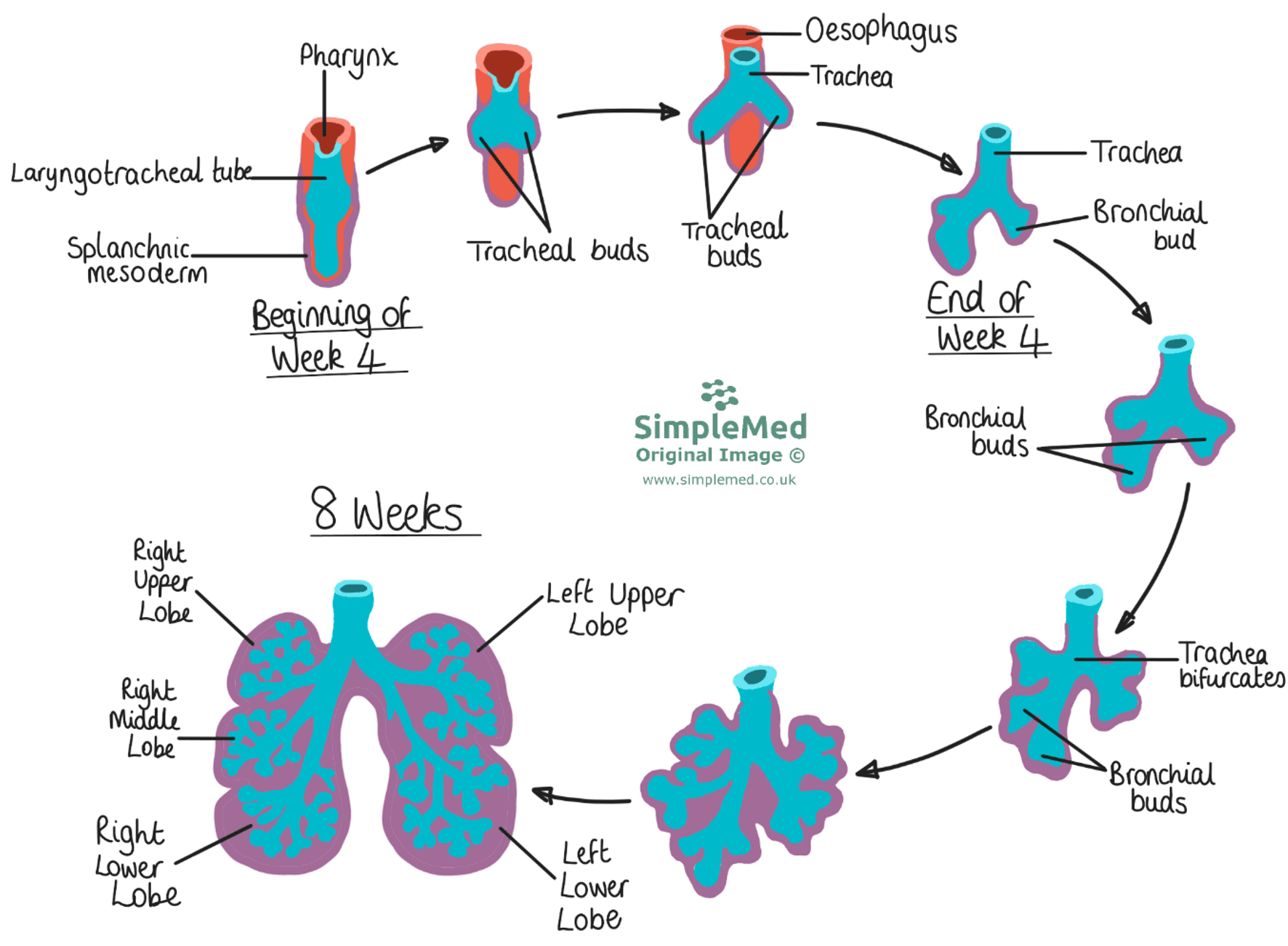
Diagram - The development of the lungs from the beginning of week 4 to week 8 of gestation
SimpleMed original by Dr. Maddie Swannack
A tracheoesophageal fistula is an abnormal connection between the trachea and the oesophagus.
Embryologically, this occurs because the respiratory diverticulum and the foregut are continuous, and if there is a defect in the formation of the tracheoesophageal septum this connection persists.
There are multiple different types, which are shown in the simplified diagram below.
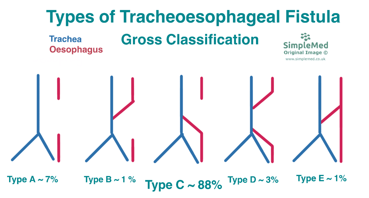
Diagram - The 5 types of tracheoesophageal fistula. Type C is the most common by far
SimpleMed original by Dr. Maddie Swannack
This stage is known as the Pseudoglandular Stage.
The conducting portion of the airways continue to develop, forming bronchioles from the tertiary bronchi formed by the end of week 8.
During the pseudoglandular stage, branching epithelial tubes are surrounded by mesenchyme, but gas-exchanging alveolar structures have not yet formed. The respiratory epithelial lineage is endodermal, with surrounding mesodermal support; the lungs are not yet capable of gas exchange.
This stage is known as the Canalicular Stage.
Throughout this ten week period the respiratory bronchioles develop by budding off the bronchioles formed during the pseudoglandular stage. The respiratory bronchioles are the first structures in the respiratory portion of the airway.
As the alveoli have still not fully developed, there is only a very limited gas exchange surface and survival is uncommon. However, towards the end of this stage some gas exchange may be possible. In the UK, the threshold of viability is generally considered to be around 24 weeks of gestation.
This stage is known as the Terminal Sac Stage.
The alveoli begin to develop around 26 weeks gestation. Within the alveolar walls, there are two main cell types which arise:
- Type I Pneumocytes (type I alveolar cells) are thin squamous epithelial cells covering most of the alveolar epithelial surface area, providing a short diffusion distance for gas exchange.
- Type II Pneumocytes (type II alveolar cells) are cuboidal epithelial cells that produce surfactant. Surfactant production begins before there is necessarily enough to support stable breathing after birth.
Surfactant is made up of a mix of phospholipids and proteins, and plays a key role in reducing surface tension in the lung. Due to its hydrophilic and hydrophobic parts it lines the surface of the fluid lining inside the alveoli reducing the surface tension within the alveoli. This means that the alveoli are able to expand at smaller pressures. See our article here for more information about surfactant.
During the second and third trimesters the fetus also ‘practices’ breathing movements. The fetus will inhale and exhale the amniotic fluid it is surrounded by. This helps to condition the respiratory musculature for life, and helps to encourage the growth and development of the lungs.
Newborn respiratory distress syndrome can occur when the lungs produce insufficient surfactant, particularly after premature birth. Surfactant production may already have begun; onset of production and a sufficient functional supply are different developmental milestones.
Without the surfactant, the alveoli need higher pressures to expand which the baby cannot easily generate, resulting in difficulty breathing and signs of respiratory distress. If premature birth is unavoidable, the mother may be given glucocorticoids (steroids), typically when preterm birth is threatened, which stimulate the production of surfactant in the fetus and improve breathing after birth.
Edited by: Dr. Ben Appleby
Reviewed by: Dr. Thomas Burnell
- 10468
