

SimpleMed - Learning Medicine, SimplifiedSimpleMed - Learning Medicine, Simplified
Hit enter to search or ESC to close

Get in Touch

Contact Us

Use our contact form.

Introduction to the Breast

Share:

By Dr. Tom Bradley

Next Lesson - Benign Conditions of the Breast

Reproductive System


Contents

Contents

  1. The Breast
  2. Physiological Changes
  3. Clinical Presentations
    • Pain
    • Palpable Masses
    • Nipple Discharge
    • Skin Changes
  4. Breast Imaging: Mammography and Mammogram abnormalities
  5. Quiz
Abstract

Abstract

  • The breasts are reproductive organs in women with the function of lactation to feed newborn children.
  • Men also have breasts, but they have no function and are much less susceptible to disease.
  • Issues with the breast can present in a number of different ways, from skin changes to pain and palpable masses.
  • Imaging of the breast is done via an ultrasound scan in younger women and either a mammogram or ultrasound scan in older women.
Core

Core

The Breast

In this article the main focus will be on the female breasts as disease is much more prevalent in women. However, it is important to note that men can still suffer breast disease, but because their breast structure is simpler, it is less likely.

The breasts are a pair of organs on the anterior side of the thoracic region that lie superficial to the pectoral fascia covering the pectoralis major muscle. They are comprised of a deep network of mammary glands surrounded by adipose and suspensory retinacula tissue (also known as interlobular stroma). They are generally circular in nature with a central nipple surrounded by the areola (a small section of pigmented skin surrounding the nipple). The areolae contain sebaceous glands which secrete lipoid fluid to prevent cracking of the nipple, and act as a target for the baby to aim for during breastfeeding.

The suspensory retinaculum is a network of fibrous connective tissue (ligaments) covering the breast to help support it and prevent ptosis (drooping) by anchoring the breast to the chest wall. They also function to keep the secretory lobules separate. They are sometimes known as Cooper’s ligaments.

The mass of adipose tissue in the breast makes up most of its contents and supports the mammary glands.

Breasts contain mammary glands which are responsible for lactation. Mammary glands are formed by multiple lobules that secrete milk into the lactiferous ducts, which is then stored in the lactiferous sinuses before the milk is ejected through pores in the nipple during breast feeding.

There is a small potential space called the retromammary space between the posterior surface of the breast and the pectoral fascia which can be utilised during surgery.
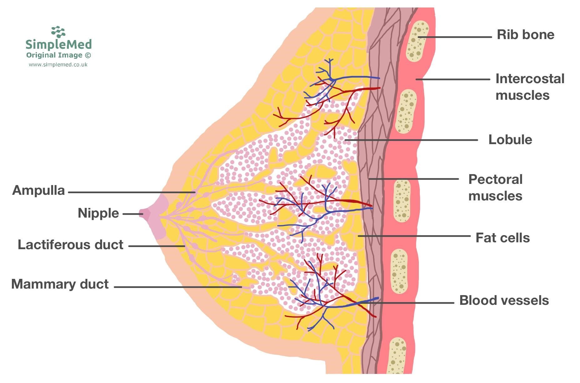
 

Structure of the Breast SimpleMed

Diagram - The anatomical structure of the breast

SimpleMed original by Dr. Maddie Swannack


The breast is supplied by a variety of blood vessels:

  • The mammary branches of the internal thoracic artery (from the subclavian artery) supply the medial side of the breast.
  • The lateral thoracic and thoracoacromial artery (from the axillary artery), and intercostal arteries supply the lateral side of the breast.

The venous supply to the breast is equivalent to the arterial supply.

Lymphatic drainage of the breast is to the axillary (majority), parasternal and the posterior intercostal nodes. These play an important role in the pathophysiology of breast cancer as the axillary nodes are a common site for initial metastasis.

The anterior and lateral cutaneous branches of the 4th to 6th intercostal nerves innervate both sensory and autonomic function in the breast.


Physiological Changes

Before puberty the breast tissue contains very few lobules, and so are identical in both males and females. At the beginning of puberty, often before the menstrual period begins, there is an increase in both the number of lobules and the volume of the interlobular stroma (adipose and suspensory tissue), and this is known as thelarche. Swelling and tenderness can occur in this time.

During pregnancy there is anincrease in both size and number of lobules, and a decrease in the volume of stroma. This occurs to prepare the breast for lactation and these are known as secretory changes.

After the period of breastfeeding atrophy of the lobules occurs, reducing both size and number of lobules, and so reduces the amount of milk produced in the breast.

In young women the breasts mainly consist of dense fibrous suspensory tissue with some adipose tissue. During aging the suspensory tissue is replaced by adipose tissue, a key point in investigating breast disease. When investigating younger women, ultrasound scans are used as mammograms are not as effective due to the high density of the tissue. In older women, the breasts are less dense, meaning that mammography is a viable option.


Clinical Presentations

Pain

Pain in the breast is known as mastalgia. The majority of breast pain is known as cyclical mastalgia and is related to hormonal changes in the menstrual cycle. If the pain is non-cyclical and located to one specific area, it may indicate injury, ruptured cysts or inflammation. Whilst rare, mastalgia may be the initial presentation of breast cancer, meaning that non-cyclical mastalgia with other symptoms of cancer should be referred.


Masses

Palpable masses are very common in breasts. Many will be at least 2cm in size before they are able to be felt, depending on both the size of the breast and how superficial the mass is. Differentials for masses include the normal nodularity of breasts, cysts, fibroadenomas, lipomas and invasive carcinomas. Red flag features for invasive carcinomas are lumps that are hard, craggy and fixed. All breast masses should be investigated and referred to a specialist, as it is impossible to know for sure the origin of the mass without imaging.


Nipple Discharge

Nipple discharge can occur with a range of different conditions and even stimulation of the normal breast. The discharge can be milky (galactorrhoea) due to a high level of prolactin, a result of endocrine disorders such as a pituitary adenoma, or it could be due to high levels of oestrogen, for example as a side effect of medications like the oral contraceptive pill. Bloody or serous discharge can be a sign of benign lesions such as an intraductal papilloma, or malignant lesions, especially if it occurs spontaneously and unilaterally. This should always be investigated.


Skin Changes

Skin changes can sometimes be seen in the breast if it becomes infected (e.g. red and swollen), or if the breast becomes oedematous. Poor lymphatic drainage to the axilla from either metastasis or lymph node clearances can lead to an appearance known as peau d’orange. This is when the cooper’s suspensory ligaments restrict the swelling and pull the skin back at their attachments, causing lots of little dimples on the surface of the breast like an orange skin.

 

Oranges SimpleMed

Image - The skin of oranges showing the dimpling on the surface of oranges

Public Domain Source [Public domain]


Breast Imaging

As already mentioned, ultrasound scans and mammograms are the most frequently used imaging strategies for breast tissue, with ultrasound being used more commonly in younger women with denser breast tissue.

Mammograms are radiological images taken of breasts in both a craniocaudal and mediolateral oblique view, allowing the differentiation of any pathology from the normal breast tissue more accurately.

In the UK, women between the ages of 50-71 are invited for mammography every three years to try and detect small, invasive tumours before they are palpable. As with all screening and imaging, there is a risk of over investigating benign breast lumps, which puts patients through unnecessary biopsy and operations.
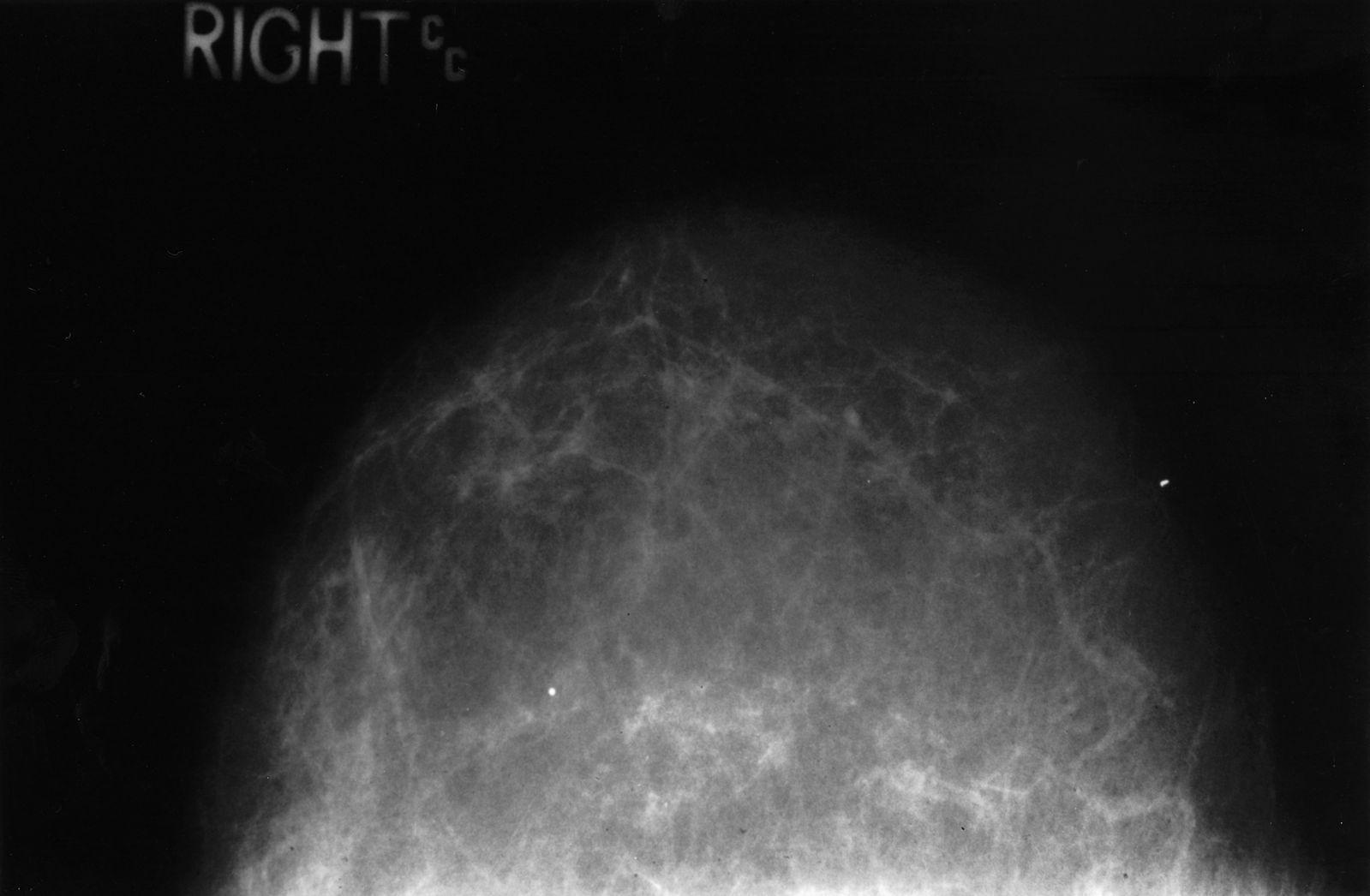
 

Mammogram of Normal Fatty Breast SimpleMed

Image - Mammogram of normal fatty breast tissue

Public Domain Source [Public domain]


Mammographic abnormalities can appear as densities or calcifications in the breast tissue. Densities are suggestive of invasive carcinomas, fibroadenomas and cysts. Calcifications are suggestive of either benign changes like fat necrosis, or malignant ones such as ductal carcinoma in situ. This is covered more in our breast cancer article.


Edited by: Dr. Maddie Swannack

Reviewed by: Dr. Thomas Burnell

Quiz

Quiz

Preview the Introduction to the Breast quiz

In this article

The breasts are reproductive organs in women with the function of lactation to feed newborn children.

Free account

Keep reading for free

Create a free account to continue this article and save your revision progress.

Your account includes:

  • Thousands of free practice questions
  • Tools to track your revision progress
Sign up free Already have an account? Log in

No payment. No spam. Just free medical revision.

SimpleMed+

Want More?

Access more on SimpleMed+

Lock it in with practice questions, the full question bank and revision tools that track what you know.

Practice now See what's in SimpleMed+
7016
simplemedlonglogo.png
  • Home
  • Terms & Conditions
  • Privacy Policy
  • Contact

The medical information on this site is provided as an information resource only, and is not to be used or relied on for any diagnostic or treatment purposes. This information is intended for medical education, specifically to biological/medical learning inclined individuals, and does not create any doctor-patient relationship, and should not be used as a substitute for professional diagnosis and treatment.

©2026 Lili's Ark Limited, UK Company #15424953
S+

Search

  • Pre-Clinical
    • Cardiovascular System
      • 1. Anatomy of the Cardiovascular System
      • 2. The Cardiac Cycle
      • 3. Pulses and Pressures
      • 4. Control of Cardiac Output
      • 5. Cardiac Conduction and Contraction
      • 6. Embryology of the CVS
      • 7. Congenital Heart Defects
      • 8. Hypertension
      • 9. Electrocardiograms
      • 10. Electrocardiogram Pathology
      • 11. Cardiovascular Drugs
      • 12. Murmurs
      • 13. Special Circulations
      • 14. Heart Failure
      • 15. Artery and Vein Disease
      • 16. Acute Coronary Syndromes
      • 17. Haemodynamic Shock
    • Musculoskeletal System
      • The Human Body
        • 1. Anatomical Terms
        • 2. Structure of the Muscles
        • 3. Muscles in Action
        • 4. Connective Tissue
        • 5. Structure of Bones
        • 6. Basics of Bone Anatomy
        • 7. Joints
        • 8. The Spine
        • 9. Dermatomes and Myotomes
        • 10. The Brachial Plexus
        • 11. The Lumbar Plexus
        • 12. The Sacral Plexus
      • Clinical Conditions
        • 1. Conditions of the Foot
        • 2. Conditions of the Ankle
        • 3. Conditions of the Lower Leg
        • 4. Conditions of the Knee
        • 5. Conditions of the Thigh
        • 6. Conditions of the Hip
        • 7. Conditions of the Lower Back
        • 8. Conditions of the Back and Neck
        • 9. Conditions of the Shoulder and Clavicle
        • 10. Conditions of the Arm
        • 11. Conditions of the Elbow
        • 12. Conditions of the Forearm and Wrist
        • 13. Conditions of the Hand
      • Musculoskeletal Anatomy
        • 1. Muscles of the Gluteal Region
        • 2. Muscles of the Thigh
        • 3. Key Areas of the Lower Limb
        • 4. Bones of the Lower Limb
        • 5. Bones of the Foot
        • 6. Muscles of the Lower Leg
        • 7. Bones of the Upper Limb
        • 8. Muscles of the Shoulder
        • 9. Muscles of the Upper Arm
        • 10. Bones of the Hand
        • 11. Muscles of the Forearm
        • 12. Key Areas of the Upper Limb
    • Cell Physiology & Biology
      • 1. An Introduction to the Cell
      • 2. Introduction to DNA
      • 3. Structure of Proteins
      • 4. Protein Function in Oxygen Transport
      • 5. Enzymes and Regulation of Protein Function
      • 6. Molecular Techniques for Medics
      • 7. Post-translational Modification and Collagen Biosynthesis
      • 8. Protein Targeting
      • 9. Biological Membranes
      • 10. G-Protein Coupled Receptor
      • 11. Membrane Transport and Intracellular Calcium Regulation
      • 12. Membrane Potentials
      • 13. Action Potentials
      • 14. Autonomic Nervous System Introduction
    • Metabolism
      • 1. Nutrition, Diet and Body Weight
      • 2. Energy Reactions in Cells
      • 3. Dietary Energy Production
      • 4. Glycolysis
      • 5. Oxygen-dependent Pathway
      • 6. Lipid Metabolism and Transport
      • 7. Protein and amino acid metabolism
      • 8. Alcohol Metabolism
      • 9. Oxidative Stress
      • 10. Energy Storage
      • 11. Temperature Regulation
      • 12. Metabolic and Endocrine Regulation
    • Endocrinology
      • 1. An Introduction to Endocrinology
      • 2. Control of Appetite
      • 3. The Adrenal Glands and Adrenal Disorders
      • 4. HPA and Growth Hormone
      • 5. Pituitary Disorders
      • 6. The Thyroid Gland
      • 7. The Endocrine Pancreas
      • 8. Diabetes Mellitus
      • 9. Calcium Metabolism
    • Haematology
      • 1. Haematopoiesis and the Spleen
      • 2. Full Blood Count
      • 3. Anaemia
      • 4. Vitamin B12 and Folate
      • 5. Iron Metabolism
      • 6. Haemoglobinopathies
      • 7. When Haematopoiesis goes wrong
      • 8. Haematology in Systemic Disease
    • Genetics
      • 1. Mitosis and Meiosis
      • 2. Genotype, Phenotype and Inheritance
      • 3. Gene Expression
      • 4. Introduction to Gene Mutation
      • 5. DNA Repair and Cancer
    • Infection
      • 1. An Introduction To Infection
      • 2. The Infection Model
      • 3. Biofilms
      • 4. Innate Immunity
      • 5. Adaptive Immunity
      • 6. Gram Staining and Infection Investigations
      • 7. Sepsis
      • 8. Antibiotics
      • 9. Streptococci
      • 10. Hepatitis
      • 11. Human Immunodeficiency Virus
      • 12. The Immunocompromised Host
      • 13. Travel-related Infections
    • Pathology
      • 1. An Introduction to Pathology
      • 2. The Basics of Pathology
      • 3. Epithelium, Skin and Exocrine Glands
      • 4. Acute Inflammation
      • 5. Chronic Inflammation
      • 6. Regeneration and Repair of Tissues
      • 7. The Lymphatic System
      • 8. Haemostasis and Clotting
      • 9. Thrombosis and Embolism
      • 10. Atherosclerosis
      • 11. Cellular Adaptations
      • 12. Introduction to Neoplasia
      • 13. Pathogenesis of Neoplasms
      • 14. Behaviour of Neoplasms
    • Population & Social Science
      • 1. Introduction to Population Science
      • 2. Epidemiological Studies
      • 3. Models of Health
      • 4. Beliefs of the Public
      • 5. Health Economics
      • 6. Evidence-Based Practice
      • 7. Patient Safety and Quality Control
      • 8. Summary and Glossary
    • Patient-Centred Care
      • 1. Lifestyle Medicine
      • 2. Childhood Immunisations
      • 3. Sepsis in Paediatrics
      • 4. Child Safeguarding
      • 5. Ageing
      • 6. Falls
      • 7. Men's Health
      • 8. Palliative Care
    • Reproductive System
      • 1. Anatomy of the Man
      • 2. Anatomy of the Woman
      • 3. The Pelvic Floor
      • 4. Development of the Reproductive System
      • 5. Puberty
      • 6. The Menstrual Cycle and Menstrual Abnormalities
      • 7. Gametogenesis
      • 8. Sex, Infertility and Contraception
      • 9. Physiology of the Mother
      • 10. Diseases of the Mother in Pregnancy
      • 11. Physiology of the Fetus
      • 12. The Placenta
      • 13. Labour and Delivery
      • 14. The Postpartum Period
      • 15. Infections of the Reproductive Tract
      • 16. Pelvic Inflammatory Disease
      • 17. The Menopause
      • 18. Malignancies of the Reproductive Tract
      • 19. Introduction to the Breast
      • 20. Benign Conditions of the Breast
      • 21. Malignant Breast Disease
    • Respiratory System
      • 1. Anatomy of the Respiratory System
      • 2. Development of the Respiratory System
      • 3. Ventilation and Lung Mechanics
      • 4. Properties of Gas Exchange
      • 5. Carbon Dioxide Transport
      • 6. Chemical Control of Breathing and Plasma pH
      • 7. Lung Function Testing
      • 8. Pulmonary Embolism
      • 9. Asthma
      • 10. Chronic Obstructive Pulmonary Disease
      • 11. Pneumonia and other Lower Respiratory Tract Infections
      • 12. Bronchiectasis and Cystic Fibrosis
      • 13. Tuberculosis
      • 14. Pneumothorax and Pleural Effusion
      • 15. Lung Cancer
      • 16. Respiratory Failure
    • Urinary System
      • 1. Anatomy of the Kidneys, Bladder and Urethra
      • 2. Development of the Urinary System
      • 3. The Nephron
      • 4. The Glomerulus – Tubular Reabsorption and Secretion
      • 5. Control of Plasma Volume
      • 6. Control of Plasma Osmolarity
      • 7. Control of Potassium
      • 8. Micturition
      • 9. Diuretics
      • 10. Glomerular Filtration Rate and the Measurement of Kidney Function
      • 11. Hyponatraemia
      • 12. Urinary Incontinence
      • 13. Glomerular Disease
      • 14. Diabetic Nephropathy
      • 15. Acute Kidney Injury
      • 16. Chronic Kidney Disease
      • 17. Urinary Tract Obstruction
      • 18. Urinary Tract Infections
      • 19. Prostate Pathology
      • 20. Male Urological Conditions
      • 21. Urological Malignancy
    • Gastrointestinal System
      • 1. Development of the Gastrointestinal Tract
      • 2. Anatomy and Physiology of the Foregut
      • 3. Anatomy and Physiology of the Midgut
      • 4. Anatomy and Physiology of the Hindgut
      • 5. Upper Gastrointestinal Pathology
      • 6. Hepatic, Biliary and Pancreatic Pathology
      • 7. Intestinal Pathology
      • 8. Distal Gastrointestinal Tract Pathology
      • 9. Gastrointestinal Malignancy
      • 10. Gastroenteritis and Enterocolitis
      • 11. Gastrointestinal Emergencies
    • Head & Neck
      • 1. Introduction to Head & Neck Anatomy
      • 2. Blood Supply of the Head and Neck
      • 3. Lymph Nodes and Neck Lumps
      • 4. The Skull
      • 5. The Temporomandibular Joint
      • 6. The Meninges
      • 7. Cranial Nerves I-VI
      • 8. Cranial Nerves VII-XII
      • 9. Development of the Head and Neck
      • 10. Autonomics in the Head and Neck
      • 11. Anatomy of the Ear
      • 12. Ear Pathology
      • 13. The Nose
      • 14. The Oral Cavity and Pharynx
      • 15. The Eye
      • 16. The Larynx
      • 17. Head and Neck Cancers
      • 18. Imaging of the Head and Neck
      • 19. Fascial Planes, Muscles and Triangles
    • Immunology
      • 1. An Introduction to Hypersensitivity
      • 2. Type 1 Hypersensitivity
      • 3. Food Allergy
      • 4. Introduction to Autoimmunity
      • 5. Rheumatoid Arthritis
      • 6. Systemic Lupus Erythematosus (SLE)
      • 7. Immunological Therapies | Monoclonal Antibodies
    • Neuroanatomy & Psychiatry
      • 1. Basics of Neuroanatomy
      • 2. Cells and Components of the Nervous System
      • 3. Arterial Supply to the Brain
      • 4. Development of the Nervous System
      • 5. Production and Circulation of CSF and Related Disorders
      • 6. Somatosensory System
      • 7. Motor System
      • 8. Visual System
      • 9. Higher Cortical Function
      • 10. Reticular Formation
      • 11. Movement Disorders
      • 12. Infections of the Central Nervous System
      • 13. Dementia
      • 14. Delirium
      • 15. Headaches
      • 16. Stroke
      • 17. Raised Intracranial Pressure (ICP)
      • 18. Subarachnoid Haemorrhage
      • 19. Anxiety
      • 20. Mood Disorders
      • 21. Psychosis
    • Embryology
      • 1. The Basics of Embryology
      • 2. Development of the Gastrointestinal Tract
      • 3. Development of the Reproductive Systems
      • 5. Development of the Respiratory System
      • 6. Development of the Head and Neck
      • 8. Embryology of the Cardiovascular System
    • Pharmacology
      • 1. Research Methods in Pharmacology
      • 2. Pharmacokinetics
      • 3. Pharmacodynamics
      • 4. Pharmacovigilance and Pharmacogenetics
      • 5. Safe Prescribing
      • 6. Hypertension and Heart Failure
      • 7. Cardiac Arrhythmias
      • 8. Antiplatelets, Anticoagulants and Thrombolysis
      • 9. Hyperlipidaemias
      • 10. Diuretics and Renal Pharmacology
      • 11. Pharmacology of Insulin
      • 12. Reproductive and Post-reproductive Health
      • 13. Antibiotics
      • 14. Antivirals
      • 15. Cancer Chemotherapy
      • 16. Anaesthetics
      • 17. Opioids
      • 18. Epilepsy and Anti-epileptic Drugs
      • 19. Parkinson's Disease and Myasthenia Gravis
      • 20. NSAIDs
      • 21. Immunosuppression and Disease-modifying Therapy
      • 22. Pharmacology of Airway Control
      • 23. Drugs Affecting Acid Secretion
      • 24. Antiemetics and Laxatives
      • 25. Poisoning and STOPP/START
  • OSCE
    • Index Conditions for OSCE exams
    • Cardiovascular System Examination
    • Shoulder Examination
    • Hand Examination
    • Venepuncture
    • Blood Pressure
    • Respiratory System OSCE Examination
    • Peripheral Vascular OSCE Examination
    • Varicose Vein OSCE Examination
    • Deep Vein Thrombosis (DVT) OSCE Examination
    • Abdominal OSCE Examination
    • Rectal (PR) OSCE Examination
    • Stoma OSCE Examination
    • Hernia OSCE Examination
    • Upper Limb Neurological OSCE Examination
    • Lower Limb Neurological OSCE Examination
    • Cranial Nerve OSCE Examination
    • Cerebellar OSCE Examination
    • GALS Screening OSCE Examination
    • Elbow OSCE Examination
    • Hip OSCE Examination
    • Knee OSCE Examination
    • Ankle and Foot OSCE Examination
    • Spine OSCE Examination
    • Thyroid Status OSCE Examination
    • Diabetic Foot OSCE Examination
    • Cushing's Syndrome OSCE Examination
    • Acromegaly OSCE Examination
    • Ear, Otoscopy and Hearing OSCE Examination
    • Nasal OSCE Examination
    • Eye and Vision OSCE Examination
    • Fundoscopy (Ophthalmoscopy) OSCE Examination
    • Visual Acuity OSCE Assessment
    • Anterior Segment Eye OSCE Examination
    • Pigmented Skin Lesion OSCE Examination
    • Rash and Non-Pigmented Skin Lesion OSCE Examination
    • Breast OSCE Examination
    • Testicular OSCE Examination
    • Neck Lump OSCE Examination
    • Lymphoreticular OSCE Examination
  • Question Bank
  • About
  • How to Use SimpleMed
  • SimpleMed Lecture Series
  • DON'T PANIC - Guide to the Wards
  • SimpleMed Plus
Your privacy Allow anonymous analytics to help us improve SimpleMed. Privacy policy
