

Next Lesson - Pathogenesis of Neoplasms
Core
Any nucleated cell has the potential to form a neoplasm - an abnormal growth of cells that persists after the stimulus is removed. This happens through accumulation of genetic alterations that change the appearance and behaviour of the cell. Tumours are any clinically detectable lump or swelling, and can be defined as either neoplastic or non-neoplastic.
Neoplasms are classified according to their behaviours and morphology. Broadly they can be classified into benign and malignant. Malignant neoplasms are commonly known as cancers.
This type of tumour is non-invasive and remains confined to its site of origin. They are often well circumscribed and may be encapsulated by a thin layer of connective tissue, with a pushing outer margin. Under the microscope the cells are typically well differentiated and resemble the tissue from which it originates.
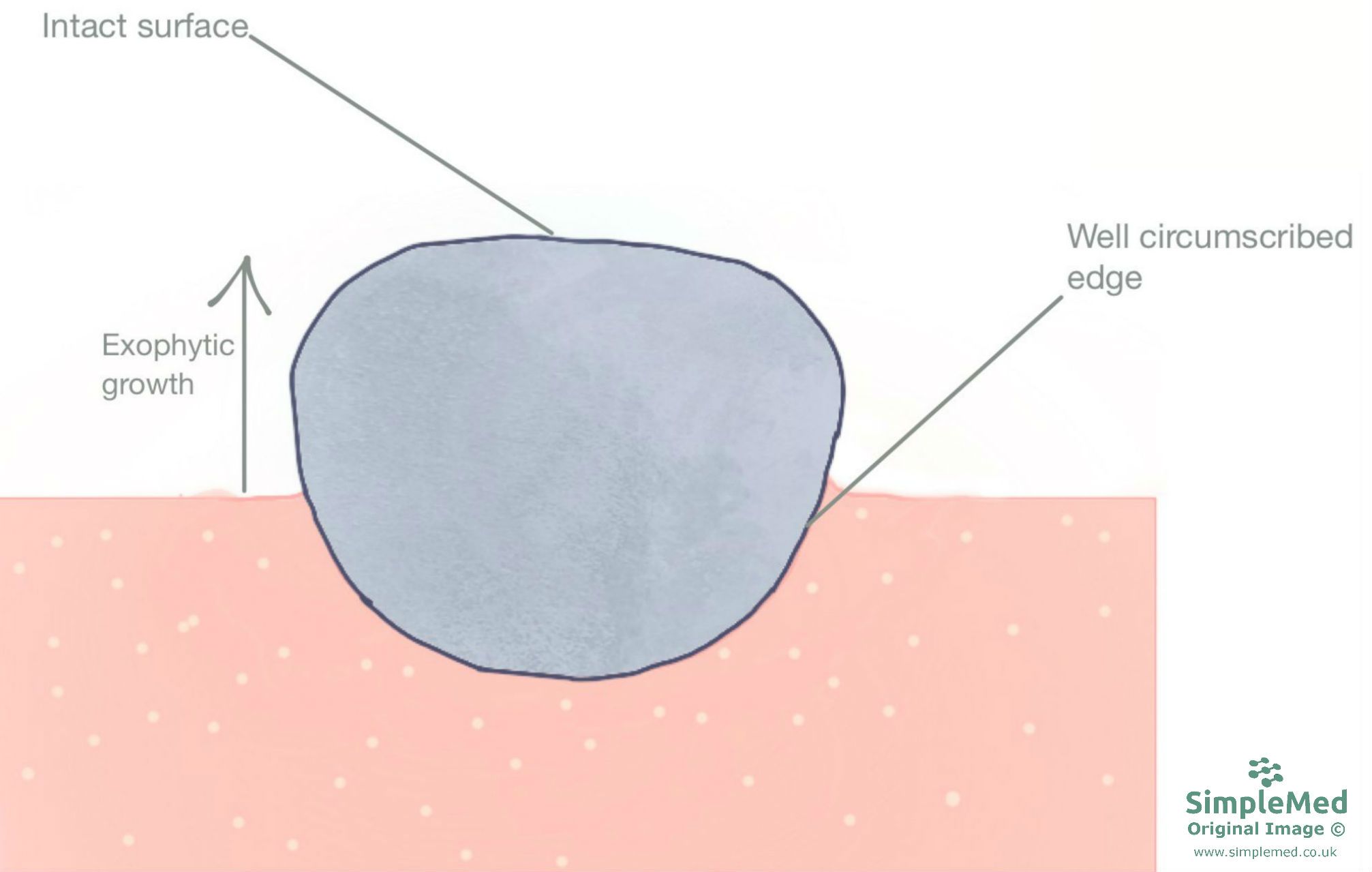
Diagram - The growth of a benign neoplasm. It is regular and well circumscribed
SimpleMed original by Dr. Emily Smith
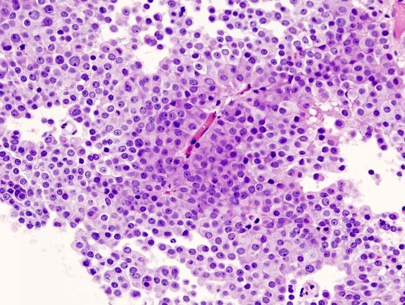
Image - A benign neoplasm under the microscope. The cells show nuclear changes, but still resemble the original tissue
Creative commons source by No machine-readable author provided. KGH assumed (based on copyright claims). [CC BY-SA 3.0 (http://creativecommons.org/licenses/by-sa/3.0/)]
The ability to invade surrounding tissues and spread to different sites (metastasise) are key defining factors of malignant neoplasms. They often grow more rapidly than benign tumours and may have irregular edges, no clear margin, with invasion into surrounding tissues. This type of neoplasm often has central necrosis due to inadequate perfusion when it outgrows its blood supply.
The degree of differentiation varies between malignant tumours. Well-differentiated cancers retain features of their tissue of origin; poorly differentiated tumours may show little resemblance. Atypical nuclei may show enlargement, hyperchromasia (dark staining), chromatin clumping and variation in size and shape (pleomorphism). Increased or abnormal mitotic figures can also be present. These findings support diagnosis but are not universal features of every cancer.
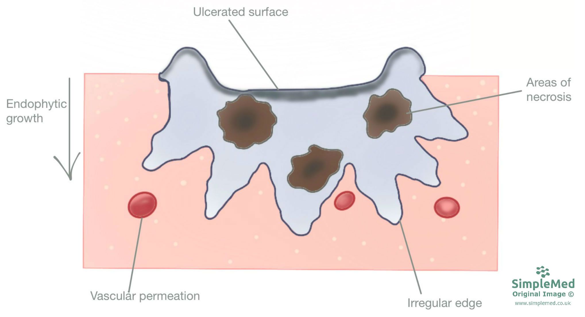
Diagram - The growth of a malignant neoplasm. It is invading into the surrounding tissues, giving it an irregular edge. The tumour is growing fast, resulting in ulceration and areas of necrosis
SimpleMed original by Dr. Emily Smith
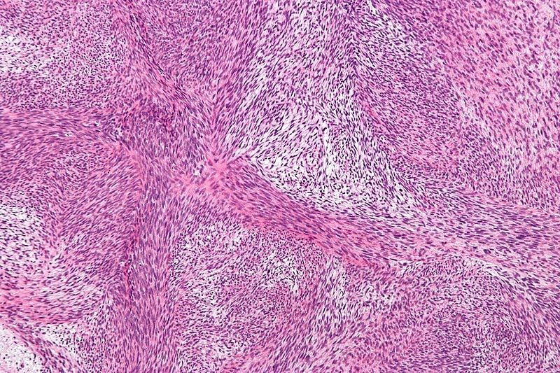
Image - A malignant peripheral nerve sheath tumour under the microscope. This example illustrates abnormal spindle-cell morphology; malignant tumours vary in their resemblance to normal tissue.
Creative commons source by Nephron [CC BY-SA 3.0 (https://creativecommons.org/licenses/by-sa/3.0)]
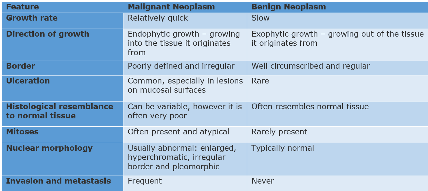
Table - Differences between benign and malignant neoplasms
SimpleMed original by Dr. Emily Smith
Nomenclature of Tumours (Tumour naming)
The naming of tumours often depends on the origin tissue, and whether the tumour is benign or malignant.
Epithelial Tumours
- Benign Tumours
- Benign tumours of epithelium are classified as either a papilloma or an adenoma and require the name of the tissue or cell it originates from.
- A papilloma is a benign tumour of non-glandular epithelium and is prefixed by the cell type, e.g. stratified squamous papilloma.
- An adenoma is a benign tumour of glandular epithelium e.g. colonic adenomas.
- Malignant Tumours
- A malignant tumour of non-glandular epithelium is called a carcinoma prefixed by the cell type e.g. squamous cell carcinoma.
- A malignant tumour of glandular epithelium is called an adenocarcinoma, e.g. colonic adenocarcinoma.
Connective Tissue Tumours
- These tumours have a prefix associated with each tissue as shown in the table below, for example cartilage has the prefix ‘chondro-‘.
- Benign
- This type of tumour has the suffix ‘-oma’.
- Malignant
- This type of tumour has the suffix ‘-sarcoma’.
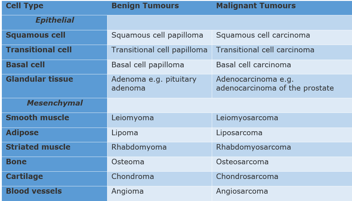
Table - Nomenclature of benign and malignant tumours
SimpleMed original by Dr. Emily Smith
Despite these rules of nomenclature, there are some tumours which don’t follow them. These tumours are often named after a person.
- Burkitt’s lymphoma - a type of B cell lymphoma
- Hodgkin’s lymphoma - malignant lymphoma
- Ewing’s sarcoma - malignant neoplasm of bone
- Kaposi’s sarcoma - malignant neoplasm of vascular endothelium
In-situ and Invasive Carcinomas
A carcinoma in-situ refers to a collection of neoplastic epithelial cells that have not yet invaded through the basement membrane, therefore, it can’t access routes of metastasis such as lymphatics and blood vessels. If a carcinoma is detected at this point (for example as part of a screening programme) it can often be completely excised, with an excellent prognosis.
An invasive carcinoma is one that has broken through the basement membrane. This means it can metastasise.

Diagram - The progression of cells from a normal state through to a malignant state
SimpleMed original by Dr. Emily Smith
Staging and Grading of Tumours
Staging and grading allows measurement of the overall burden of a tumour, and therefore the prognosis. The higher the grade or stage then the worse the prognosis.
Grading is used to determine the behaviour of the malignancy from histological features of the tissue. Generally, tumours are graded 1-4 with 1 being well-differentiated (low grade) and 4 being undifferentiated (high grade). Higher grade tumours tend to grow rapidly and spread faster than lower grade tumours.
Tumour grade is determined by:
- Degree of differentiation, and how much it resembles normal tissue
- Appearance of nuclei - size, hyperchromasia, pleomorphism
- Presence of mitoses
The stage of a tumour is determined by the size of the primary tumour and extent of metastasis.
Staging involves assessment of:
- Tumour size
- Node involvement
- Metastasis to other organs

Diagram - TNM staging. For example, if a patient has a large primary tumour, local lymph node metastasis and no organ metastasis, their staging would be written as T3N1M0
SimpleMed original by Dr. Emily Smith
The aim of cancer screening is to identify individuals with the disease in early stages or those most at risk. It allows them to start treatment early to get the best outcomes possible.
In the UK there are screening programmes for cervical, breast and colorectal cancer.
Cervical Cancer Screening
In England, everyone with a cervix aged 25 to 64 should be offered cervical screening. The sample is tested first for high-risk human papillomavirus (HPV). If HPV is detected, the same sample is examined for cell changes that may require further assessment or treatment. Following a negative high-risk HPV result, routine invitations are every five years; abnormal results or relevant history may require earlier follow-up. Screening helps prevent cervical cancer by identifying risk and treatable precancerous changes.
Breast Cancer Screening
This screening programme is routinely offered to women aged 50 to 71, with invitations every three years. It involves having a mammogram, an x-ray of the breasts, to detect changes such as asymmetrical densities or calcifications that may indicate cancer. Any abnormalities can be assessed with more imaging, biopsy and fine needle aspiration.
Bowel Cancer Screening
In England, bowel cancer screening is routinely offered to people aged 50 to 74 every two years. A home faecal immunochemical test (FIT) is sent by post to check a stool sample for blood. A result requiring further investigation leads to specialist assessment, usually including colonoscopy; blood in the sample does not itself diagnose cancer.
Edited by:Dr. Bethany Turner
Reviewed by: Dr. Marcus Judge
- 15583

.jpg)
