

Next Lesson - Protein Targeting
Core
Post-translational Modification
Every protein forms a unique three-dimensional structure that allows the protein to be active, but some proteins require further modification after the process of translation so they can form their final activated state.
Post-translational modification involves the enzymatic and covalent modification of proteins and is an important process in cell signalling. Protein modifications occur at the C- or N-terminus of the polypeptide chain, or on the side chains of the amino acids present.
There are hundreds of ways proteins can be modified, but in this article we are only going to discuss the most common mechanisms of post-translational modifications of proteins.
Phosphorylation is the most common mechanism of modifying a protein post-translation, especially enzymes. The process of phosphorylation is reversible and involves adding a phosphate group to specific amino acids such as serine, threonine and tyrosine; the phosphate group is donated from ATP.
Phosphorylation is catalysed by enzymes, these include protein kinases which transfer the terminal phosphate group of ATP to the hydroxyl group present on serine, threonine and tyrosine amino acid residues.
Phosphorylation can be reversed and this involves protein phosphatases, which catalyse the removal of a phosphoryl group from the protein molecule through hydrolysis.
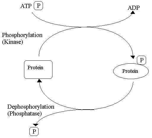
Diagram - The process of phosphorylation and dephosphorylation
Creative commons source by Petaholmes [CC BY-SA 4.0 (https://creativecommons.org/licenses/by-sa/4.0)]
Why phosphorylation of proteins is highly effective:
- It adds two negative charges to the modified enzyme or protein. This disrupts the electrostatic interactions allowing new interactions to form, which can affect the activity of the enzyme and the substrate binding.
- The phosphoryl group can form hydrogen bonds.
- The rate of phosphorylation and dephosphorylation can change, either taking less than a second or many hours. This allows the number of phosphorylated proteins to change depending on the needs of the physiological processes taking place.
- As ATP is used as an energy currency and in phosphorylation, this links the regulation of metabolism and the energy status of the cell together.
- Phosphorylation can lead to highly amplified effects within the cell as one single activated kinase can phosphorylate many target proteins in a short space of time. The activated proteins may also be enzymes, which can go on to catalyse further reactions. Therefore, the initial signal can be amplified exponentially throughout the cell through a cascade of kinases in a short time period.
Proteins can be phosphorylated abnormally and this can lead to diseased states. Diseases such as Alzheimer’s disease, Parkinson’s disease and cancer have been linked to abnormal protein phosphorylation.
Another mechanism of post-translational modification is glycosylation, and this process involves the covalent attachment of a carbohydrate to a protein. It is one of the most complicated processes of post-translational modification due to the large number of enzyme-dependent steps involved. The donor molecule is a glycosyl group, which is a mono- or oligosaccharide that forms a glycosidic bond with the acceptor molecule, which in this case is a protein.
There are a number of subtypes of glycosylation which we will discuss further:
N-linked glycosylation is a specific type of glycosylation takes place in the endoplasmic reticulum and involves the attachment of a mono- or oligosaccharide to a nitrogen atom present in the asparagine amino acid residue. N-linked glycosylation requires energy and usually occurs to proteins bound for the membrane or proteins that are going to be secreted.
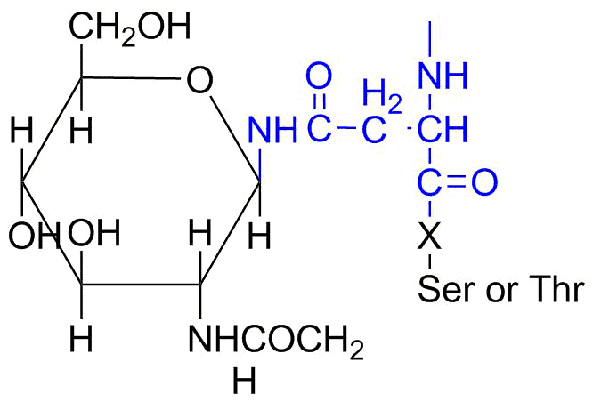
Diagram - N-linked glycosylation with an asparagine residue in blue
Creative commons source by Lizziechka [CC BY-SA 4.0 (https://creativecommons.org/licenses/by-sa/4.0)]
O-linked glycosylation takes place in the Golgi apparatus and involves glycans being attached to the hydroxyl groups of serine or threonine amino acid residues. This process occurs after N-linked glycosylation and folding of the protein. O-linked glycosylation has been found to be important in the production of proteins released in mucus secretions and proteoglycans, which are a component of the extracellular matrix.
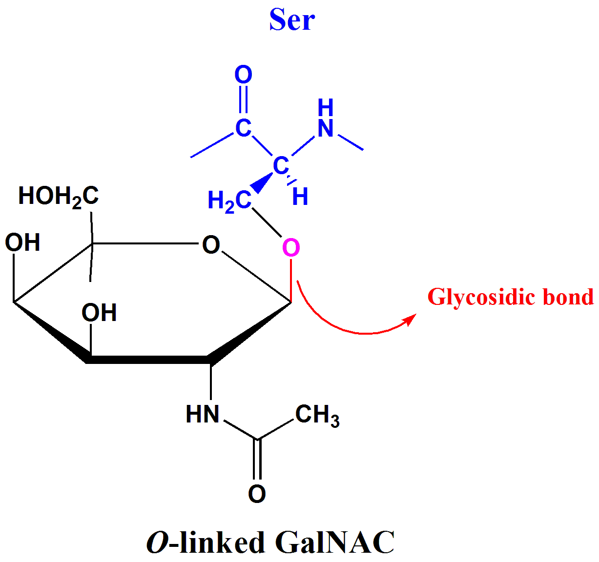
Diagram - O-linked glycosylation with a serine residue in blue
Public Domain source by Tpirojsi
There are other types of glycosylation such as S-linked glycosylation and C-linked glycosylation, but we are not going to discuss these types in this article.
Why glycosylation is an important type of post-translational modification:
- Glycosylation helps in ensuring that proteins fold correctly, it also provides protein stability, helping the protein carry out its function.
- Glycosylation has been shown to be important in how proteins interact with other molecules.
- For example, how a ligand interacts with its complementary receptor or how the receptor interacts with the cell signalling mechanisms inside the cell.
- How these examples interact has been shown to be due to glycosylation, with the process of glycosylation affecting the biological response.
- Glycosylation plays an important role in determining the blood group of a person. The blood group is determined by the presence or absence of glycosyltransferases. These enzymes catalyse the final stage of the transfer of a saccharide molecule to the H antigen present on the outer surface of the red blood cell, and the final result of the carbohydrates added determines the person’s blood group.
It is easy to get confused about the difference between glycosylation and glycation, so we shall go through the differences now:
- Glycation is when a sugar molecule is attached to a protein not under enzymatic control, whereas glycosylation is the attachment of a sugar to a protein under enzyme control using activated sugar donors.
- Glycation often occurs in the blood as simple sugars, such as glucose and galactose, are found here and they can attach to haemoglobin. This results in the formation of glycated haemoglobin which can increase the number of free radicals within the red blood cell leading to damage.
- Glycated haemoglobin or HbA1c can be used as a diagnostic tool for diabetes mellitus as the level of HbA1c can be used to assess how well a patient with diabetes mellitus is controlling their glucose levels. (Check out our article on Diabetes Mellitus).
Acetylation is another example of post-translational modification and it is an important process that occurs in cell biology. In proteins, acetylation usually involves the addition of an acetyl group (CH3CO-) to an amino group, such as the free amino group at the N-terminal or the side chain of a lysine residue, and is catalysed by enzymes.
There are two forms of protein acetylation: N-terminal acetylation and lysine acetylation.
N-terminal acetylation has been shown to be irreversible, and is important for the function and regulation of a variety of proteins. The process involves the addition of an acetyl group to the free amino group present at the N-terminal of the polypeptide chain.
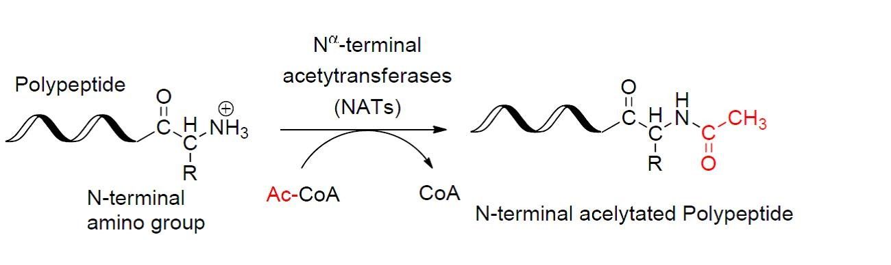
Diagram - The process of N-terminal acetylation
Creative commons source by Ybs.Umich [CC BY-SA 4.0 (https://creativecommons.org/licenses/by-sa/4.0)]
Acetylation of lysine residues involves acetyl-CoA which donates its acetyl group to lysine. The acetyl becomes attached to an amino group present on the side chain of a lysine residue.
An example of lysine acetylation is the acetylation of histones and p53. Lysine residues present at the N-terminal of the polypeptide tail extending from the core of the histone protein are acetylated and the acetylated histones leads to the chromatin to be less condensed, therefore higher levels of transcription. The acetylation of p53 is important for its activation and once activated, the protein can suppress the cell cycle, allow for any damaged DNA to be repaired or begin the process of cell apoptosis.
Disulphide bonds are strong bonds formed by the oxidation of the thiol groups (R-SH) of two cysteine residues, with the formation of disulphide bonds occurring in the lumen of the endoplasmic reticulum.
Disulphide bonds are an important in stabilising the tertiary structure of single polypeptide chains and in stabilising multi-subunit proteins such as antibodies and insulin. The majority of proteins that are secreted into the extracellular space contain disulphide bonds, while cytosolic proteins do not contain this type of bond. If a protein contains a high number of disulphide bonds, the molecule will be more resistant to external forces, e.g. heat and detergents, and less likely to denature.
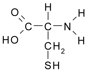
Image - Structure of cysteine
Creative commons source by bigblue0092 [CC BY-SA 4.0 (https://creativecommons.org/licenses/by-sa/4.0)]
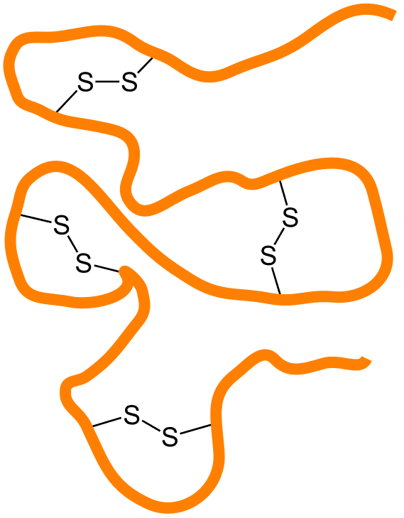
Image - Polypeptide chain with disulphide bonds
Creative commons source by Jü [CC BY-SA 4.0 (https://creativecommons.org/licenses/by-sa/4.0)]
Many proteins can be modified post-translation by specific proteolysis of the polypeptide chain to produce the final active form of the protein. This can include the removal of the signal peptide, removal of the N-terminal methionine and/or the conversion of an inactive protein to its active form.
For some proteins, the first synthesised version of the protein is called the preproprotein, which is then cleaved to produce the proprotein, then after further modification or cleavage, the final functional form is produced.
For example, albumin, a protein found in blood, is synthesised as preproalbumin and then the signal peptide is removed to give proalbumin. To form the final functional form that is albumin, the N-terminal 6 residues are removed from proalbumin.
Proteins may contain a signal peptide that is usually present at the N-terminal, and this polypeptide sequence allows the protein to be directed to an organelle or to be secreted out of the cell. Once the protein passes through a membrane such as the endoplasmic reticulum, the signal peptide is removed by an enzyme.
Removal of the N-Terminal Methionine
Methionine aminopeptidase removes the initiator methionine from many newly synthesised proteins. Cleavage depends strongly on the side-chain size of the second amino acid: a small adjacent residue, such as glycine or alanine, favours access to the first peptide bond, whereas a bulky residue tends to prevent cleavage. The initiator methionine is therefore retained in some proteins.
Cleavage of Precursor Proteins
Some proteins are synthesised in the form of their precursors, such as zymogens and prohormones, and are then cleaved to give rise to the final active form of the protein.
One example of this process is the synthesis of proteases, which are synthesised in their inactive form so they can be stored in cells safely without causing destruction to the organism. The inactive proteases are stored in the cell and when required they are released, after which they are activated in the correct location and/or context. An example of a zymogen is trypsinogen, which is cleaved to form the activated protease trypsin by causing a slight change to the conformation of the protein and completing the active site so the enzyme is fully activated.
Another example of this process is the synthesis of insulin. The hormone is synthesised as preproinsulin, which then has the signal peptide removed to produce proinsulin. Disulphide bridges then form between the A and B chains to provide stability to the protein molecule before the C chain is removed by a specific endopeptidase, producing mature insulin. Insulin and free C chains are packaged into secretory granules by the Golgi apparatus, and these granules accumulate in the cytosol of pancreatic beta cells in the islets of Langerhans before secretion.
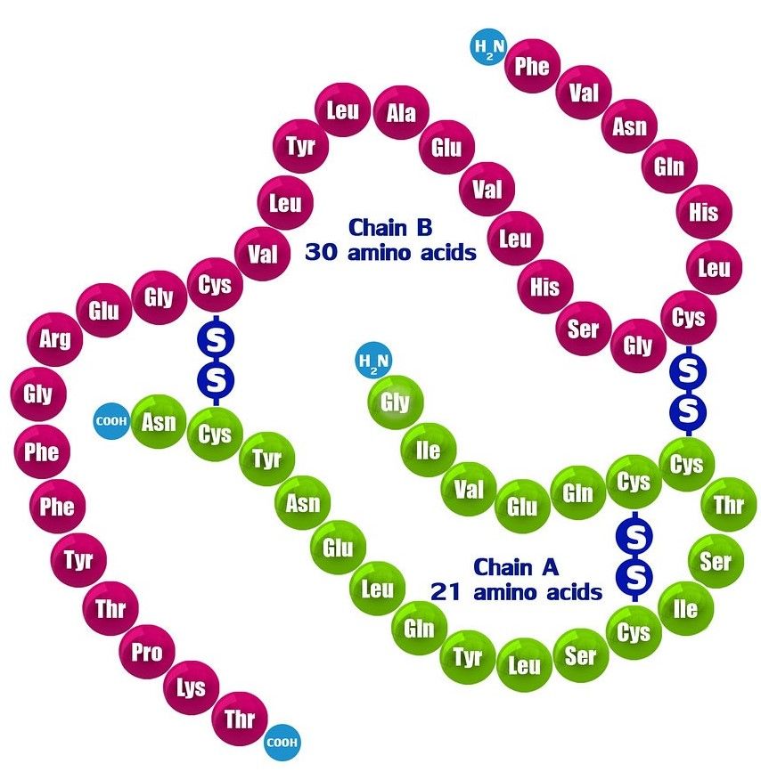
Diagram - The structure of mature insulin
Creative commons source by Zappys Technology Solution, edited by Dr. Peter Parkinson [CC BY-SA 4.0 (https://creativecommons.org/licenses/by-sa/4.0)]
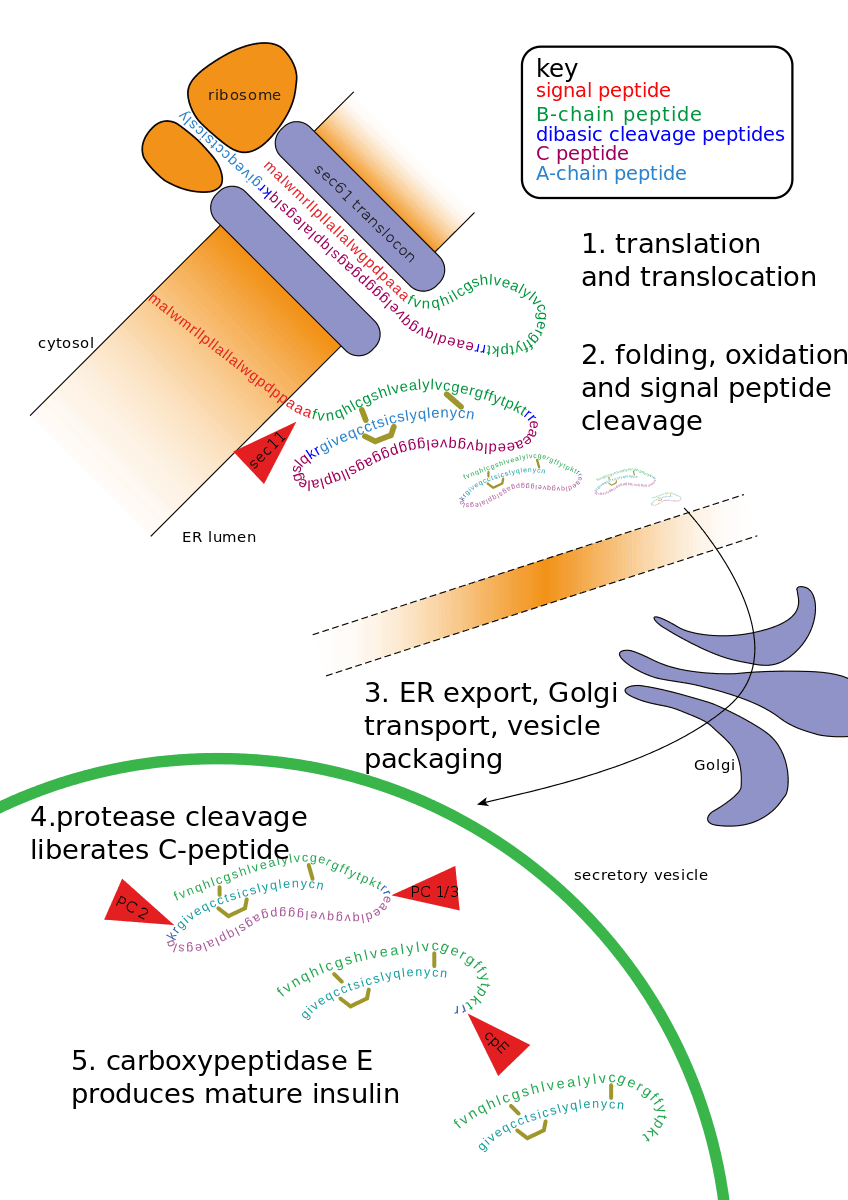
Diagram - Process of insulin production
Creative commons source by Fred the Oyster [CC BY-SA 4.0 (https://creativecommons.org/licenses/by-sa/4.0)]
The majority of polypeptide hormones found in the human body are firstly produced as large precursor polypeptides called polyproteins which are then cleaved to produce smaller polypeptide molecules. An example of this is pro-opiomelanocortin (POMC) and this large precursor contains many different smaller polypeptides. Depending on the tissue in the human body, the cleavage of this polyprotein can give rise to a variety of hormones such as adrenocorticotropic hormone and beta-melanocyte-stimulating hormone.
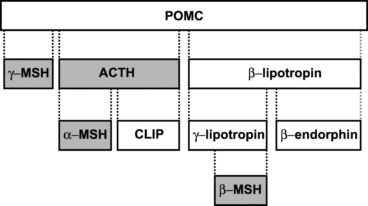
Diagram - The derivatives of pro-opiomelanocortin
Public Domain source by Klaus Hoffmeier
As mentioned at the beginning of the article, there are many ways in which a protein can undergo post-translational modification. Here are some other forms of post-translational modification:
- Lipidation - attachment of lipids to protein and usually these proteins are destined to part of the cell membrane.
- Alkylation - attachment of an alkyl group such as methyl and ethyl to amino acid residue.
- Hydroxylation - attachment of an oxygen atom to side chain of a lysine or proline.
Constitutive and Regulatory Secretory Pathways
When a protein destined for secretion into the extracellular environment is produced, it can be delivered to the plasma membrane via one of two pathways: constitutive secretory pathway or regulatory secretory pathway.
Proteins such as immunoglobins, collagen, albumin and plasma membrane receptors are synthesised in the cell and packaged into vesicles. The vesicles then fuse with the plasma membrane in an unregulated manner and are released into the extracellular space. This is an example of constitutive secretory pathway as they are constantly produced.
Proteins involved in the regulatory secretory pathway include insulin, ACTH and trypsin. This pathway involves the protein being synthesised in the cell then packaged into a storage vesicle. Once a hormone or neurotransmitter binds to a receptor on the plasma membrane of the cells, it leads to a signalling pathway occurring inside the cell to result in the vesicles containing the protein fusing with the plasma membrane and releasing the protein into the extracellular space.
Collagen is a fibrous protein found in connective tissue such as tendons, ligaments, bone and cartilage. The protein is also a part of the loose connective tissue which helps to provide structure to the internal organs.
The fibrous protein is produced by cells called fibroblasts, which are found in connective tissue. As well as synthesising collagen, these cells also produce ground substance, a shapeless gel-like matrix that is found between cells and fibres in connective tissue.
The basic unit of the fibrous protein collagen is tropocollagen. Collagen is formed of three polypeptide chains that form a right-handed triple helix structure. The triple helical structure allows the protein to have high tensile strength, be non-compressible and non-extensible, therefore giving strength to connective tissue where the protein is found. The fibrous protein is stabilised by hydrogen bonds between the polypeptide chains.
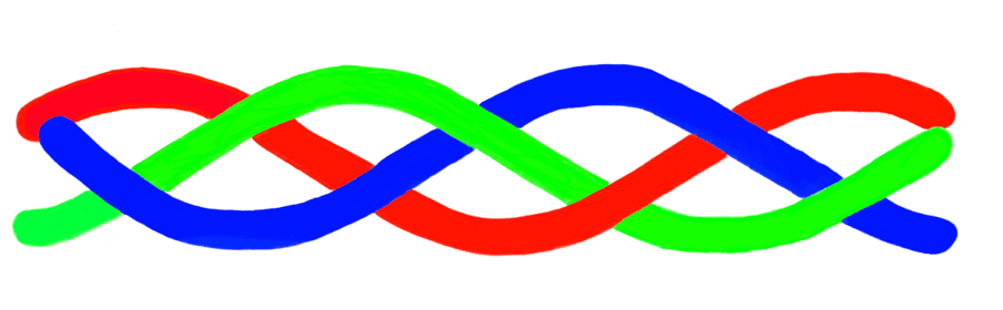
Image - The triple helical structure of collagen
SimpleMed original by Dr. Peter Parkinson
Every third amino acid in the polypeptide chain of tropocollagen is glycine with proline or hydroxyproline making up majority of the other positions in the sequence of amino acids. Glycine's side chain is a single hydrogen atom and this allows glycine to fit into the middle of the helix because the side chain is small enough to fit in the middle of the helix.
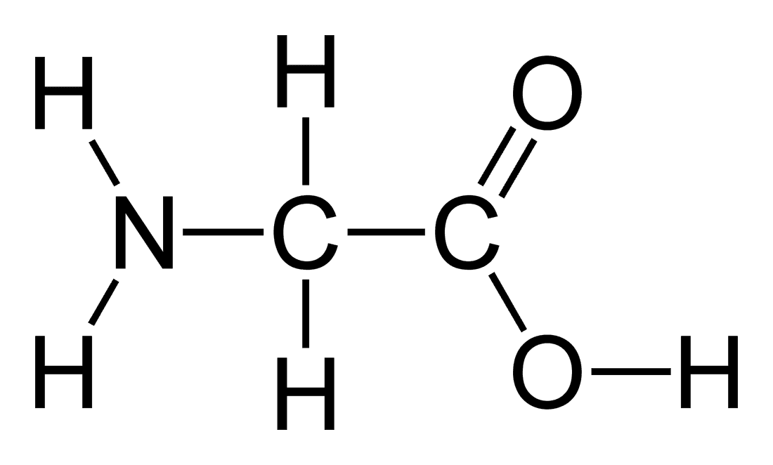
Image - The structure of glycine
Public Domain Source by Benjah-bmm27
Collagen is a great example of post-translational modification as there are a number of ways in which the protein is modified. We are now going to discuss the steps involved in the synthesis and modification of collagen.
- The polypeptide chains are synthesised at ribosomes on the endoplasmic reticulum (ER) and then the chains enter the lumen of the ER.
- Once the polypeptide chains have entered the lumen of the ER, the signal sequence is cleaved by signal peptidase.
- Proline and lysine residues are hydroxylated by prolyl hydroxylase and lysyl hydroxylase. The hydroxylation increases the hydrogen bonding between polypeptide chains and therefore helps to stabilise the triple helix structure.
- Whilst in the lumen of the ER, there is the addition of N-linked oligosaccharides.
- In the ER lumen, galactose, or glucose linked to galactose, is added to hydroxylysine residues. This collagen O-linked glycosylation occurs before triple-helix formation.
- When three polypeptide chains align, there is the formation of disulphide bonds.
- In the lumen of the ER, the triple helical shape of procollagen forms from the C- to N-terminus.
- After triple-helix formation, procollagen passes through the Golgi apparatus and is packaged into secretory vesicles for transport to the plasma membrane.
- At the plasma membrane, procollagen is secreted out of the cell via the process of exocytosis.
- Around 150-250 amino acids at the N- and C-terminus do not form the triple helical shape and once the procollagen molecule is secreted out of the cell, these amino acids at each terminus are removed by the enzyme procollagen peptidase to form the molecule tropocollagen.
- After the collagen molecules associate with each other laterally, covalent cross-linking between the molecules occurs to form fibrils. This process is catalysed by lysyl oxidase which requires vitamin B6 and Cu2+ ions for its activity.
- Finally, the fibrils aggregate together to form collagen fibres.
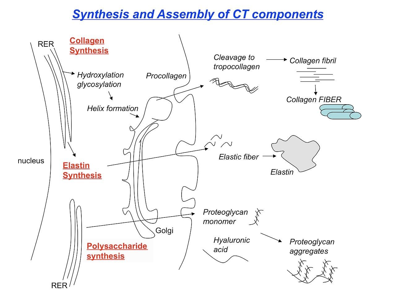
Diagram - The process of collagen synthesis and the post-translational modification it goes under
Collagen synthesis diagram by Mfigueiredo. Source. Licensed under CC BY-SA 3.0 Unported.
Scurvy is a rare disease in modern times and is more associated with sailors who suffered from the condition in history.
Collagen prolyl hydroxylases hydroxylate proline residues, while separate lysyl hydroxylases hydroxylate lysine residues. Both enzyme groups require vitamin C and Fe2+ for their activity. Hydroxyproline helps stabilise the collagen triple helix; hydroxylysine provides sites for glycosylation and contributes to collagen cross-linking. Vitamin C deficiency impairs these hydroxylation reactions, producing fragile connective tissue. Symptoms of scurvy include easy bruising, tiredness, irritability, swollen and painful joints, and swollen or bleeding gums.
The condition is treated with oral vitamin C and sees improvement within days, with a full recovery in weeks.
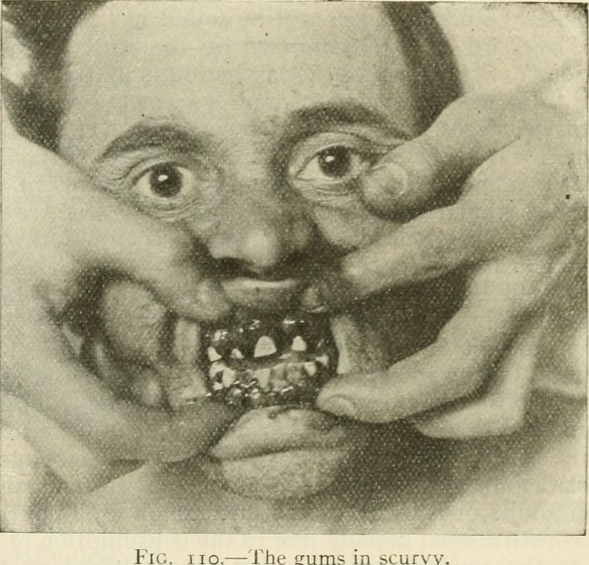
Image - A patient suffering from swollen, bleeding gums due to scurvy
Historical image from Modern surgery, general and operative (1919), via Internet Archive Book Images. Source. The source marks the image CC0 (Public Domain Dedication).
EDS is a group of genetic connective tissue disorders due to a genetic mutation. The form of EDS that the patient will have depends on which of the genes involved in the structure, processing or production of collagen is affected. The patient will have symptoms such as skin hyperelasticity, joint dislocations, scoliosis, aortic dissection and osteoarthritis.
At the moment, there is no cure for the syndrome, so treatment for a patient with EDS is supportive.
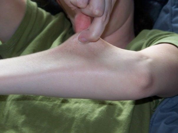
Image - Skin hyperelasticity in a patient with Ehlers-Danlos Syndrome
Creative commons source by Ellen C. Kelleher [CC BY-SA 4.0 (https://creativecommons.org/licenses/by-sa/4.0)]
Edited by: Dr. Thomas Burnell
Reviewed by: Dr. Marcus Judge
- 21002



_V.1.svg)


