Next Lesson - Lymph Nodes and Neck Lumps
Abstract
- The main blood supply to the head and neck comes from the common carotid arteries, and the main drainage is by the internal and external jugular veins.
- The external carotid artery supplies branches to the head and neck that are extra-cranial.
- It is the internal jugular vein that is used to assess jugular venous pressure.
Core
The brain could be considered to be the most important structure within the head and neck region. It requires 0.75-1L of blood every minute, and can consume between 20-50% of the oxygen in blood (depending on brain activity). This means that the arterial supply to the head and neck, and the subsequent venous drainage, needs to be able to handle a large volume of blood effectively.
It is important to remember that the vascular system in the human body is, for the most part, symmetrical - there is a left and a right vessel unless otherwise stated. There are also some normal anatomical variants e.g. not every individual may have bifurcation of the common carotid artery at the level of C4, it may occur at C3 or C5.
There are some abbreviations of vessel names used in this article:
- CCA = Common Carotid Artery
- ECA = External Carotid Artery
- ICA = Internal Carotid Artery
- IJV = Internal Jugular Vein
- EJV = External Jugular Vein
Blood travelling to the brain begins in the heart. This oxygenated blood travels towards the head via the common carotid and vertebral arteries.
The arch of the aorta has three branches: the left subclavian artery, the left common carotid artery, and the brachiocephalic trunk. The brachiocephalic trunk then divides into the right common carotid artery and the right subclavian artery. Both common carotid arteries ascend in a carotid sheath, a fibrous tissue sleeve that contains the common carotid artery (medially), the internal jugular vein (laterally), and the vagus nerve (posteriorly).
At the level of C4 (the superior border of the thyroid cartilage) the common carotid bifurcates into the internal and external carotid arteries. At this level, the internal and external carotid arteries look similar, and so it can be difficult to tell them apart. At the level of C4, the internal carotid artery will appear more bulbous due to the presence of the carotid sinus at its origin, with the carotid body located at the bifurcation, and the external carotid artery will exit the carotid sheath. The other main difference between the internal and external carotid arteries is the lack of branches from the ICA in the neck. The ICA ascends up the neck and enters the base of the skull via the carotid canal and continues up through the cavernous sinus. It then forms an anastomosis with the vertebral arteries named the Circle of Willis, which supplies the structures of the brain, and this will be covered in the neuroanatomy unit found here.
The ECA is the major blood supply to the extra-cranial structures of the head and neck (i.e. things on the outside of the skull). The eight branches it gives off can be remembered by the following mnemonic and are shown on the image below.

Table - Mnemonic for the branches of the external carotid artery
SimpleMed original by Dr. Ethan Kavanagh

Diagram - Branches of the external carotid artery
SimpleMed original by Dr. Maddie Swannack
The maxillary and superficial temporal arteries are the terminal branches of the ECA, meaning this is where the ECA is said to end, and arise high up in the parotid gland.
The important branches of the maxillary artery are:
- Middle Meningeal Artery - susceptible to cause extradural haemorrhage if there is trauma to the side of the head (temporal region).
- Sphenopalatine Artery - contributes to the anastomosis that supplies the nasal cavity and, in a small number of nosebleeds (particularly in the elderly) is the culprit vessel. It is also located quite far back within the nasal cavity, meaning posterior epistaxis can be more difficult to control with simple pressure alone and often requires more advanced management.
The superficial temporal artery is pulsatile just anterior to the ear.
The ECA does not supply the front of the skull nor the forehead - this is where the ICA contributes. Once inside the skull, it gives rise to the ophthalmic artery which runs into the orbit and then it itself gives rise to the supraorbital and supratrochlear arteries which then join the other branches of the ECA.
The posterior circulation of the brain is supplied by the vertebral arteries. The vertebral arteries ascend through the transverse foramina of the cervical vertebrae (with the exception of C7) and enter into the subarachnoid space between C1 (also called the atlas) and the occipital bone. Both vertebral arteries then pass through the foramen magnum, curve around the medulla, and join to form the basilar artery. The basilar artery then contributes to the Circle of Willis via the posterior cerebral arteries, which connect with the ICA through the posterior communicating arteries, to supply the brain.
Inside the skull, there are dural folds that exist on the sagittal plane (posterior to anterior - separates the two hemispheres of the brain) and on the transverse plane (left to right - cups the base of the brain). Within these dural folds, between the periosteal and meningeal layers of dura, exist spaces called dural venous sinuses, which are spaces through which venous blood flows.
The image below shows the different sinuses in the skull relating to the dural folds and shows how they converge at the confluence of sinuses, travel round the sigmoid sinus and descend through the floor of the skull to become the internal jugular veins. This vein joins the carotid sheath to travel through the neck.

Diagram - The dural venous sinuses
SimpleMed original by Dr. Maddie Swannack
Both the left and the right IJVs descend down the neck and join with the subclavian veins to form the brachiocephalic veins (which then go on to form the superior vena cava).
As the IJV descends, it receives blood from a number of different vessels. These vessels include:
- Facial Vein
- Maxillary Vein
- Lingual Vein
- Occipital Vein
- Superior and Middle Thyroid Veins
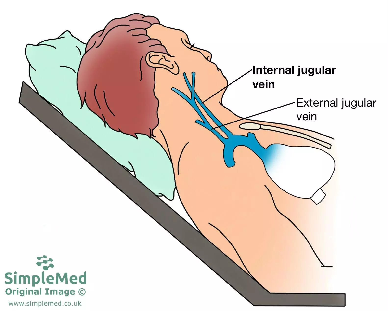
Learning the path of the internal jugular vein is important, as it is used on the right side to assess the jugular venous pressure (JVP), which can be used to assess the pressure of the venous return to the right atrium. For more information on the JVP, see this article.

Diagram - The position for measuring the jugular venous pressure
SimpleMed original by Dr. Bethany Turner
The external jugular veins drain the scalp and face, as well as some of the deeper structures within the face. It is formed by the connection of the posterior auricular vein (draining the area of the scalp superior and posterior to the ear) and the posterior branch of the retromandibular vein (which is formed by the maxillary and superficial temporal veins and drains the face). The EJV descends down the neck and feeds into the subclavian vein.
Edited by: Dr. Maddie Swannack
Reviewed by: Dr. Thomas Burnell
- 18458

