

Next Lesson - Gametogenesis
Core
The Hypothalamic-Pituitary-Ovarian Axis
The menstrual cycle is regulated by the hypothalamic-pituitary-ovarian (HPO) axis. The hypothalamus releases gonadotrophin-releasing hormone (GnRH) which stimulates the anterior pituitary gland to release the gonadotrophins (hormones that stimulate the ovaries or testes). These are luteinising hormone (LH) and follicle-stimulating hormone (FSH). LH and FSH in turn stimulate the ovaries to produce oestrogen, progesterone and inhibin (a hormone that reduces FSH release).
Oestrogen and progesterone can either positively or negatively feed back on the anterior pituitary and hypothalamus in order to control the axis. Inhibin release is prompted by FSH. Inhibin then reduces further release of FSH. Together with oestrogen, this fall in FSH means that usually only one follicle (a fluid-filled sac containing a developing egg) keeps developing each cycle. This is the dominant follicle, which goes on to release its egg.
GnRH is released in a pulsatile way (meaning it is not continuously released). This is because GnRH receptors that are continuously exposed to GnRH become desensitised (they stop responding). Continuous GnRH first increases LH and FSH release, then suppresses their release. As a result, oestrogen and progesterone production falls. GnRH agonist drugs use this effect.
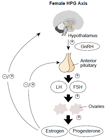
Diagram - The Hypothalamic-Pituitary-Ovarian axis
Creative commons source by Lu Kong, Ting Zhang, Meng Tang and Dayong Wang
[CC BY-SA 4.0 (https://creativecommons.org/licenses/by-sa/4.0)]
The menstrual cycle can be described as two linked cycles: one in the ovaries (the ovarian cycle) and one in the endometrium (the uterine cycle). The menstrual cycle is classically described as lasting 28 days, but this can vary from person to person.
The ovarian cycle is split into two phases: the follicular and luteal phases. The follicular phase is variable in length, whereas the luteal phase lasts around 14 days in most cycles.
The follicular phase begins on day 1 and involves the development of the ovarian follicle (containing the ovum). In a 28-day cycle, on about day 14, the follicle ruptures and the ovum is released from the ovary. The ovum is then collected by the fallopian tube (the tube that carries the egg from the ovary to the uterus). It may be fertilised as it travels along the tube.
Over the next 14 days, the luteal phase occurs, and the follicle develops into the corpus luteum which will help to maintain an early pregnancy if fertilisation occurs. If fertilisation does not occur, the corpus luteum atrophies and becomes the non-functioning corpus albicans.
The endometrial cycle has three phases: menses, the proliferative phase, and the secretory phase.
Menses is the bleeding which begins on day 1 and represents the shedding of the endometrium in the absence of implantation taking place. This lasts 5 to 7 days on average.
The proliferative phase runs from the end of menses until ovulation (about day 14 in a 28-day cycle). It is when the endometrium lining is thickened in preparation for implantation under the influence of oestrogen.
After ovulation, the secretory phase occurs; this is where the endometrium lining is at its thickest and contains glandular epithelium. This occurs under the influence of progesterone.
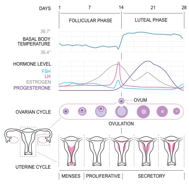
Diagram - A graph detailing the changes in hormones, endometrial changes, and ovarian changes throughout the menstrual cycle. Note: Inhibin levels not shown on the graph
Creative commons source by Isometrik [CC BY-SA 4.0 (https://creativecommons.org/licenses/by-sa/4.0)]
In the first few days of the menstrual cycle the levels of ovarian hormones are very low with a low level of inhibin. This means there is very little inhibition of the hypothalamus and anterior pituitary which causes FSH levels to rise. FSH binds to the granulosa cells (the cells lining the follicle) of the developing ovarian follicle and stimulates it to grow. The theca interna (the inner layer of cells around the follicle) also develops. The follicle then makes oestrogen using both cell types. LH stimulates the theca cells to make androgens (male-type steroid hormones, such as androstenedione). These pass into the granulosa cells, where FSH stimulates the enzyme aromatase to convert them to oestrogen. This is called the two-cell, two-gonadotrophin model. The growing follicle also makes inhibin.
In the mid-follicular phase, the follicle produces increasing amounts of oestrogen, which mainly exerts a negative feedback effect on the hypothalamus and anterior pituitary. Sustained high oestrogen levels later in the follicular phase switch to positive feedback, leading to a marked LH (and smaller FSH) surge. This occurs because in the presence of a high oestrogen concentration, the pituitary makes more GnRH receptors and becomes more sensitive to each pulse of GnRH. This means more LH is released per GnRH pulse.
Before ovulation, oestrogen rises rapidly. The dominant follicle keeps growing even as FSH levels fall, because it makes more FSH receptors and so becomes more sensitive to FSH. The high oestrogen leads to a surge in LH production. The surge of LH causes the granulosa cells of the follicle to produce progesterone. Prompted by the LH surge, ovulation occurs. The mature oocyte (egg cell) is released from the ovary into the peritoneal cavity (the space in the abdomen around the organs). It is then picked up by the fimbriae (the finger-like ends) of the fallopian tube.
After ovulation, the follicle becomes ‘luteinised’ (it changes into the corpus luteum). The corpus luteum mainly secretes progesterone. It also secretes oestrogen and a form of inhibin called inhibin A. LH production is now suppressed as progesterone from the corpus luteum exerts a negative feedback effect. This causes further maturation of oocytes to be put on hold.
Without fertilisation, there is no further rise in LH, meaning the corpus luteum will regress spontaneously. This leads to a large fall of oestrogen and progesterone, meaning the negative feedback on the HPO axis is removed. FSH levels then rise to initiate the next cycle, with LH also increasing.
If fertilisation occurs, the syncytiotrophoblast (the outer cell layer of the developing embryo, which helps to form the placenta) produces human chorionic gonadotrophin (HCG) which acts to sustain the corpus luteum. This allows the corpus luteum to continue to produce oestrogen and progesterone which supports the pregnancy. Eventually the placenta develops and can produce enough oestrogen and progesterone to control the HPO axis throughout the pregnancy. These high levels of oestrogen and progesterone result in negative feedback on the HPO axis, preventing further maturation and release of oocytes.
The endometrium is sensitive to the ovarian hormones: oestrogen and progesterone. Oestrogen causes the proliferation (growth) of the endometrium. Oestrogen and progesterone then make its glands develop and secrete.
The endometrium is split into two layers:
- Functional layer - the layer that proliferates and sheds at the end of the cycle.
- Basal layer - the layer where the cells in the functional layer grow from.
At the start of the proliferative phase, there are few glands in the functional layer of the endometrium. Throughout the proliferative phase, glands begin to coil and form in the thickened endometrium ready for implantation if fertilisation occurs. By the end of the proliferative phase, the endometrium is typically 8 to 12 mm thick. The secretory phase begins with the endometrium at its maximum thickness with very pronounced coiled glands. In the late secretory phase, the glands have a characteristic saw-tooth appearance.
The next phase of the uterine cycle is the menses. This is the phase during which the functional layer sheds, causing menstrual bleeding (a period). The basal layer stays behind and regrows the functional layer in the next cycle.
Amenorrhoea is the absence of menstruation.
Causes can be split into primary or secondary amenorrhoea.
- Primary amenorrhoea is the failure to establish menarche (the first period). Definitions vary. The World Health Organization uses no periods by 16 years of age in someone with normal secondary sexual characteristics (such as breast development), or by 14 years if these are absent. Other authorities use 15 and 13 years.
- Secondary amenorrhoea is the cessation (stopping) of periods after they have started. It is defined as no periods for 3 months in someone with previously regular monthly cycles, or for 6 months in someone who was previously menstruating.
Amenorrhoea is normal (physiological) before puberty, during pregnancy and breastfeeding (lactation), and after the menopause.
Pathological causes of amenorrhoea can occur at any level of the HPO axis (the hypothalamus, pituitary gland, or ovaries) or with the outflow tract (the uterus, cervix or vagina). The cervix is the neck of the uterus.
The likely location of the pathology can be suggested by looking at the levels of LH and FSH in the blood. Low LH and FSH suggest an issue with the hypothalamus or pituitary gland, whereas high LH and FSH suggest primary ovarian failure (the ovaries themselves are not working). Outflow tract causes usually have normal hormone levels.
There are also structural causes of amenorrhoea where there is a mechanical obstruction of blood flow:
- Imperforate Hymen - a condition occurring when a thin layer of connective tissue (hymen) covers the vaginal entrance, and this has not perforated at the onset of menstruation. Blood collects in the vagina behind the hymen (haematocolpos). This causes primary amenorrhoea, often with cyclical (monthly) lower abdominal pain. The collected blood can become infected.
- Cervical Stenosis - narrowing of the cervix. The os (opening) of the cervix is not open, meaning blood cannot flow from the uterus into the vaginal canal.
- Transverse Vaginal Septum - a wall of tissue that forms across the vagina during its development. It blocks menstrual flow, so blood collects above it. It typically presents with abdominal pain and amenorrhoea.
- Agenesis or Hypoplasia of the Genital Tract - underdevelopment (hypoplasia) or lack of development (agenesis) of any part of the genital tract can stop the movement of blood.
- Asherman’s Syndrome - an acquired condition where scar tissue (intrauterine adhesions) forms in the uterus, often after procedures inside the uterus (instrumentation), such as surgery to remove pregnancy tissue (surgical evacuation), leading to reduced or absent menstruation.
- Uterine Fibroids - rarely, a fibroid (leiomyoma, a non-cancerous growth of the uterine muscle wall) that obstructs the outflow of menstrual blood, for example one in the cervix, can cause blood to collect in the uterus (haematometra).
Menorrhagia or heavy menstrual bleeding (HMB) is excessive menstrual blood loss that interferes with a woman’s physical, social, emotional and/or material quality of life. This is the definition used by NICE. An older definition was blood loss of more than 80 ml per period. This is no longer considered useful, because blood loss is rarely measured in practice.
Menorrhagia is a common presenting complaint. In about half of women with menorrhagia, no cause is found. Possible causes range from minor to very serious:
- Bleeding tendency, such as von Willebrand Disease
- Hypothyroidism (an underactive thyroid gland)
- Iatrogenic (caused by medical treatment), such as due to anticoagulation or insertion of a copper intrauterine device (IUD).
- Uterine polyps
- Uterine fibroids (non-cancerous growths of the muscle wall of the uterus)
- Adenomyosis (endometrial tissue growing into the muscle wall of the uterus) and endometriosis (endometrial-like tissue growing outside the uterus)
- Endometrial cancer
Irregular menstrual bleeding is another common presentation in gynaecology (the medical care of the female reproductive system).
Oligomenorrhoea is defined as infrequent menstruation with cycles lasting more than 35 days (having 4 to 9 periods per year). Oligomenorrhoea can present as a change in a patient’s usual bleeding pattern. Progestogen-only hormonal contraception (methods containing a synthetic form of progesterone) can also change bleeding patterns. It can cause unscheduled bleeding or absent periods.
Other causes include:
- Polycystic ovary syndrome (PCOS) - a condition of irregular ovulation leading to irregular periods. The NHS website now calls it polyendocrine metabolic ovarian syndrome (PMOS).
- Pelvic inflammatory disease (PID), an infection of the uterus, fallopian tubes or ovaries
- Hyperthyroidism (an overactive thyroid gland)
- High levels of prolactin (the hormone that stimulates milk production) - this can have many causes including breastfeeding or the presence of a prolactin-secreting tumour.
Intermenstrual or Post-Coital Bleeding
Intermenstrual bleeding (IMB) is bleeding unrelated to a period that occurs within a regular cycle.
Post-coital bleeding (PCB) is bleeding unrelated to a period that occurs after sex.
Causes of IMB and PCB range from benign (harmless) to malignant (cancerous), so it is important to investigate thoroughly.
Postmenopausal bleeding is bleeding from the vagina after the menopause. The menopause is usually diagnosed in women over 45 who have not had a period for more than a year. Postmenopausal bleeding is not usually serious, but it should always be checked because it can be a sign of cancer. NICE recommends a suspected cancer pathway referral (an urgent referral to check for cancer) for endometrial cancer (cancer of the lining of the uterus) in anyone aged 55 and over with unexplained postmenopausal bleeding not caused by hormone replacement therapy (HRT). For those under 55, a referral should be considered.
Causes of IMB and PCB include:
- Hormonal contraception, such as progestogen-only methods, which can cause unscheduled bleeding
- Sexually Transmitted Infections (STIs)/Pelvic Inflammatory Disease (PID)
- Cervical ectropion (also called cervical eversion) - the glandular (columnar) cells that line the inside of the cervical canal are present on the outer surface of the cervix. Their fine blood vessels tear easily, for example during sex, so they can bleed. Ectropion is a common, normal (physiological) finding in adolescents and in pregnancy.
- Cervical cancer
- Endometrial polyps or cancer
- Ovarian cysts

Image - A cervical ectropion as seen on speculum examination (a speculum is an instrument that holds the vagina open so the cervix can be seen)
Creative commons source by Gynpath.ru [CC BY-SA 4.0 (https://creativecommons.org/licenses/by-sa/4.0)]
Dysmenorrhoea is pain, typically cramping in nature, that occurs shortly before or during menses, or both. Dysmenorrhoea can be primary or secondary.
Primary dysmenorrhoea is period pain with no underlying pelvic disease. During menstruation the uterus makes prostaglandins (local chemical messengers). These make the myometrium (the muscle wall of the uterus) contract. The contractions reduce blood flow to the uterus and cause pain. Primary dysmenorrhoea usually begins 6 to 12 months after menarche, once cycles are regular. It can be treated: a non-steroidal anti-inflammatory drug (NSAID) is usually offered first, unless it is unsuitable.
Secondary dysmenorrhoea is caused by an underlying pelvic pathology, such as endometriosis, adenomyosis, fibroids, endometrial polyps or pelvic inflammatory disease. It can also follow insertion of an IUD, or be caused by obstructed menstrual flow. It often starts after several years of painless periods.
PMS is a cyclical disorder occurring in the luteal phase of the menstrual cycle (the time between ovulation and the start of the period). Symptoms such as mood swings, feeling anxious, bloating, abdominal pain, breast tenderness, and headaches are common and can be debilitating. Symptoms ease as the period starts or soon after, and a symptom-free week follows. To count as PMS, the symptoms must significantly affect daily life, work or relationships. Its most severe form is called premenstrual dysphoric disorder (PMDD), which presents with severe mood symptoms.
Investigation and Management of Menstrual Disorders
When a patient presents with menstrual disorders, the first step is to take a thorough history. The areas of focus are similar irrespective of what the presentation is:
- Normal cycle - length, heaviness, regularity
- Age at onset of menstruation
- Sexual health history - any risk of STIs or pregnancy
- Family history of menstrual disorders - some conditions like endometriosis have genetic links.
An examination may follow, depending on the history. Initial investigations depend on the problem:
- Pregnancy test - a urine pregnancy test is one of the first tests in amenorrhoea.
- Hormone Profile - FSH and LH.
- Karyotype (a test of the chromosomes) - may be needed in primary amenorrhoea when secondary sexual characteristics are absent. For example, short stature with high FSH and LH suggests Turner’s syndrome (where a girl has one X chromosome instead of two).
- Thyroid Function Tests - hypothyroidism can cause menorrhagia and hyperthyroidism can cause oligomenorrhoea. In heavy menstrual bleeding, NICE advises thyroid tests only if there are other signs or symptoms of thyroid disease.
- Full Blood Count - rule out anaemia which may be making symptoms worse.
Further imaging can include pelvic ultrasounds, CT scans, or MRIs.
Hysteroscopy is a procedure where a camera is inserted into the uterus to look for any abnormalities and obtain biopsies. Hysteroscopy can be done as an outpatient in clinic, or under general or regional anaesthesia.
Laparoscopy is similar to hysteroscopy, but a camera is inserted into the abdomen, so the outer surfaces of the abdominal and pelvic viscera (internal organs) can be seen. This helps in diagnosing conditions like endometriosis.
Hysteroscopies and laparoscopies both have the benefit of being able to be therapeutic (i.e. you can treat the condition there and then) as well as being diagnostic.
The overall goal in the management of menstrual disorders is to manage the underlying condition. It is very important in gynaecology to create a patient-centred management plan. The mainstay of pharmacological management is the use of hormones such as gonadotrophins, progesterone, combined oral contraceptive pill, or hormone replacement therapy. Symptoms can be controlled with supportive measures like analgesia. Depending on the condition, a surgical approach may be needed.
It is also important to consider the patient’s fertility as this will affect whether contraceptive options are appropriate for the patient. Is this patient considering pregnancy at the moment? Are they likely to want a pregnancy soon?

Image - Endometrial tissue in the peritoneum causing endometriosis as seen on laparoscopy
Public Domain Source by Hic et nunc [Public domain]
Edited by: Dr. Maddie Swannack
Reviewed by: Dr. Thomas Burnell and Dr. Marcus Judge
- 5505



