

Next Lesson - Cartilage: Structure, Growth and Repair
Core
The simplest way of understanding connective tissue is to take a complex definition and break it down bit-by-bit.
"a tissue of mesodermal origin that consists of various cells (such as fibroblasts and macrophages) and interlacing protein fibres embedded in a chiefly carbohydrate ground substance, that supports, ensheathes, and binds together other tissues, and that includes loose and dense forms (such as adipose tissue, tendons, ligaments, and aponeuroses) and specialised forms (such as cartilage and bone)."
First, three words in the definition that may be new: to ensheathe is to wrap around, adipose tissue is fat tissue, and an aponeurosis (plural: aponeuroses) is a flat, sheet-like tendon. Now let's break the definition down:
"A tissue of mesodermal origin" -
This means that the cells came from the mesoderm. This happened during gastrulation (the stage in week 3 of development, in the embryo, when the three germ layers form). The three germ layers are the ectoderm (outer layer), the mesoderm (middle layer) and the endoderm (inner layer). Most connective tissue comes from the mesoderm. Some connective tissue of the face and head has a different origin: neural crest cells (cells that arise from the ectoderm beside the developing neural tube) contribute to its cartilage and bone.
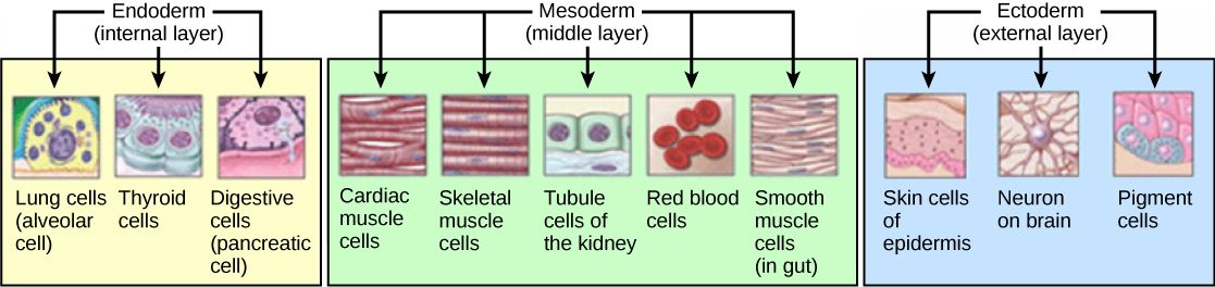
Figure: The germ layers present during the gastrulation stage of embryonic development and what develops from each. Most connective tissue structures develop from the mesoderm.
Creative Commons Source CNX [CC BY-SA 3.0 (https://creativecommons.org/licenses/by-sa/3.0)]
"that consists of various cells (such as fibroblasts and macrophages)" -
The cells present in connective tissue are:
Fixed cells - do not move within the tissue:
- Fibroblasts - the most common cell in connective tissue, their role is to synthesise the extracellular matrix, collagen and ground substance, producing the structural framework of connective tissue.
- Other fixed cells: Mast cells (immune cells whose granules release histamine and other chemical signals), Macrophages (cells that engulf pathogens and debris, and show pieces of them (antigens) to other immune cells), Adipocytes (fat-storing cells) and Mesenchymal stem cells (stem cells that can become other connective tissue cells).
Wandering cells - able to exit and enter tissues, these are immune cells that monitor the tissues for infection:
- Leucocytes (white blood cells), including lymphocytes, monocytes, eosinophils and basophils (white cells with granules), and plasma cells (cells that make antibodies).
"and interlacing protein fibres" -
The fibres present in connective tissue are:
These fibres are flexible and have a high tensile strength (they resist being pulled apart). Collagen is the most common protein in the body, and at least 28 types have been identified. Type I makes up over 90% of the body's collagen. The structure of type I collagen is a triple helix of three alpha chains (protein chains wound around each other), as seen below:
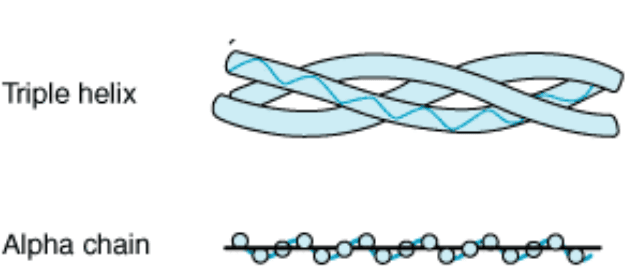
Figure: The structure of collagen, formed from a triple helix of alpha chains.
Fibroblasts secrete procollagen, which is then converted to collagen molecules outside the cell and aggregated to form the final collagen fibrils. Vitamin C is essential for the hydroxylation (adding hydroxyl, or OH, groups) of the amino acids proline and lysine residues in procollagen. This step is crucial for stable collagen formation. A deficiency in vitamin C can lead to scurvy, which is characterised by tiredness, swollen or bleeding gums, red or blue spots on the skin, poor wound healing and impaired bone formation. Abnormal production or processing of type I collagen causes osteogenesis imperfecta (brittle bone disease, a genetic condition in which the bones break easily).
Reticular fibres are thin fibres made of type III collagen. They are secreted by fibroblasts, including the fibroblast-like reticular cells of lymph nodes, and are also called reticulin fibres. The fibres branch and cross-link (join to one another) to form a fine supporting mesh in soft tissues such as liver, bone marrow, and the tissues and organs of the lymphatic system.
Elastic fibres allow tissues to recoil (spring back) after being stretched or distended (swollen). Elastin is the main component of elastic fibres, forming a core that is surrounded by a mantle of microfibrils (very fine fibres) rich in fibrillin. Elastic fibres occur in most connective tissues but to varying degrees. They are particularly important in the artery walls and the dermis (the deeper layer of the skin). Marfan syndrome is an autosomal dominant disorder (a child can inherit it even if only one parent has it) caused by a defect in the fibrillin gene, which reduces the production of fibrillin and makes the elastic tissue abnormal. People with Marfan syndrome are often tall and slim, with abnormally long, thin limbs, fingers and toes (arachnodactyly) and loose, very flexible joints. The walls of the aorta (the main artery of the body) can be weak, so the aorta may enlarge and, in severe cases, split (rupture).
"embedded in a chiefly carbohydrate ground substance" -
Ground substance is a viscous, slippery substance with a high water content, composed primarily of proteoglycans.
Proteoglycans are very large molecules. Each consists of a core protein with glycosaminoglycans (GAGs, long chains of repeating sugar units) attached. These GAGs attract water and form a gel that permits diffusion but resists compression. The sugar chains of the GAGs are why the definition above describes ground substance as "chiefly carbohydrate".
Hyaluronic acid (also called hyaluronan) is a GAG with no sulfate groups, unlike chondroitin sulfate and keratan sulfate. It forms very large proteoglycan aggregates by binding to proteoglycans (such as aggrecan, the main proteoglycan of cartilage) via link proteins. These highly hydrophilic (water-attracting) complexes help cartilage resist compression while remaining flexible. In cartilage, the main GAGs within proteoglycans are chondroitin sulfate and keratan sulfate.

Image: Shows the arrangement of fibroblasts within an extracellular matrix of fibrous proteins and ground substance.
Creative Commons Source Sagearbor [CC BY-SA 4.0 (https://creativecommons.org/licenses/by-sa/4.0)] modified by Dr. Marcus Judge
"that supports, ensheathes, and binds together other tissues" -
There are a number of roles of connective tissues - here are the 6 key ones:
- Support and connection - connects cells to form tissues, tissues to form organs and organs to form the body.
- Transportation - the surrounding matrix provides a medium through which nutrients can diffuse, and blood (a fluid connective tissue) carries nutrients, waste and chemical messengers around the body.
- Protection - provides a cushion between tissues.
- Storage - adipose tissue (fat tissue), a type of connective tissue, stores surplus energy as fat.
- Defence against infection - immune cells within the tissue attack microbes that breach the epithelial surface.
- Wound healing - fibroblasts lay down new collagen to repair damaged tissue.
"and that includes loose and dense forms (such as adipose tissue, tendons, ligaments, and aponeuroses) and specialised forms (such as cartilage and bone)" -
Connective tissue can be:
Loose - contains many cells, few collagen fibres and much ground substance, so it has a viscous (thick and sticky) consistency. It is found mainly directly beneath epithelia (the sheets of cells that line the body surfaces) and around blood vessels. These are places where bacteria or viruses that have breached the epithelial surface can be attacked and destroyed by the large number of immune cells present in loose connective tissue.
Dense - contains few cells, almost all of which will be fibroblasts (for fibre formation), many collagen fibres and little ground substance. Dense connective tissue can be further split into:
- Regular dense - Collagen fibres are arranged in parallel bundles and are densely packed, with fibroblasts between the bundles. It is designed to withstand stress in a single direction. It is seen in tendons, ligaments and aponeuroses.
- Irregular dense - Collagen fibres are arranged in bundles orientated in various directions, with fibroblasts between the bundles. It is designed to withstand stress in multiple directions. It is seen in the submucosa (the layer beneath the lining) of the intestine and in the deeper (reticular) layer of the dermis.
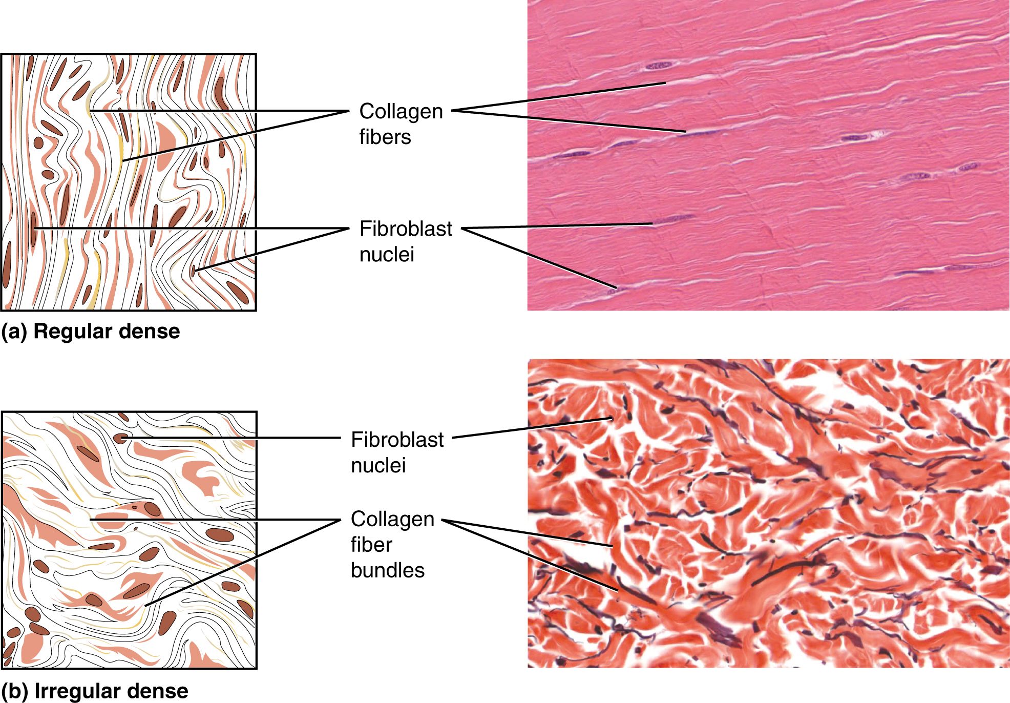
Image: The difference between regular and irregular dense connective tissue. Notice the parallel bundles of regular dense vs the seemingly random arrangement of irregular dense.
Creative Commons Image Source OpenStax College [CC BY 3.0 (https://creativecommons.org/licenses/by/3.0)]
Examples of connective tissues within the body:
Tendons
In tendons (connective tissue that connects muscles to bones) the collagen bundles lie in a parallel, densely packed formation in line with the tensile (pulling) force exerted by the muscle. Rows of elongated flattened fibroblasts lie between the collagen bundles. The bundles transmit the muscle's force to the bone and so cause movement.
Ligaments
Ligaments (connective tissue that connects bone to bone) are mostly dense regular connective tissue. Their collagen bundles are densely packed and mostly parallel, but they undulate (follow a wavy course) and are arranged in fascicles (small bundles), separated by loose connective tissue. A few ligaments, such as those between the vertebrae of the spine, are elastic and contain many elastic fibres.
Aponeuroses
An aponeurosis is a flat sheet of dense regular connective tissue. The bundles of fibres within one layer run parallel, and they are often arranged at about 90 degrees to the bundles in the adjacent layers. They are commonly found covering large areas of the body and providing structural support to areas such as the abdomen and back.
Dermis
The dermis (the layer of skin beneath the epidermis) is mostly dense irregular connective tissue. This makes up its thick, deeper reticular layer, while the thinner, more superficial papillary layer is loose connective tissue. The bundles of collagen are densely packed but irregularly arranged, such that they are orientated in multiple directions. The skin can therefore resist forces in multiple directions to prevent tearing. The elastic fibres let the skin stretch a little and spring back to its original shape after it is bent or folded.
- 16980



