

Next Lesson - Lung Function Testing
Core
The pH of plasma is important because having a high or low pH can affect the ability of certain proteins to function. For example, haemoglobin molecules function best at pH around 7.4, meaning that the pH of plasma needs to be kept between 7.35 and 7.45 to allow adequate oxygen transport.
In clinical practice, this is measured on an arterial blood gas (ABG). The interpretation of this test is explained at the end of the article (it is important to understand the physiology behind the control of plasma pH to be able to interpret an ABG).
Hypercapnia occurs when there is a rise in the partial pressure of carbon dioxide (pCO2) above 6kPa, whilst hypocapnia occurs when there is a reduction in the partial pressure of carbon dioxide below 4.7kPa.
Hypoxaemia means abnormally low oxygen in arterial blood. Hypoxia means inadequate tissue oxygenation; it can occur even without hypoxaemia. The normal reference range for the partial pressure of oxygen (pO2) on an arterial blood gas is 11-13kPa.
When there is an increased ventilation rate, the pO2 will increase and the pCO2 will fall as more oxygen is inhaled and more carbon dioxide is expired. The opposite can be seen when the ventilation rate decreases.
In times of hypoxia, the ventilation rate will increase (hyperventilation) as an attempt to increase the pO2, but this leads to a decrease in the pCO2.
Based on the oxygen-haemoglobin disassociation curve, the oxygen saturation of haemoglobin falls much more steeply below 8kPa. This means that at any partial pressure of oxygen above 8kPa, haemoglobin is pretty much fully saturated. However, due to the co-operative binding seen with haemoglobin (change in structure of haemoglobin as more oxygen molecules bind to make it easier to fully saturate), below 8kPa there is a steep drop in percentage saturation of haemoglobin.
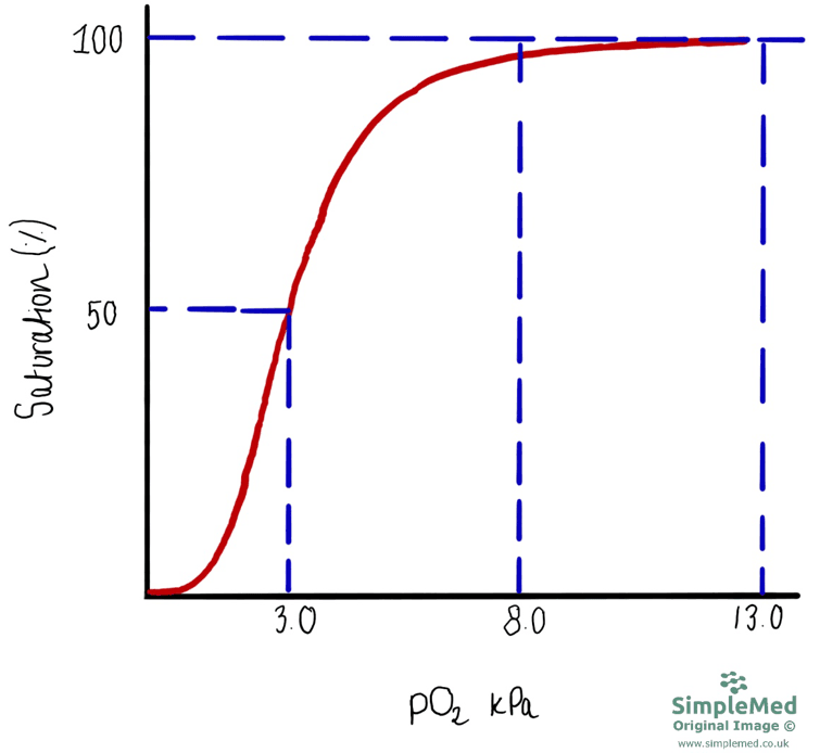
Diagram - Graph of the oxygen-haemoglobin dissociation curve. Saturation falls more steeply below approximately 8 kPa and also changes along the flatter upper portion of the curve.
SimpleMed original by Dr. Peter Parkinson
Once the pCO2 begins to increase, the pH of the plasma will begin to fall, causing acidosis. When the pCO2 begins to decrease, the pH of the plasma will increase, resulting in alkalosis.
Structures and mechanisms within the body can be affected by changes to pH of the plasma. Enzymes can lose normal function and may denature at extremes of pH, which can affect processes that rely on enzymes such as aerobic respiration and gluconeogenesis.
Once the pH of the plasma is above 7.6, the distribution of free calcium ions changes, with less ionised calcium available due to increased binding to proteins. This increases neuronal excitability, making neurones more likely to depolarise and can cause tetany (involuntary muscle contraction).
Hypoventilation can cause a build up of carbon dioxide in the blood. Carbon dioxide reacts with water to form carbonic acid, which dissociates into hydrogen ions (H+) and hydrogen carbonate ions, lowering blood pH. This is known as respiratory acidosis. Conditions such as type 2 respiratory failure, e.g. severe chronic obstructive pulmonary disease (COPD), drug overdose and neuromuscular disease, can lead to respiratory acidosis.
Hyperventilation removes carbon dioxide faster than it is produced, lowering blood pCO2. The carbonic acid/bicarbonate equilibrium shifts towards carbon dioxide and water, reducing free H+ and raising pH. This causes respiratory alkalosis; it does not remove all carbon dioxide from the blood. Conditions such as panic attacks can cause respiratory alkalosis.
Breathing helps regulate arterial oxygen and carbon dioxide tensions. Peripheral chemoreceptors in the carotid and aortic bodies respond to reduced arterial pO2, increased pCO2 and reduced pH. Their ventilatory response to falling oxygen becomes stronger below approximately 8 kPa.
Peripheral chemoreceptors contribute to the graded ventilatory response to changes in arterial carbon dioxide and pH.
Central chemoreceptors in the brainstem respond to changes in the pH of surrounding extracellular fluid and CSF. Carbon dioxide crosses the blood-brain barrier readily and forms hydrogen ions locally. The rapid movement of blood hydrogen and hydrogen carbonate ions is much more limited, so acute changes in arterial carbon dioxide strongly influence central chemoreceptor stimulation. This is limited permeability, not an absolute inability of ions to cross.
The blood-brain barrier is formed by specialised brain capillary endothelial cells. The choroid plexus produces and helps regulate the composition of CSF, including hydrogen carbonate transport. Tight junctions between its epithelial cells form the distinct blood-CSF barrier.
The pH of CSF depends on the balance between hydrogen carbonate and dissolved carbon dioxide. Carbon dioxide equilibrates readily, while regulation of CSF hydrogen carbonate occurs more slowly. The system is therefore not wholly independent of plasma hydrogen carbonate, particularly during sustained acid-base disturbances.
If arterial pCO2 rises, there will be a short delay before the CSF pCO2 increases. This change will be detected by central chemoreceptors and will lead to an increase in ventilation to attempt to remove the excess carbon dioxide. This is a form of negative feedback.
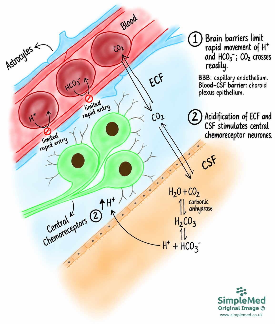
Diagram - Central chemoreceptors respond to local extracellular fluid and CSF pH changes after carbon dioxide crosses the endothelial blood-brain barrier. Rapid hydrogen and hydrogen carbonate movement is limited. Choroid plexus epithelium forms the separate blood-CSF barrier.
SimpleMed original by Dr. Peter Parkinson

Diagram - The homeostatic control of plasma pH and the responses made to changes to pH
Creative commons source by OpenStax College [CC BY-SA 4.0 (https://creativecommons.org/licenses/by-sa/4.0)]
In people with healthy lungs, this system works really well as changes in pCO2 are detected centrally and the ventilation rate changed accordingly.
However, in people with chronic lung conditions, like COPD, increased levels of CO2 become normal as their lungs are not very good at getting rid of CO2. If the system worked in these patients as it does in a healthy patient, they would be persistently hyperventilating, which would be very uncomfortable.
In reality, with cases of chronic hypercapnia, the CSF pH prompts the choroid plexus to persistently pump in large amounts of HCO3- ions, meaning that the pH becomes normal. This brings the ratio of carbon dioxide to hydrogen carbonate into normal range (even though both of these results are high), and this means that the pH is normal. This returns the breathing rate of this patient to normal, and ‘resets’ the system around a higher tolerated pCO2.
This ‘resetting’ of the central chemoreceptors to accept much higher pCO2 as ‘normal’ means that the patient depends on their oxygen sensitive peripheral chemoreceptors to modify their ventilation rate if needed. For example, in a patient with COPD, their central chemoreceptors may have ‘reset’, but their peripheral chemoreceptors still detect a low pO2, so the ventilation rate is increased. This is known as the ‘hypoxic drive’, and is why, in some COPD patients, giving high levels of oxygen for long periods of time can be dangerous. Raising the pO2 reduces peripheral chemoreceptor stimulation, which can contribute to reduced ventilation.
Metabolic Acidosis and Alkalosis
The body produces acids in various processes: lactic acid during anaerobic respiration and acidic ketones from gluconeogenesis of fatty acids and ketogenic amino acids.
Hydrogen carbonate ions react with the H+ ions produced by the body and when in excess, it can lead to the depletion of HCO3- ions. As the concentration of H+ ions increases, the ratio between pCO2 and HCO3- ions increases, resulting in a decrease in plasma pH. This is deemed as metabolic acidosis. The body tries to counteract the decrease in plasma pH by increasing ventilation to decrease pCO2 and bring the plasma pH into the normal range.
Persistent vomiting causes loss of gastric hydrogen and chloride ions and can produce metabolic alkalosis. Compensatory hypoventilation raises carbon dioxide, but is limited by the need to maintain oxygenation. Normally the kidneys can excrete excess hydrogen carbonate. However, volume and chloride depletion and hypokalaemia favour renal hydrogen carbonate retention and can maintain the alkalosis, rather than allowing effective renal correction.
Renal Production and Control of Hydrogen Carbonate Ions
The pH of the plasma depends on the ratio of the concentration of HCO3- ions to the pCO2 of the blood. The kidneys are responsible for controlling the concentration of HCO3- ions by altering the excretion of the ion.
Hydrogen carbonate ions are filtered at the glomerulus of the kidneys and enter the filtrate, but the majority of these ions are recovered at the proximal convoluted tubule (PCT). PCT cells excrete H+ ions into the lumen as they transport sodium ions into the cell. The H+ ions react with HCO3- ions present in the filtrate, resulting in carbon dioxide and water being formed. The carbon dioxide diffuses into the PCT cells and the reverse reaction occurs, producing H+ and HCO3- ions inside the PCT cell. The HCO3- ions can then be secreted into the extracellular fluid (ECF) before moving into the plasma.
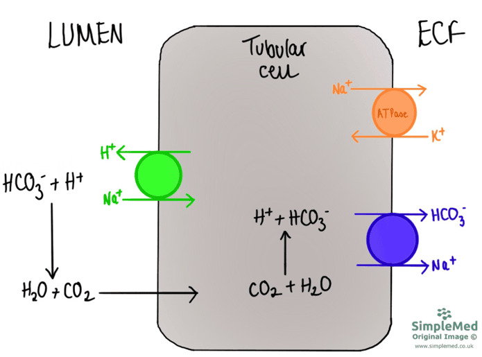
Diagram - The movement of hydrogen ions out of proximal convoluted tubule (PCT) cells and movement of hydrogen carbonate ions into the extracellular fluid from PCT cells
SimpleMed original by Dr. Peter Parkinson
Kidneys also produce HCO3- ions from amino acids, in the proximal tubule cells of the kidney, but this process takes a while. Glutamine (an amino acid) is broken down to ammonium (which further breaks down into H+ ions and ammonia) and α-ketoglutarate. The ammonia molecules diffuse into the filtrate and are able to bind with H+ ions present in the lumen to form ammonium, which cannot diffuse across the cell membrane, trapping the ammonium in the filtrate. This is a form of buffering in the urine to prevent the urine from becoming too acidic, as this would damage the cells of the urinary tract. The α-ketoglutarate is converted to HCO3- ions inside the cells of the PCT, which can be secreted into the ECF, before being transported into the blood.
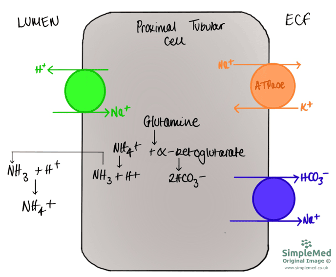
Diagram - The generation of ammonium and hydrogen carbonate ions from glutamine
SimpleMed original by Dr. Peter Parkinson
In the distal convoluted tubule and collecting ducts, H+ ions are also actively secreted into the lumen via α-intercalated cells. These H+ ions bind with ammonia and phosphate ions; these molecules are then excreted in urine. Phosphate ions act in the same as as ammonia, by buffering H+ ions.
Inside the α-intercalated cells, the reaction of carbon dioxide and water to H+ ions and HCO3- ions is in favour of the forward reaction as H+ ions are excreted into the lumen. Hydrogen carbonate ions are secreted into the ECF by α-intercalated cells as chloride ions are moved into the α-intercalated cells.
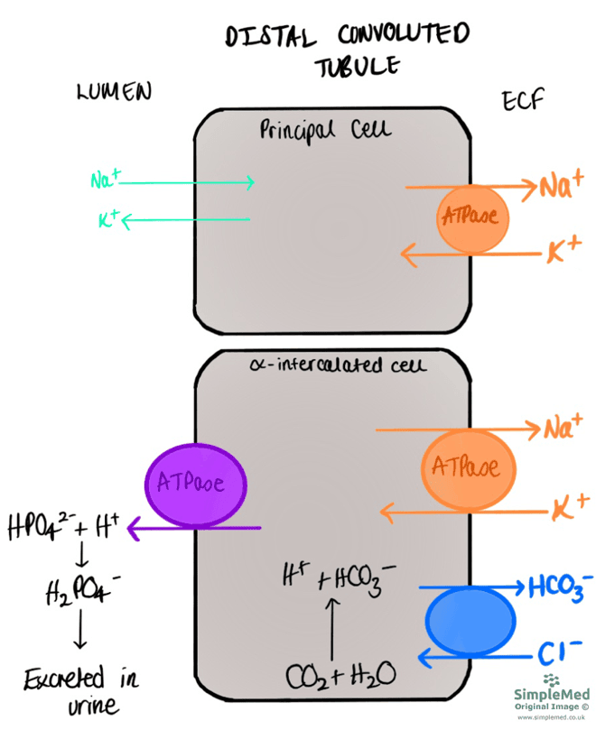
Diagram - The movement of phosphate ions out of α-intercalated cells in the distal convoluted tubule
SimpleMed original by Dr. Peter Parkinson
When there is an increase in the pCO2 (for example secondary to hypoventilation), the concentration of H+ ions secreted into the urine will increase, causing the pH of the blood to decrease. The kidneys will begin to excrete less HCO3- ions, so there are more HCO3- ions present in the blood. This will help the pH to go back to normal, and is a form of compensation (see later).
If the pCO2 begins to decrease (for example secondary to hyperventilation), the pH of the plasma will begin to increase. The kidneys will then excrete more HCO3- ions to try to bring the pH of the plasma back within normal range. However, this change can take quite a long time, so any compensation (change in HCO3-) seen in these patients is a chronic change rather than an acute one.
Potassium Ions and Acid Base Disturbances
There is a relationship between potassium ions and the plasma pH.
To regulate the amount of H+ in the plasma, the body can move it in or out of cells in exchange for K+ ions. This means that any changes in plasma pH can cause changes in K+ levels. This swap is known as the reciprocal cation shift.
Acidosis can increase serum potassium through movement of potassium out of cells, particularly in mineral metabolic acidosis. The effect is smaller in organic acidoses and is not a universal one-for-one hydrogen/potassium exchange. Serum potassium also depends on insulin, renal function, urinary losses and volume status, so acidosis does not invariably cause hyperkalaemia.
Alkalosis can favour movement of potassium into cells. In persistent vomiting, gastric acid and chloride loss can cause metabolic alkalosis, while volume depletion stimulates aldosterone-related renal potassium loss. These mechanisms can contribute to hypokalaemia, but renal potassium excretion also depends on volume status, renal function and other factors; alkalosis alone does not invariably cause increased potassium loss.
This reciprocal cation shift can also be put into practice where the potassium abnormalities come before the pH changes:
- Hyperkalaemia can contribute to metabolic acidosis by reducing renal ammonia excretion and therefore net acid excretion. In type 4 renal tubular acidosis, impaired aldosterone production or action also reduces potassium and hydrogen ion excretion. This association depends on the underlying renal and hormonal disturbance; hyperkalaemia does not invariably cause acidosis through urinary hydrogen carbonate loss.
- Hypokalaemia can promote and maintain metabolic alkalosis, including through increased renal hydrogen carbonate reabsorption and hydrogen ion excretion. The acid-base outcome depends on the underlying illness, volume status and renal function; a low potassium concentration alone does not establish metabolic alkalosis.
Diabetic ketoacidosis (DKA) is a key condition when considering the relationship between pH and potassium disturbance: it causes both metabolic acidosis and systemic depletion of potassium ions. In DKA, ketones are produced as a source of fuel, and because ketones are acidic, this causes K+ ions to move out of the cells as blood pH falls due to increased ketoacid levels and reduced insulin, which normally drives potassium into cells. However, with there being increased concentration of glucose present in the lumen of nephrons, this increases the osmolality and K+ ions are trapped in the lumen as well. This results in increased concentration of K+ ions being excreted in urine and a depletion systemically.
Compensation and The Anion Gap
As previously discussed, the plasma pH depends on the ratio between concentration of hydrogen carbonate ions and the partial pressure of carbon dioxide.
If there are changes in the pCO2, the kidney can compensate by alternating the excretion of HCO3- ions. In respiratory acidosis, the kidneys will compensate by excreting less HCO3- ions and in respiratory alkalosis, the kidneys will compensate by excreting more HCO3- ions. However, it can take the kidneys several days to compensate, so this is not an immediate management strategy.
In a patient with compensated respiratory acidosis, the arterial blood gas will show: high pCO2, raised [HCO3-] with a relatively normal plasma pH.
In a patient with compensated respiratory alkalosis, the arterial blood gas will show: low pCO2, low [HCO3-] with a relatively normal plasma pH.
The anion gap is the difference between the concentrations of cations and anions in the blood. It includes the concentrations of sodium and potassium ions as the cations, and the concentrations of chloride and hydrogen carbonate ions as the anions.
The normal anion gap is usually around 8 to 12 mmol/L when potassium is excluded from the calculation (local reference ranges may vary). This reflects the presence of unmeasured anions in normal plasma.
It can be helpful to use the anion gap to work out the cause of an abnormality, for example:
- Changes in potassium concentration have little direct effect on the anion gap, as potassium contributes minimally to the calculation.
- In compensated respiratory acidosis, renal compensation leads to a raised HCO3- concentration, and the anion gap is typically unchanged.
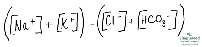 Image - The anion gap equation
SimpleMed original by Dr. Peter Parkinson
It is important to remember that the anion gap is calculated using a limited number of measured ions, but it does reflect the build-up of unmeasured anions such as lactate or ketoacids, which increase the gap.
The anion gap is increased if HCO3- ions are replaced by another anion; this is seen when there is metabolic production of an acid such as diabetic ketoacidosis and lactic acidosis.
With a renal cause of metabolic acidosis, the anion gap can be normal. During the excretion of HCO3- ions, they are replaced by chloride ions in a 1:1 ratio, meaning the anion gap will be unaffected.
In metabolic alkalosis, the kidneys try to compensate by reducing H+ ion excretion and HCO3- recovery. Metabolic alkalosis is also accompanied by systemic volume depletion, this leads to the kidneys increasing the recovery of sodium ions and HCO3- ions whilst excreting H+ ions. This means that the resorption of anions and cations is balanced, so the anion gap will not change.
Arterial Blood Gas Interpretation
As mentioned at the start of this article, all of this information is provided on an arterial blood gas (ABG). It is important to remember that pCO2 and HCO3- results can be outside the normal range due to pathology relating to their formation or removal, or because they have compensated for a change in the other variable to get the pH closer to the normal range.
This is one method that can be used to interpret the results of an ABG:
First look at the pH - is it low (below 7.35) indicating acidosis, or high (over 7.45) indicating alkalosis?
If Acidotic:
- Look at the pCO2 - is it high (respiratory cause of the acidosis) or low (compensating for a metabolic cause)
- Look at the HCO3- - is it low (metabolic cause of the acidosis) or high (compensating for a respiratory cause)
If alkalotic:
- Look at the pCO2 - is it low (respiratory cause of the alkalosis) or high (compensating for a metabolic cause)
- Look at the HCO3- - is it high (metabolic cause of the alkalosis) or low (compensating for a respiratory cause)
Things to remember with ABGs:
- Remember that changes in HCO3- reflect renal handling and metabolic processes. Renal compensation takes time, but a low bicarbonate does not by itself indicate whether a disorder is acute or chronic.
- Remember that compensation is done to get the pH back into the normal range. This means that there are two levels of compensation:
- Partial compensation occurs when both variables are outside the normal range, and the pH has not been pushed back into the normal range.
- Full compensation occurs when both variables are outside the normal range, but the pH is within normal range.
- It is always important to interpret the ABG in the clinical context of the patient - this may help determine whether the cause is chronic or acute.
Edited by: Dr. Maddie Swannack
Reviewed by: Dr. Thomas Burnell
- 7791

