

By Dr. Thomas Burnell and Dr. Bethany Turner
Next Lesson - Embryological Development of the CVS
Core
When an action potential (AP) reaches the cardiac muscle, it travels down the T-tubules (deep invaginations of the myocyte cell membrane).
The AP causes the opening of L-type calcium channels that are found on the cell surface membrane, causing an influx of calcium ions into the sarcoplasm.
Calcium within the cell binds to ryanodine receptors on the sarcoplasmic reticulum (SR) surface resulting in the release of calcium ions from the sarcoplasmic reticulum. This is calcium induced, calcium release (CICR).
The calcium that has been released, binds to troponin C causing a conformational change in tropomyosin to expose binding sites on actin. The myosin heads bind to these sites. This then allows the sliding filament mechanism to occur and for the cardiac muscle to contract.
After contraction, most calcium is pumped back into the sarcoplasmic reticulum by sarcoplasmic reticulum calcium-ATPase (SERCA), while the rest is transported out of the cell across the surface membrane by Ca2+-ATPase and the Na+/Ca2+ exchanger.
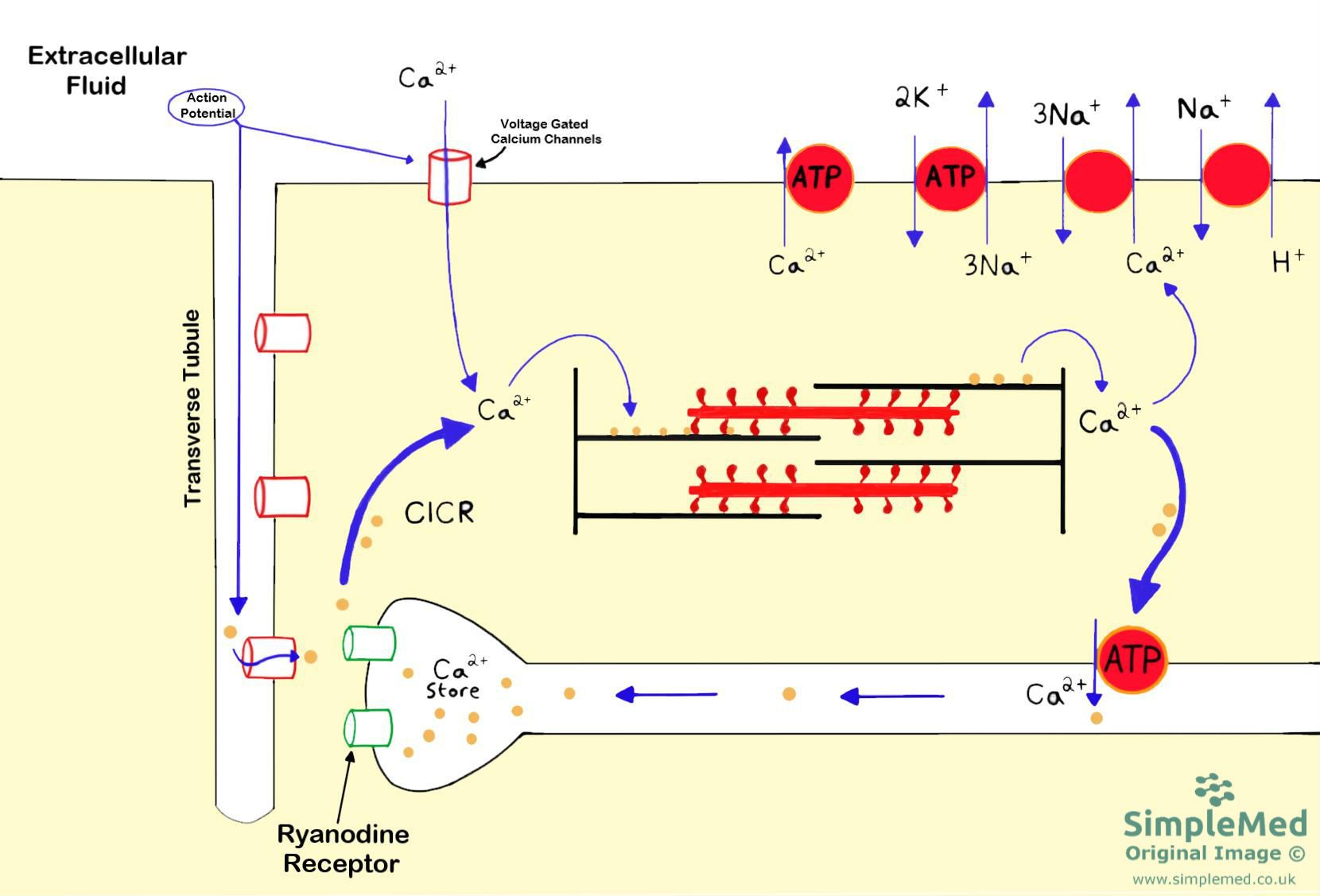
Diagram - The events involved in muscle contraction
SimpleMed original by Dr. Thomas Burnell
The conduction system in the heart is outlined in the Cardiac Cycle article. This explains how the contraction of the heart is co-ordinated. Here, we will discuss the regulation of the electrical activity in the heart.
Sino-Atrial Node Action Potential
The cells within the Sino-Atrial (SA) node slowly depolarise, triggering a wave of depolarisation which will spread through the conduction system of the heart, causing it to beat.
The pacemaker potential is the gradual depolarisation between action potentials. Central SA node cells have no stable resting membrane potential. Hyperpolarisation activates HCN channels (hyperpolarisation-activated, cyclic nucleotide-gated channels), which conduct both Na+ and K+. They carry the funny current (If), a net inward current at diastolic pacemaker voltages. If contributes to the pacemaker potential alongside calcium currents and a declining outward potassium current; it is not the name for the whole depolarising phase.
Calcium entry through T-type and L-type channels contributes as the membrane approaches threshold. The action-potential upstroke in central SA node cells is carried mainly by L-type Ca2+ channels. Repolarisation follows as these calcium channels inactivate and outward current through voltage-gated K+ channels increases.
Repolarisation returns the membrane towards its most negative voltage in the cycle, the maximum diastolic potential. Outward potassium current then declines and inward pacemaker currents bring the membrane towards threshold again. There is no stable resting phase between beats.
Cardiac sympathetic nerve endings release chiefly noradrenaline, while adrenaline reaches the heart chiefly through the circulation after release from the adrenal medulla. Both can stimulate β1-adrenoceptors in SA node cells. The resulting increase in cyclic AMP (cAMP) enhances pacemaker activity, helping the cells reach threshold sooner and increasing heart rate.
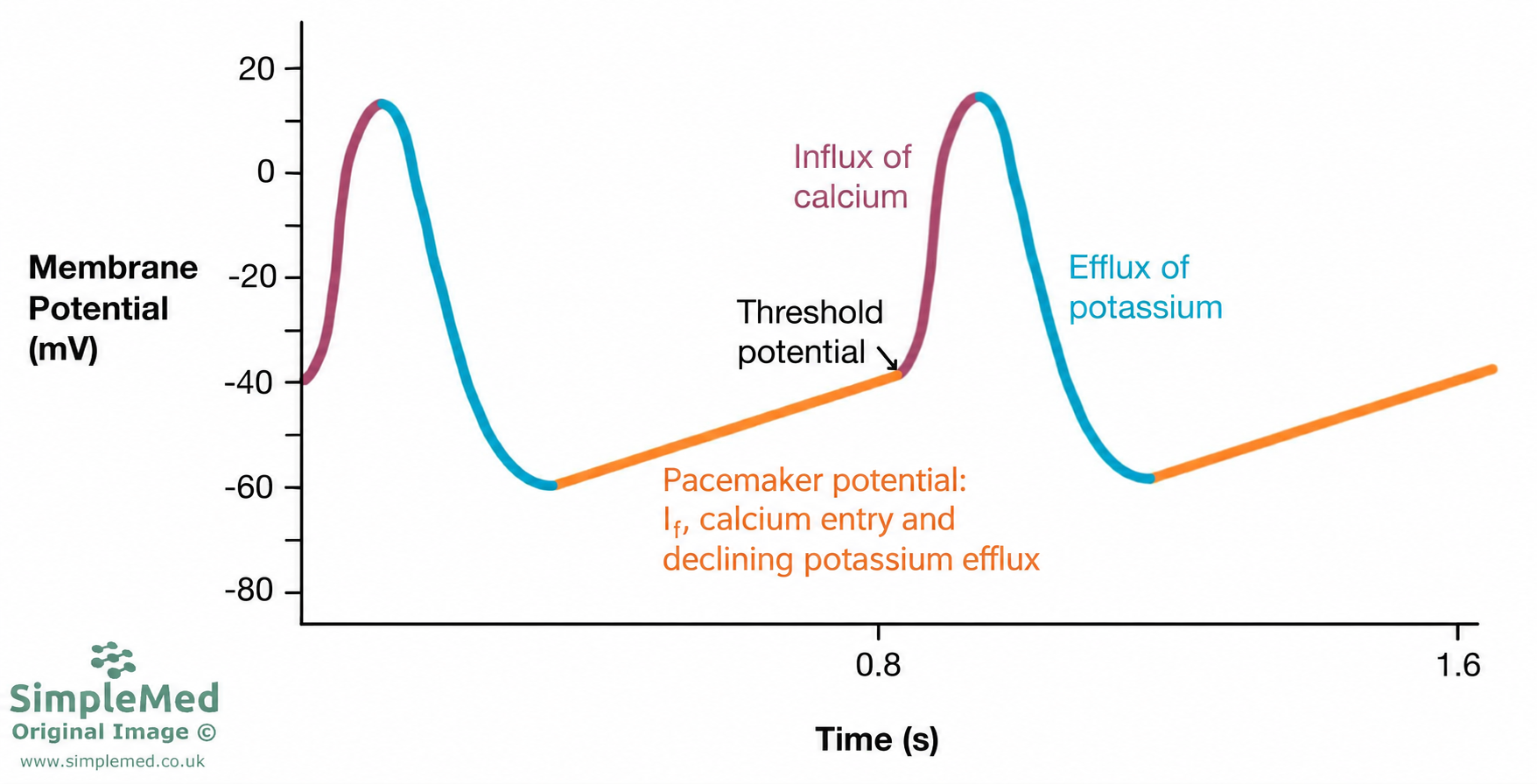
Diagram - SA node membrane potential. The orange phase is the pacemaker potential, the red upstroke is mainly calcium-dependent, and the blue phase is repolarisation towards the maximum diastolic potential. The pacemaker phase involves mixed Na+/K+ If, calcium entry and declining outward potassium current.
SimpleMed original by Dr. Bethany Turner
When the myocytes in the ventricles receive an AP from the conduction system, a special membrane potential is generated.
When depolarisation reaches the myocytes, the threshold is reached (around -80mV) and the voltage-gated Na+ channels (VGNC) open to allow an influx of Na+ ions into the cell. This is the upstroke of the AP (the orange line on the diagram below).
The AP peaks at around +20mV, where the VGNC close and there is opening of some VGKC. This results in transient repolarisation, with some K+ ions moving out the cell (small blue line).
L-type voltage-gated Ca2+ channels open and Ca2+ ions move into the cell. The influx of Ca2+ ions causes a plateau as the efflux of K+ ions is counteracted (purple line). This calcium binds to troponin C allowing muscle contraction to take place.
Once muscle contraction has occurred, there is inactivation of the Ca2+ channels and more VGKCs open to allow more K+ ions out of the cell and cause repolarisation (final blue line).
The membrane returns to a stable resting potential of about −90 mV, maintained mainly by potassium conductance. The myocyte remains at rest until the next depolarising impulse arrives.
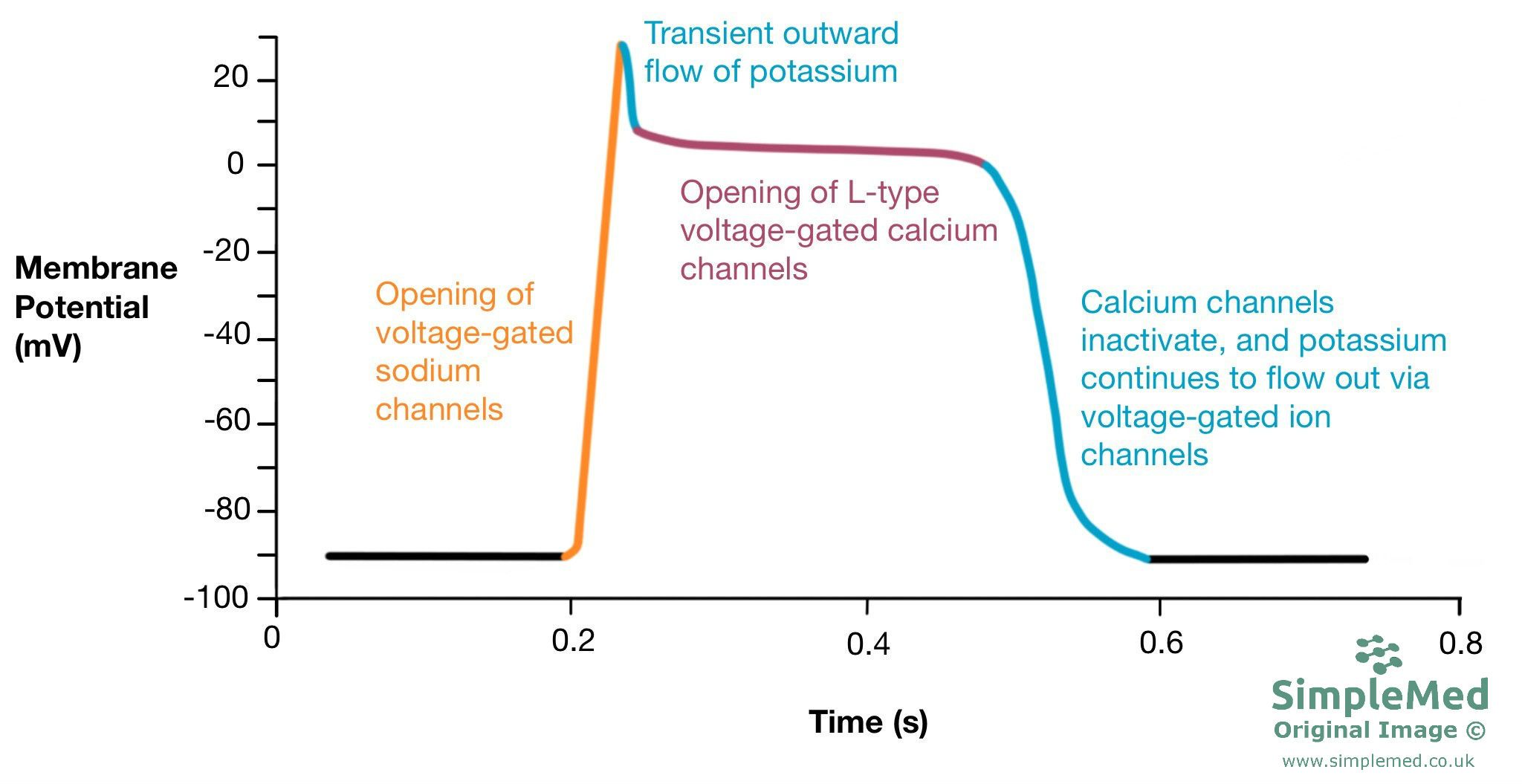
Diagram - The changes in the membrane potential in cardiac myocytes. The orange line represents the influx of sodium when the action potential arrives at the myocyte. There is a slight outward movement of potassium which causes a lowering of the membrane potential, represented by the blue line. This is followed by the influx of calcium into the cell which causes the membrane potential to plateau, represented by the purple line. The final blue line shows the return to the resting membrane potential by the outflow of potassium
SimpleMed original by Dr. Bethany Turner
There are changes to the ventricular action potential if a patient has hyperkalaemia or hypokalaemia:
- In hypokalaemia, there is a delay in repolarisation. This is because K+ channels do not work effectively as there is a decreased K+ ion concentration outside the cell.
- This delay in repolarisation results in increased excitability of the myocytes which increases the risk of fibrillation.
- In hyperkalaemia, there is less of a concentration gradient of K+ across the myocyte cell membrane.
- This leads to the equilibrium potential of K+ becoming less negative (closer to 0). As a result, the cell depolarises slightly.
- Some VGNCs in the cell membrane become inactivated due to the slight depolarisation, and this causes a slower upstroke in the ventricular AP.
- This slows the heart rate and can stop the heart if the K+ ion concentration is very high outside the cell.
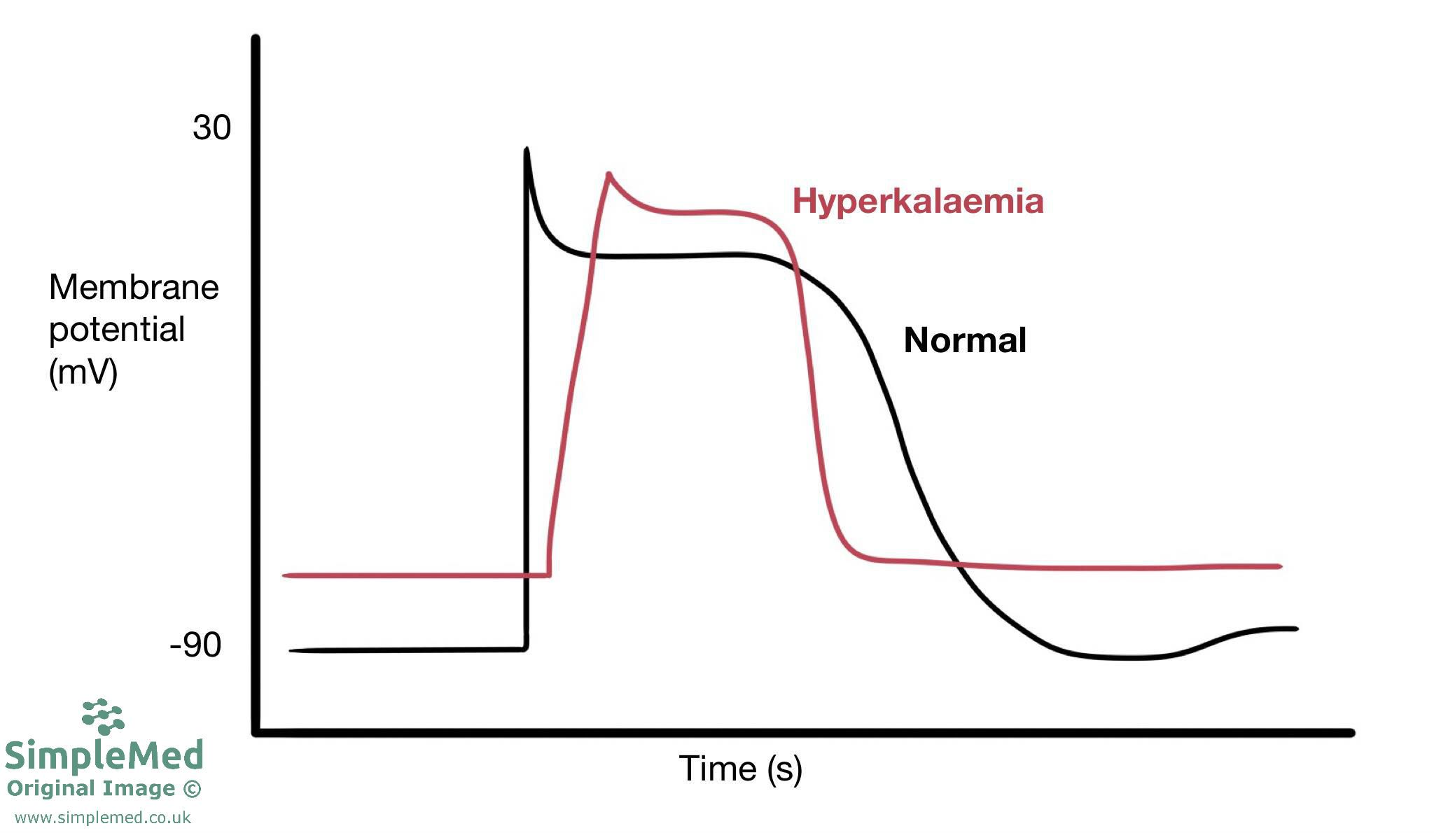
Diagram - The solid line shows the normal ventricular AP. The dotted line shows the ventricular action potential in hyperkalaemia. The upstroke is less rapid in hyperkalaemia
SimpleMed original by Dr. Bethany Turner
Edited by: Dr. Ben Appleby
Reviewed by: Dr. Marcus Judge
- 24447
