

By Dr. Marcus Judge and Dr. Ethan Kavanagh
Next Lesson - Gram Staining and Infection Investigations
Core
To understand this article, we advise that you read the Innate Immune Response, or have a rough understanding of it.
As outlined in the previous article, the innate response is rapid, but it is not specific to a particular microbe. It does not build a specific memory of past infections. The adaptive immune system fills this gap. So why does the adaptive immune response not start immediately fighting bacteria on infection?
While the innate immune response is coordinating the early ‘front line’ response against the invaders, the adaptive response must wait for its cells to become activated. Certain types of cells, called antigen presenting cells (APCs), are found throughout the body where they sample their environment for foreign antigens. APCs are a diverse group. They include macrophages and B-lymphocytes. Monocytes are white blood cells that circulate in the blood and mature into macrophages in the tissues. Dendritic cells are cells in tissues such as the skin that present antigens to T-lymphocytes. Langerhans cells are the dendritic cells of the skin.
An antigen presenting cell, e.g. a macrophage, will phagocytose a foreign pathogen, as part of the innate immune response. The macrophage takes in the pathogen and degrades it within lysosomes. It then displays a small sample of the antigen on its surface, held on a molecule called a Major Histocompatibility Complex (MHC). This display is called "presenting" the antigen (hence the name antigen presenting cell). This process can be broken down into 3 simple steps of: Capture (phagocytosis), Processing (digestion down to antigen fragments) and Presentation (using an MHC).
The next step is T cell activation. This occurs when the T cell receptor of a T-lymphocyte binds the antigen-displaying MHC on the surface of the macrophage. Other antigen presenting cells can do this too, and dendritic cells are especially good at activating naïve T-cells. Very few naïve T-cells can recognise any one antigen, so the right T-cell has to meet an antigen presenting cell that is displaying its antigen. It then has to multiply into enough cells to act. This is what delays activation of the adaptive immune response. Once activated, helper T-lymphocytes help B-cells to produce specific antibodies. They signal to the B-cells using cytokines (chemical messenger proteins released by immune cells). T lymphocytes also begin to multiply and to mount their own attack against the invading pathogen. We’ll come back to T- and B-lymphocytes, as we need to discuss how the APCs do their job.
MHC class I and class II are the two main groups of cell-surface molecules that present antigenic peptides to T cells. In humans, MHC molecules are called human leucocyte antigens (HLA). The MHC class III region of the genome (our genetic code) encodes other immune proteins, including complement components (blood proteins that help to destroy microbes). It also encodes inflammatory mediators (chemical signals that drive inflammation). The class III region does not make a third class of peptide-presenting surface molecules.
The two main classes differ in where they are found and which T cells they present to:
MHC class I molecules, including HLA-A, HLA-B and HLA-C, are expressed on almost all nucleated cells. They usually present peptides from proteins made inside the cell. These include normal self proteins and proteins from intracellular pathogens (microbes that live inside our cells). The peptides are shown to CD8 T cells (CD stands for “cluster of differentiation”, a numbered marker on the surface of T cells). The T-cell receptor recognises the peptide–MHC complex and CD8 acts as a co-receptor (a second surface molecule that helps the T cell grip the MHC molecule). Specialised antigen-presenting cells can also cross-present extracellular antigens (antigens from outside the cell) on class I, which is explained further below.
MHC class II molecules are characteristically expressed by professional antigen-presenting cells (cells whose main job is to present antigens to T cells), including dendritic cells, macrophages and B cells. Other cells, including epithelial cells (cells that line surfaces), can also carry class II molecules, and inflammation can trigger these cells to make more of them. In the conventional pathway, antigen taken up from outside the cell is processed into peptides and presented to CD4 T cells. Uptake may involve phagocytosis or other forms of endocytosis; phagocytosis is not confined to professional antigen-presenting cells. The T-cell receptor recognises peptide–MHC II and CD4 acts as a co-receptor. Co-stimulatory signals (extra activating signals that the T cell needs as well as antigen recognition) and cytokines help determine T-cell activation and subsequent differentiation.
Key facts about MHCs (they matter in medicine because they affect infections, transplants and autoimmune disease):
- The MHC molecules have a broad specificity (each one can bind many different peptides) so that a single cell can present multiple antigens from the same microbe to increase the immune response.
- The specific HLA molecules on a person's MHCs affect how well the body deals with certain infections, e.g. HIV. Some people with HIV carry HLA molecules (such as HLA-B57 and HLA-B27) that are linked to good control of the virus and slow disease progression. This is because these HLA molecules allow an effective T-cell response. These HLA molecules present key viral peptides. The virus needs these peptides to survive, so it cannot easily change (mutate) them. People whose HLA molecules only present non-crucial peptides (which the virus can mutate freely) are less likely to have this protection.
- A mismatch in MHC molecules between a donor and recipient in an organ transplant can cause rejection of the organ. If the organ transplanted is bone marrow, a mismatch in MHC molecules can lead to graft versus host disease (GvHD). GvHD can also happen when the MHC types match. This is because donor and recipient differ in other (minor) antigens. In GvHD, the new white blood cells produced by the donor marrow attack the host’s tissues.
- Having certain MHC molecules can predispose individuals to certain autoimmune conditions e.g. type 1 diabetes (the immune system destroys the insulin-making beta cells of the pancreas). The main high-risk types are HLA-DR3 and HLA-DR4, usually inherited with the neighbouring genes HLA-DQ2 and HLA-DQ8 (as the pairs DR3-DQ2 and DR4-DQ8).
A helpful way of remembering which class of MHC binds to which CD receptor is:
When you multiply the MHC class and CD number together you should get 8:
MHC Class I binds to CD8+ (1x8)
MHC Class II binds to CD4+ (2x4)
Fighting off intracellular microbes: MHC Class I
An intracellular organism, for example a virus invading a cell to take over its replication machinery, can infect any nucleated cell. For this reason, MHC class I is found on almost all nucleated cells.
An APC can present antigens on both MHC class I and MHC class II molecules. Cross-presentation specifically refers to the ability of some APCs to present extracellular (phagocytosed) antigens on MHC class I, allowing activation of CD8+ T cells.
So, if an APC (e.g. a macrophage) displaying an epitope on an MHC molecule arrives at a naïve T-cell, what happens next? (An epitope is the part of an antigen that the immune system recognises. An antibody attaches to it directly, whereas a T-cell recognises a short peptide fragment of it displayed on an MHC molecule.)
If the epitope is displayed on an MHC class I molecule, a CD8+ T cell binds it and becomes an activated cytotoxic T-cell. This ‘killer’ cell then kills infected body cells that display the same antigen on MHC class I.
Fighting off extracellular microbes: MHC Class II
Once a CD4+ T-cell binds an MHC class II molecule that is presenting its antigen, the T-cell can differentiate into a Th1, Th2 or Th17 cell. Class II molecules present antigen that the cell has taken up from outside. This route therefore covers extracellular microbes, and also microbes that have been engulfed by macrophages and survive inside them.
Th1 cells activate macrophages (using a cytokine called interferon-gamma). This helps the macrophages to kill microbes that try to survive inside them, such as mycobacteria (the group of bacteria that includes the cause of tuberculosis). This is why Th1 cells are linked to cell-mediated immunity, summarised below.
Th17 cells have several roles, but the main one we need to know is recruiting neutrophils (bringing them to the site of infection) using a cytokine called interleukin (IL) 17. This also helps to defend the skin and mucosal surfaces (such as the lining of the gut and airways) against extracellular bacteria and fungi.
Th2 cells have a few different responses:
- Increases the number of eosinophils to help kill parasites (via IL-5)
- Helps B-cells to produce antibodies (via IL-4). Antibodies help phagocytes to take up microbes, and they activate complement (a group of blood proteins, made mainly in the liver, that help to destroy microbes)
- Helps B-cells to switch to making IgE (immunoglobulin E) antibodies (via IL-4). IgE attaches to mast cells (tissue cells that release histamine, a chemical that makes blood vessels leaky and causes itching and swelling). When an allergen (the substance that triggers the allergy) binds the IgE, the mast cells release chemicals that cause local inflammation and our allergic reactions
A Quick Summary so far
Cell-mediated immunity:
- This fights off intracellular microbes such as viruses, some bacteria (those that live inside cells) and some protozoa (single-celled parasites)
- Cell-mediated immunity is an immune response that does not involve antibodies. It involves the activation of phagocytes, antigen-specific cytotoxic T-lymphocytes, and the release of various cytokines in response to an antigen
- The cells involved are naïve CD4+ T-cells, which differentiate into Th1 cells, and naïve CD8+ T-cells, which differentiate into cytotoxic T-cells
- Attack mechanisms are macrophages and cytotoxic T cells
Humoral immunity:
- This fights off extracellular microbes (microbes that live outside cells) such as many bacteria, parasites and worms. Fungi are a mixed case, because Th1 and Th17 cell responses are central to defence against fungi such as Candida (the yeast that causes thrush)
- Humoral immunity is the aspect of immunity that is mediated by large molecules (macromolecules) found in the fluids outside cells. These include secreted antibodies, complement proteins and certain antimicrobial peptides (small proteins that kill microbes)
- The cells involved are CD4+ T-cells, which differentiate into Th2 cells (these help B-cells to make antibodies) and Th17 cells (these bring neutrophils to the site of infection)
- Attack mechanisms are antibodies, complement, and phagocytosis
Primary and Secondary Immune Antibody-based Response
If you are unlucky enough to have had chickenpox, the good news is that you are very unlikely to get it again. This is because, if the varicella zoster virus has invaded before, the body has developed a memory against it. On re-exposure, the body mounts a memory-based reaction called the secondary immune response, which can destroy the virus before it causes disease or symptoms. The virus is not completely removed, though: it stays in nerve ganglia (clusters of nerve cells) and can reactivate years later as shingles.
The primary response is your body’s first response to a new pathogen that it has never encountered before. The main antibody made early in the primary response is IgM (immunoglobulin M). Each binding site of IgM grips its target less tightly (it has lower affinity) than IgG (immunoglobulin G), because IgM is made early, before the body has had time to fine-tune its antibodies. However, IgM is a large molecule with many binding sites, so overall it can still bind a target firmly (this overall binding strength is called avidity). It takes around 7-10 days for antibody levels to peak in a primary immune response, which is slow.
On the second exposure to the same pathogen, the secondary immune response is activated. This is primarily IgG based and takes less time, usually 3-5 days to reach its peak. This is a much faster and stronger response which also lasts longer than the primary immune response with IgM. This memory effect is also the basis of vaccines where the initial exposure is to an antigen or an attenuated (weakened) or dead pathogen. This builds the memory. A secondary immune response can then be mounted much more quickly when the person later meets the real pathogen. A quick secondary response is helpful because some diseases can do serious harm after just one exposure. For example, polio (a viral infection that can affect the brain and nerves) causes no symptoms in most people. Rarely, it causes paralysis, which can be life-threatening or leave permanent disability. Vaccines let us build a secondary response without having to suffer the disease.
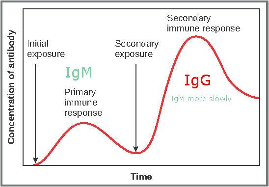
Figure: Shows the response of the adaptive immune system to a standard infection. The primary response on first exposure will be mainly IgM mediated. The secondary response on second exposure will be mainly IgG, which is made quickly because memory cells (long-lived cells that remember the antigen) activate faster than naïve cells. Some IgM is still made in the secondary immune response.
Creative commons source by OpenStax College, edited by Dr. Marcus Judge [CC BY 3.0 (https://creativecommons.org/licenses/by/3.0)]
The important thing to remember is that the secondary immune response is strongest when the same antigens that were there during the primary immune response are present. If the antigens have changed, the memory may give only partial protection. An example where this is relevant is with the influenza virus. It has certain antigens, such as haemagglutinin and neuraminidase. These are proteins on the surface of the flu virus, and they are also used to name certain flu strains (e.g. H1N1). These proteins differ slightly between flu strains. It is possible to get the flu in different years because the antigens change gradually from season to season (this is called antigenic drift). Immune memory of an earlier strain may not fully protect against a new strain. This is why the flu vaccine is updated for each winter: the World Health Organization recommends which strains to include, based on its monitoring of flu viruses around the world. The match is not always perfect, but the vaccine can still give some protection against a drifted strain.
Edited by: Dr. Ben Appleby and Dr. Marcus Judge
- 12802

