

Next Lesson - Motor System
Core
Sensation can be split into two main classifications: general sensation and special sensation.
Special sensation refers to vision, hearing, taste and smell. General sensation relates largely to touch and arises predominantly from epithelial surfaces. General sensation can be divided into somatic sensation, arising from the body wall and usually consciously perceived, and visceral sensation, arising from internal organs and often not consciously perceived.
There are a number of different modalities through which sensory information is expressed, and this is done through the type of receptor that the sensation is detected through.
Sensation is detected in different ways based on which receptor is activated.
The body can detect somatic sensation in seven main categories:
- Temperature
- Pain
- Pressure or crude touch
- Vibration
- Fine touch
- Proprioception or joint position sense
- Two point discrimination
In the central nervous system, these seven modalities are carried in two separate systems.
Temperature, pain, and pressure are carried through the spinothalamic tract.
Vibration, fine touch, proprioception and two point discrimination are carried through the dorsal column-medial lemniscus tract.
The sensory system of the body detects stimuli that have levels of gradation. This means that the body can detect temperature as cold-cool-comfortable-warm-hot, rather than just cold-hot. This variety in strength of signal detection means that the receptors are producing analogue signals.
The conversion from analogue to digital signals occurs in the primary sensory neurone, also called the first order sensory neurone, as graded receptor potentials are translated into action potentials. This is the neurone that connects the receptor to the central nervous system, as shown in the diagram below. It travels down the spinal nerve, through the dorsal root, and into the spinal cord. The cell body of the first order sensory neurone is in the dorsal root ganglion, as shown below.
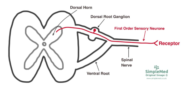
Diagram - The Path of a First Order Sensory Neurone from the Receptor to the Central Nervous System
SimpleMed original by Dr. Maddie Swannack
The conversion from analogue signals to digital signals occurs in the first order sensory neurone. The receptor detects a sensation, and the strength of the receptor activation results in a frequency of action potentials. Strong receptor activation causes high frequency of action potentials in the primary sensory neurone, and weak receptor activation causes lower frequency of action potentials. For example, bumping your head on a doorframe and being hit by a cricket ball both stimulate the crude touch receptors (along with others eg pain), but the light bump will cause weak receptor activation (leading to fewer action potentials), and the cricket ball will cause strong receptor activation (leading to a high frequency of action potentials).
Some rapidly adapting receptors, including certain mechanoreceptors, respond best to changes in signal strength. However, these receptors are rapidly adapting, meaning their frequency of firing decreases with extended stimuli, to help prevent ‘normal’ sensations (like clothes on the skin) from becoming overwhelming.
Slowly adapting receptors such as nociceptors (pain receptors) change the frequency of firing very little with continuous stimulation, explaining why pain is a persistent sensation.
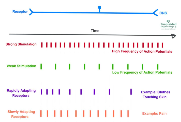
Diagram - Comparison of Action Potential Frequencies from Strong and Weak Stimulations
SimpleMed original by Dr. Maddie Swannack
A single primary sensory neurone supplies a single area of skin known as its receptive field. Each neurone has more than one receptor that inputs to it, but all of these receptors are of the same type - this makes sense because the neurone can only output digital signals through action potentials, and this cannot be used to differentiate between receptor types.
If an area of skin is supplied by a primary sensory neurone with a large receptive field, the area has a low sensory acuity, meaning its two-point discrimination is poor. This means that the two needle tips would have to be quite far apart to be detected as two separate points. The skin of the back is an example of a region with poor two point discrimination.
If an area of skin is supplied by a primary sensory neurone with a small receptive field, the area has a high sensory acuity, meaning its two point discrimination is good. This means that the two needle tips can be detected as separate even when they are placed quite close. The skin of the fingertip has very good sensory acuity.
It is also important to note that there is some overlap between the receptive fields of adjacent first order sensory neurones. This means that there is some overlap between the areas of skin supplied by one spinal nerve, and therefore some overlap between adjacent dermatomes. This is why when examining dermatomes, the best practice is to test the function of each dermatome using a spot that is central to the dermatome, as this is unlikely to have overlap with a neighbouring dermatome.
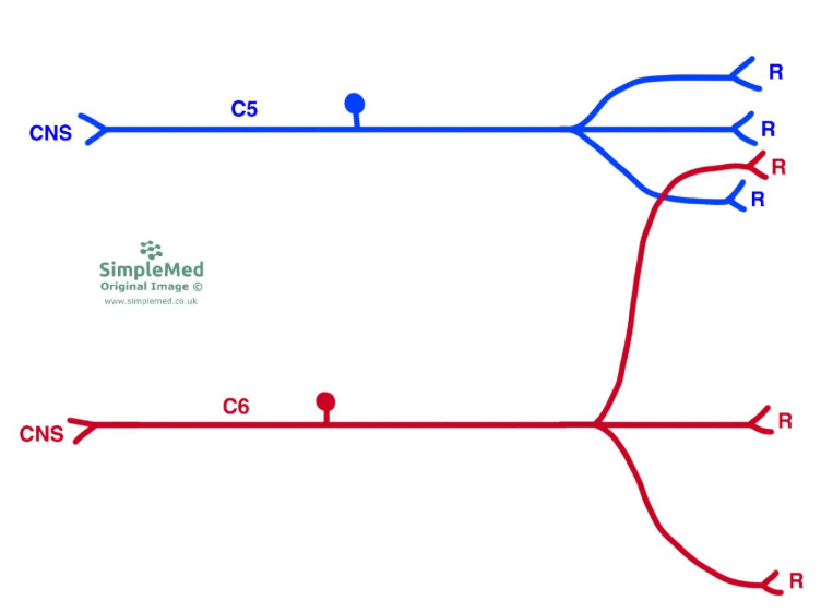
Diagram - The Receptive Fields of Two Adjacent Nerves
SimpleMed original by Dr. Maddie Swannack
Organisation of the Somatosensory System
The somatosensory system functions through the actions of a chain of three neurones, and every somatosensory impulse follows the following three laws:
The first neurone in the chain of the somatosensory system is called the first order sensory neurones or primary sensory neurones, noted with the symbol 1°.
These first order sensory neurones:
- Have their cell bodies in the dorsal root ganglion
- Communicate with a receptor
- Projects to the ipsilateral side of the CNS (on one side of the midline)
The second order neurones:
- Have their cell bodies in the spinal cord dorsal horn or medulla
- Decussate (cross the midline)
- Project onto third order neurones
The third order neurones:
- Have their cell bodies in the thalamus
- Project to the primary sensory cortex in the post-central gyrus.
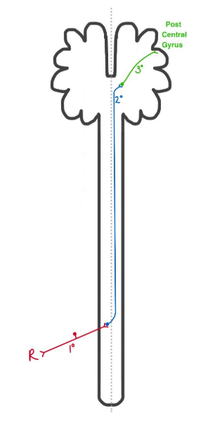
Diagram - The Basic Organisation of the Sensory Pathways in the Central Nervous System
SimpleMed original by Dr. Maddie Swannack
First order neurones are organised in a dermatomal pattern, and third order neurones are organised in a homuncular pattern (ie according to the homunculus). This means that a conversion of the signals must take place in the second order neurones.
This entire path is mapped out so that the impulses from neighbouring receptive fields are carried on neighbouring axons (ie the impulses for the sensation of the hand are carried on axons by the ones carrying impulses for the wrist). This is done to reduce the amount of ‘wiring’ needed.
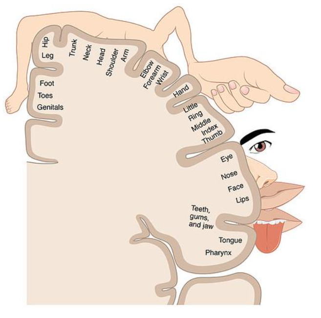
Diagram - The Primary Sensory Cortex
Licensed under Creative Commons, Source.
Pathways of the Somatosensory System
There are a variety of modalities of somatic sensation, with a modality being defined as a ‘unit’ of sensation, relying on one distinct receptor type to detect the sensation. These various modalities can be split into systems depending on which pathway their impulses follow.
The first pathway is the spinothalamic pathway. This is responsible for the following modalities:
- Temperature - thermoreceptors
- Pain - nociceptors
- Pressure or crude touch - mechanoreceptors
First order neurones of the spinothalamic pathway:
- Project onto second order neurones in the ipsilateral spinal cord dorsal horn at the level at which they enter the cord through the dorsal root (ie they enter the cord and synapse immediately).
- Cell bodies in the dorsal root ganglion (same as all first order neurones).
Second order neurones of the spinothalamic pathway:
- Cell bodies in the dorsal horn of the spinal cord.
- The axons decussate in the ventral white commissure of the spinal cord, then ascend as part of the spinothalamic tract to the spinal lemniscus (the path through the brainstem).
- They project to the thalamus.
The second order neurones are organised so that the most superior spinal levels are carried medially in the spinal cord, and progressively inferior spinal levels are added onto the lateral side of the pathway.
Third order neurones of the spinothalamic pathway:
- Thalamic neurones receiving information from more inferior parts of the body project to the medial portion of the primary sensory cortex.
- Thalamic neurones receiving information from more superior parts of the body project to the lateral portion of the primary sensory cortex.
This means that a cross occurs in the primary sensory cortex - impulses from the lower portions of the body are carried laterally in the spinal cord but project to the medial primary sensory cortex (according to the homunculus), and the higher portions of the body are carried medially in the spinal cord but project to the lateral primary sensory cortex. This means that a cross of fibres occurs. It is important to remember that this cross is NOT a decussation, as the fibres do NOT cross the midline.
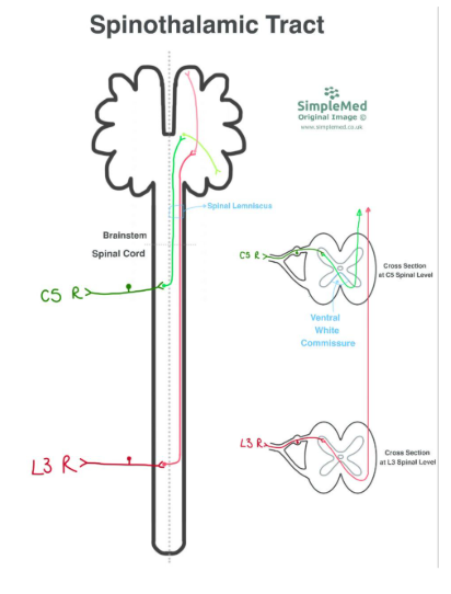
Diagram - The Spinothalamic Tract Pathway from Two Different Spinal Levels
SimpleMed original by Dr. Maddie Swannack
Information on Lissauer’s tract is very high level information and is not necessary for basic understanding of the neuroanatomy of the sensory system tracts. It is included here as a very simple explanation.
Lissauer’s tract is a white matter path in the spinal cord that allows for the ascent of first order spinal neurones of the spinothalamic tract by a few spinal levels, before they synapse with the second order neurones. With no spinal injury, this is not clinically relevant, but with a spinal injury (for example at the level C5), the spinothalamic modalities are typically lost a few spinal segments below the level of the lesion due to this ascent through Lissauer’s tract.
Dorsal Column Medial Lemniscus (DCML) Pathway
The second pathway is the dorsal column medial lemniscus pathway.
It is named like this as the dorsal columns are the regions in the spinal cord that carry these neurones, and the medial lemniscus is the corresponding region in the brainstem.
This is responsible for the following modalities:
- Vibration - mechanoreceptors
- Proprioception (joint position sense) - variety of receptors such as muscle spindle fibres
- Fine touch - mechanoreceptors
- Two-point discrimination - mechanoreceptors
First order neurones of the DCML pathway:
- Ascend ipsilaterally through the dorsal columns of the spinal cord.
- Those from the lower body (spinal level T7 and below) ascend through the gracile fasciculus (or the fasciculus gracilis in Latin) to the gracile nucleus in the medulla.
- Those from the upper body (spinal level T6 and above) ascend through the cuneate fasciculus (or the fasciculus cuneatus in Latin) to the cuneate nucleus in the medulla.
- Axons from the lowest spinal level run most medially in the dorsal columns, with axons from more superior body parts added on laterally.
Second order neurones of the DCML pathway:
- Neurones in the gracile nucleus project to the contralateral thalamus through the medial lemniscus pathway.
- Neurones in the cuneate nucleus project to the contralateral thalamus through the medial lemniscus pathway.
As shown in the image below, the decussation of the second order neurones needs to be organised so that the lateral fibres on one side of the spinal cord are still the lateral fibres of the other side once the decussation has occurred.
Third order neurones of the DCML pathway:
- Thalamic neurones receiving information from the lower half of the body via the gracile nucleus project medially into the primary sensory cortex.
- Thalamic neurones receiving information from the upper half of the body via the cuneate nucleus project laterally into the primary sensory cortex.
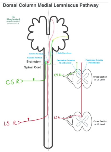
Diagram - The Dorsal Column Medial Lemniscus Pathway from Two Different Spinal Levels
SimpleMed original by Dr. Maddie Swannack
It is important to note that the DCML pathway and spinothalamic pathway organise their order of fibres differently:
The spinothalamic pathway has superior spinal levels carried medially through the spinal cord and inferior levels added on laterally.
The DCML pathway has superior spinal levels carried laterally through the spinal cord and inferior levels are added medially.
Brown-Sequard Syndrome is a very rare clinical condition that has symptoms that help to learn neuroanatomy, even if it is extremely rare in clinical practice.
It is a collection of symptoms that occur if there is a perfect hemisection of the cord, resulting from complete destruction of one side of the spinal cord at a single spinal level, and this can be due to trauma or ischaemia.
Imagine there is a complete hemisection of the spinal cord on the left side at the level T10. The following structures will be destroyed:
- The left dorsal horn
- The left ventral horn
- All other grey and white matter pathways in the left side of the cord
- The left dorsal and ventral roots.
This will lead to:
- Ipsilateral (left sided) anaesthesia affecting a single dermatome (in this case T10)
- Ipsilateral (left sided) loss of dorsal column modalities below the spinal level (in this case below T10)
- Contralateral (right sided) loss of spinothalamic modalities beginning a few segments below the spinal level (for example, below T10).
In the spinothalamic system, the first order neurones that carry impulses from nociceptors relating to pain are carried by C fibres and by faster A-delta fibres. Impulses from mechanoreceptors relating to pressure or crude touch are mainly carried by A-beta fibres.
These A fibres synapse onto inhibitory inter-neurones that contain the endorphin enkephalin and connect to the second order neurones of the C fibres. Enkephalin is similar in structure to morphine, meaning that this modulation has been used in pharmacology to develop opiate-related painkillers.
These enkephalinergic neurones can be activated by incoming impulses from mechanoreceptors, and act to inhibit the second order neurones carrying pain impulses, which explains why rubbing something sore can help relieve the pain.
These inhibitory interneurons are also acted upon by descending neurones from the brain. This means that descending pathways from the brainstem can encourage the release of enkephalin and reduce the amount of pain felt. These connections are also the reason why hypnosis can be used to modulate pain.
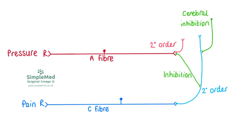
Diagram - The Inhibition of the Second Order Neurones Carrying Pain Information
SimpleMed original by Dr. Maddie Swannack
Edited by: Dr. Marcus Judge
Reviewed by: Adrian Judge
In this article
Sensation in the body can be a special sensation or general sensation, normally from epithelial surfaces.
- 733
