

SimpleMed - Learning Medicine, SimplifiedSimpleMed - Learning Medicine, Simplified
Hit enter to search or ESC to close

Get in Touch

Contact Us

Use our contact form.

The Skull

Share:

By Dr. Joshua Bray

Next Lesson - The Temporomandibular Joint (TMJ)

  Head & Neck


Contents

Contents

  1. Introduction to the Skull
  2. The Facial Skeleton
  3. The Calvaria
  4. The Cranial Floor
  5. Skull Fractures
  6. Quiz
Abstract

Abstract

  • The skull is made up of 22 individual bones joined by sutures.
  • The skull has two parts: the neurocranium and the viscerocranium.
  • The viscerocranium is made up of 14 bones: the zygomatic, lacrimal, nasal, palatine, and vomer bones, inferior nasal conchae, maxillae and mandible.
  • The neurocranium has a roof (calvaria), a floor and a cranial cavity.
  • The calvaria is arranged in 3 layers: the inner table, diploe and outer table.
  • The cranial floor is divided into 3 fossae: anterior, middle and posterior.
  • The cranial floor has a series of canals, foramina and fissures which transmit important nerves and blood vessels.
  • Fractures of the calvaria can be linear or depressed.
  • A fracture of the pterion can shear the middle meningeal artery, resulting in an extradural haemorrhage.
  • Basal skull fractures are rare but important and present with specific signs.
  • Facial fractures are common: the nasal bones, zygomatic arches and mandible are most commonly affected.
Core

Core

Introduction to the Skull

The skull, or cranium, is the bony skeleton of the head. It is made up of 22 individual bones which are joined by fibrous joints called sutures. Most of these bones form by intramembranous ossification, although parts of the cranial base develop by endochondral ossification.

The skull can be broadly divided into two parts:

  • Neurocranium (8 bones) - this is the part which encases the brain. It has a roof (calvaria), a floor and a cranial cavity (which holds the brain).
  • Viscerocranium (14 bones) - this is the skeleton of the face. It supports the soft tissue of the face and houses the eyes, oral cavity and upper respiratory tract.


The Facial Skeleton

The facial skeleton is made up of 14 bones:

  • Zygomatic Bones (2) - these are the cheekbones. They articulate with the frontal, sphenoid, temporal bones and the maxillae.
  • Lacrimal Bones (2) - these are the smallest bones of the facial skeleton, roughly the size a fingernail. They each have a groove for the nasolacrimal duct.
  • Nasal Bones (2) - these form the bony root of the nose.
  • Palatine Bones (2) - these form part of the hard palate of the oral cavity.
  • Inferior Nasal Conchae (2) - these protrude into the nasal cavity and enhance the warming and humidification of inspired air by increasing surface area.
  • Vomer Bone - this forms part of the nasal septum posteriorly.
  • Maxillae (2) - these bones make up the majority of the upper facial skeleton. It forms the upper jaw, which is fixed to the cranial base. It also forms part of the hard palate.
  • Mandible - this U-shaped bone forms the lower jaw. It is moveable as it articulates with the cranial base at the temporomandibular joint (TMJ).
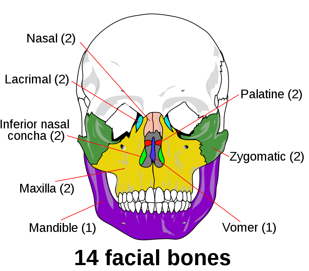
 

Osteology of the Viscerocranium SimpleMed

Diagram - Osteology of the viscerocranium

Public Domain Source by العربيَّة [Public domain]


The Calvaria

The calvaria is the roof of the neurocranium. It comprises 4 bones: frontal, occipital and two parietal bones.

The bones of the calvaria are joined by sutures. Important suture lines to note are:

  • Coronal Suture - joins the frontal bone with the two parietal bones.
  • Sagittal Suture - joins the two parietal bones in the midline.
  • Lambdoid Suture - joins the occipital bone to the two parietal bones.
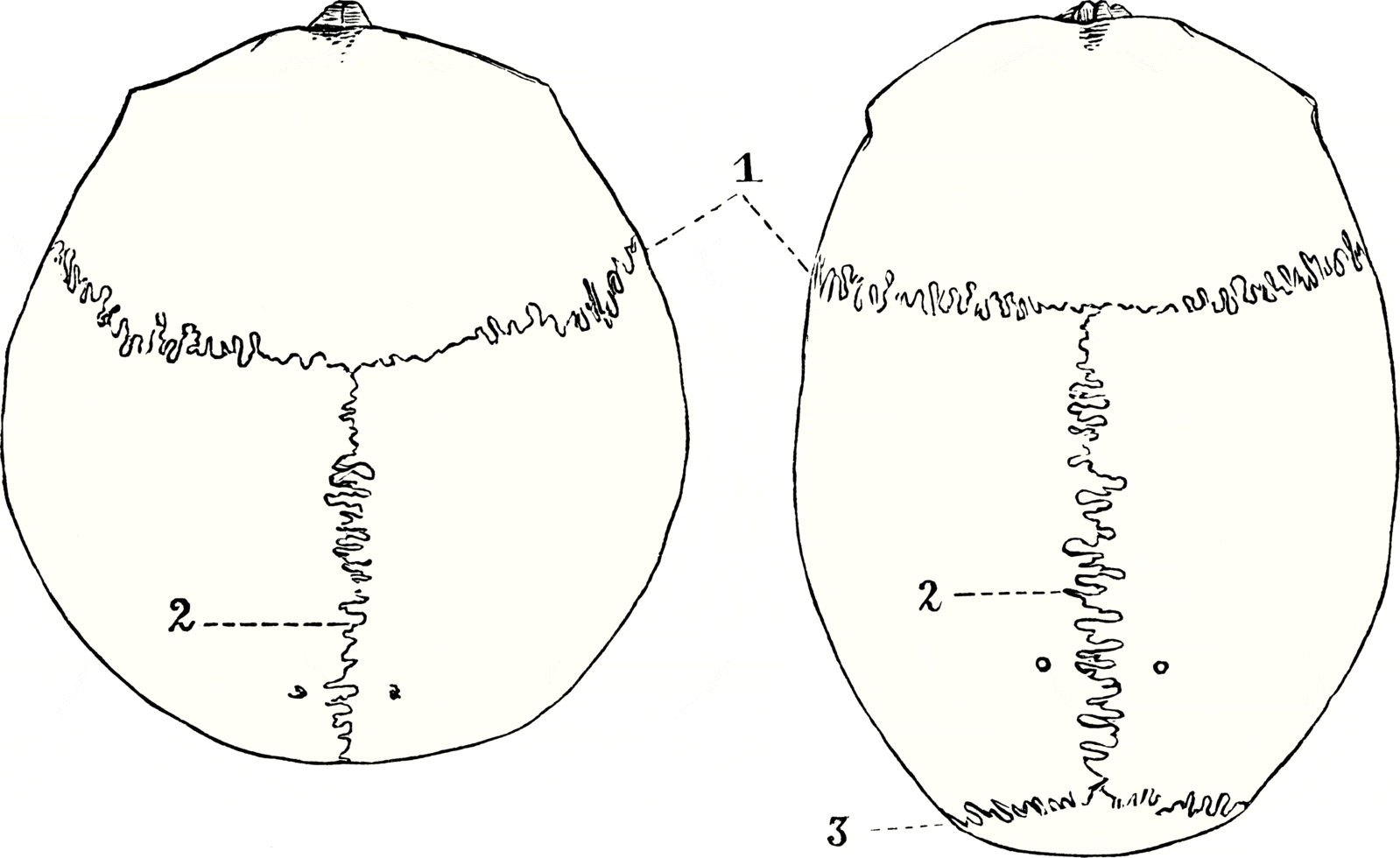
 

Sutures of the Skull SimpleMed

Diagram - Sutures of the skull: 1) Coronal; 2) Sagittal; 3) Lambdoid

Public Domain Source by Julius Kollmann [Public domain]


In infants, the skull is immature, leaving membranous gaps between ossified bone. This permits enlargement of the skull in line with the growth of the brain. These unossified areas are termed fontanelles: there is an anterior fontanelle and posterior fontanelle. These fontanelles typically fuse at 18-24 months and 1-3 months, respectively.

The areas of the adult skull which correspond to the previous locations of the anterior and posterior fontanelles are called bregma and lambda, respectively.
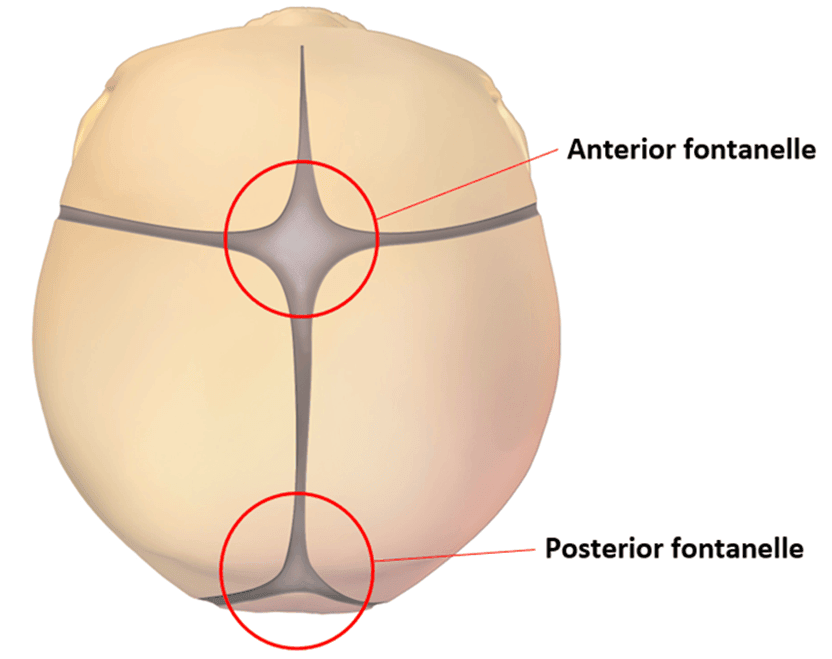
 

Anterior and Posterior Fontanelles SimpleMed

Diagram - The anterior and posterior fontanelles

Creative commons source by BruceBlaus, edited by Dr. Joshua Bray [CC BY-SA 4.0 (https://creativecommons.org/licenses/by-sa/4.0)]


The bone of the calvaria is arranged in 3 layers:

  • Inner Table - the innermost layer of cortical bone.
  • Diploe - the middle layer composed of spongy bone.
  • Outer Table - the outermost layer of cortical.

This trilaminar arrangement ensures adequate protective strength without excessive weight.

The outer table of bone is covered with periosteum (connective tissue), which is bound to the suture line. The inner table is lined by the dura mater (endosteal layer), which is continuous across the cranial cavity.


The Cranial Floor

The cranial floor is the inferior portion of the neurocranium which the brain rests upon. It is formed by parts of the frontal, ethmoid, sphenoid, temporal and occipital bones.

The cranial floor is anatomically divided into three depressions known as the cranial fossae:

  • Anterior Cranial Fossa - bound posteriorly by the lesser wings of the sphenoid. Its floor consists of the frontal, ethmoid and sphenoid bone. It holds the frontal lobe of the brain. The ethmoid bone has a projection called the crista galli, which serves as an attachment for the falx cerebri.
  • Middle Cranial Fossa - bound anteriorly by the lesser wings of the sphenoid and posteriorly by the petrous part of the temporal bones. Its floor consists of the sphenoid and temporal bones. It holds the temporal lobes of the brain. The sphenoid bone has a saddle-shaped depression called the sella turcica, which holds the pituitary gland.
  • Posterior Cranial Fossa - bound anteriorly by the petrous part of the temporal bones. Its floor is made up of the temporal and occipital bone. It accommodates the brainstem and cerebellum.


The Cranial Fossae SimpleMed

Diagram - The cranial fossae

Creative commons source by Anatomist90, edited by Dr. Joshua Bray [CC BY-SA 4.0 (https://creativecommons.org/licenses/by-sa/4.0)]


The cranial floor contains a series of canals, foramina and fissures which transmit important structures such as arteries, veins and nerves. These are summarised in the table below:
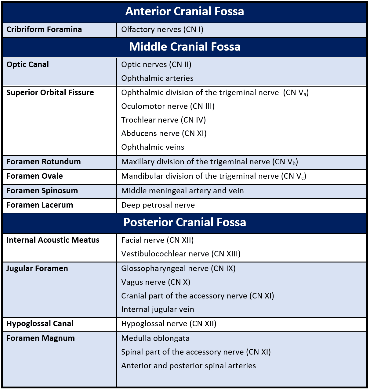


Cranial Canals, Foramina and Fissures SimpleMed

Table - Summary of the cranial foramina and their contents

SimpleMed original by Dr. Joshua Bray
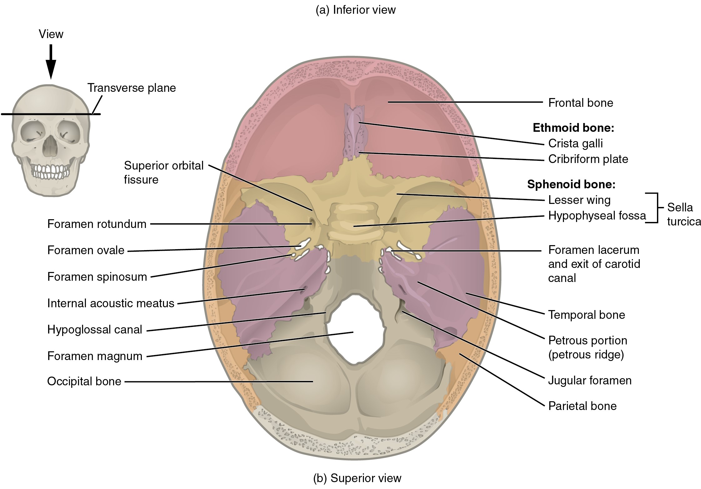


Openings of the Cranial Floor SimpleMed

Diagram - Openings of the cranial floor

Creative commons source by OpenStax College, edited by Dr. Joshua Bray [CC BY-SA 4.0 (https://creativecommons.org/licenses/by-sa/4.0)]


For more information about the cranial nerves, check out our articles: Cranial Nerves I-VI and Cranial Nerves VII-XII.


Skull Fractures

The skull is a very strong structure and so skull fractures require significant blunt trauma or a penetrating injury.

Fractures of the cranial vault can be either linear or depressed:

  • Linear - a full thickness crack in the bone. There are often multiple fracture lines radiating from the site of injury.
  • Depressed - fracture of the skull where the bony fragments displace inwards, resulting in an indentation. These fractures are more likely to result in brain injury.

The thinnest area of bone is located on the lateral aspect of the skull and is known as the pterion. Anatomically it is defined as the junction between the frontal, parietal, temporal, and sphenoid bones. Closely underlying the pterion is the middle meningeal artery. Therefore, blunt trauma to the lateral head can fracture the pterion and shear the middle meningeal artery, resulting in an extradural haemorrhage.
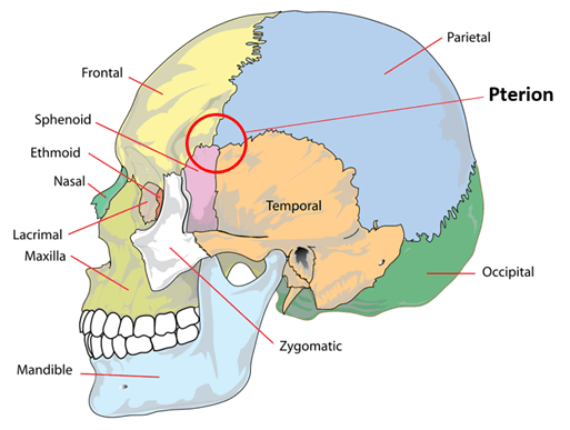
 

The Pterion SimpleMed

Diagram - Skull with the pterion circled

Public Domain Source by LadyofHats Mariana Ruiz Villarreal, edited by Dr. Joshua Bray [Public domain]


Fractures of the cranial floor (basal skull fractures) are rare but must not be missed. They classically present with very specific signs:

  • CSF Rhinorrhoea - fracture of the cribriform plate of the ethmoid bone can cause cerebrospinal fluid (CSF) to leak into the nasal cavity.
  • Periorbital Ecchymoses (‘Raccoon Eyes’) - fracture of the anterior cranial fossa causes blood to enter the orbit and collect in the periorbital tissue.
  • CSF Otorrhoea - fracture through the petrous part of the temporal bone can cause CSF to leak into the middle ear and into the ear canal.
  • Haemotympanum - fracture through the petrous bone can cause blood to collect in the middle ear.
  • Battle’s Sign - fracture through the petrous bone can result in bruising around the mastoid process.

Fractures of the facial skeleton are common. The nasal bones are most susceptible due to their prominent nature on the face. The zygomatic arches and mandible are also commonly affected. The mandible is commonly fractured at more than one site due to its ring-like structure, often involving two fracture points.


Simple Mandible Fracture X-ray SimpleMed

Image - Orthopantomogram showing a fracture through the body of the mandible (indicated by the white arrow)

Creative commons source by Coronation Dental Specialty Group [CC BY-SA 4.0 (https://creativecommons.org/licenses/by-sa/4.0)]


Reviewed by: Dr. Thomas Burnell

Quiz

Quiz

Preview the The Skull quiz

In this article

The skull is made up of 22 individual bones joined by sutures. The skull has two parts: the neurocranium and the viscerocranium.

Free account

Keep reading for free

Create a free account to continue this article and save your revision progress.

Your account includes:

  • Thousands of free practice questions
  • Tools to track your revision progress
Sign up free Already have an account? Log in

No payment. No spam. Just free medical revision.

SimpleMed+

Want More?

Access more on SimpleMed+

Lock it in with practice questions, the full question bank and revision tools that track what you know.

Practice now See what's in SimpleMed+
7368
simplemedlonglogo.png
  • Home
  • Terms & Conditions
  • Privacy Policy
  • Contact

The medical information on this site is provided as an information resource only, and is not to be used or relied on for any diagnostic or treatment purposes. This information is intended for medical education, specifically to biological/medical learning inclined individuals, and does not create any doctor-patient relationship, and should not be used as a substitute for professional diagnosis and treatment.

©2026 Lili's Ark Limited, UK Company #15424953
S+

Search

  • Pre-Clinical
    • Cardiovascular System
      • 1. Anatomy of the Cardiovascular System
      • 2. The Cardiac Cycle
      • 3. Pulses and Pressures
      • 4. Control of Cardiac Output
      • 5. Cardiac Conduction and Contraction
      • 6. Embryology of the CVS
      • 7. Congenital Heart Defects
      • 8. Hypertension
      • 9. Electrocardiograms
      • 10. Electrocardiogram Pathology
      • 11. Cardiovascular Drugs
      • 12. Murmurs
      • 13. Special Circulations
      • 14. Heart Failure
      • 15. Artery and Vein Disease
      • 16. Acute Coronary Syndromes
      • 17. Haemodynamic Shock
    • Musculoskeletal System
      • The Human Body
        • 1. Anatomical Terms
        • 2. Structure of the Muscles
        • 3. Muscles in Action
        • 4. Connective Tissue
        • 5. Structure of Bones
        • 6. Basics of Bone Anatomy
        • 7. Joints
        • 8. The Spine
        • 9. Dermatomes and Myotomes
        • 10. The Brachial Plexus
        • 11. The Lumbar Plexus
        • 12. The Sacral Plexus
      • Clinical Conditions
        • 1. Conditions of the Foot
        • 2. Conditions of the Ankle
        • 3. Conditions of the Lower Leg
        • 4. Conditions of the Knee
        • 5. Conditions of the Thigh
        • 6. Conditions of the Hip
        • 7. Conditions of the Lower Back
        • 8. Conditions of the Back and Neck
        • 9. Conditions of the Shoulder and Clavicle
        • 10. Conditions of the Arm
        • 11. Conditions of the Elbow
        • 12. Conditions of the Forearm and Wrist
        • 13. Conditions of the Hand
      • Musculoskeletal Anatomy
        • 1. Muscles of the Gluteal Region
        • 2. Muscles of the Thigh
        • 3. Key Areas of the Lower Limb
        • 4. Bones of the Lower Limb
        • 5. Bones of the Foot
        • 6. Muscles of the Lower Leg
        • 7. Bones of the Upper Limb
        • 8. Muscles of the Shoulder
        • 9. Muscles of the Upper Arm
        • 10. Bones of the Hand
        • 11. Muscles of the Forearm
        • 12. Key Areas of the Upper Limb
    • Cell Physiology & Biology
      • 1. An Introduction to the Cell
      • 2. Introduction to DNA
      • 3. Structure of Proteins
      • 4. Protein Function in Oxygen Transport
      • 5. Enzymes and Regulation of Protein Function
      • 6. Molecular Techniques for Medics
      • 7. Post-translational Modification and Collagen Biosynthesis
      • 8. Protein Targeting
      • 9. Biological Membranes
      • 10. G-Protein Coupled Receptor
      • 11. Membrane Transport and Intracellular Calcium Regulation
      • 12. Membrane Potentials
      • 13. Action Potentials
      • 14. Autonomic Nervous System Introduction
    • Metabolism
      • 1. Nutrition, Diet and Body Weight
      • 2. Energy Reactions in Cells
      • 3. Dietary Energy Production
      • 4. Glycolysis
      • 5. Oxygen-dependent Pathway
      • 6. Lipid Metabolism and Transport
      • 7. Protein and amino acid metabolism
      • 8. Alcohol Metabolism
      • 9. Oxidative Stress
      • 10. Energy Storage
      • 11. Temperature Regulation
      • 12. Metabolic and Endocrine Regulation
    • Endocrinology
      • 1. An Introduction to Endocrinology
      • 2. Control of Appetite
      • 3. The Adrenal Glands and Adrenal Disorders
      • 4. HPA and Growth Hormone
      • 5. Pituitary Disorders
      • 6. The Thyroid Gland
      • 7. The Endocrine Pancreas
      • 8. Diabetes Mellitus
      • 9. Calcium Metabolism
    • Haematology
      • 1. Haematopoiesis and the Spleen
      • 2. Full Blood Count
      • 3. Anaemia
      • 4. Vitamin B12 and Folate
      • 5. Iron Metabolism
      • 6. Haemoglobinopathies
      • 7. When Haematopoiesis goes wrong
      • 8. Haematology in Systemic Disease
    • Genetics
      • 1. Mitosis and Meiosis
      • 2. Genotype, Phenotype and Inheritance
      • 3. Gene Expression
      • 4. Introduction to Gene Mutation
      • 5. DNA Repair and Cancer
    • Infection
      • 1. An Introduction To Infection
      • 2. The Infection Model
      • 3. Biofilms
      • 4. Innate Immunity
      • 5. Adaptive Immunity
      • 6. Gram Staining and Infection Investigations
      • 7. Sepsis
      • 8. Antibiotics
      • 9. Streptococci
      • 10. Hepatitis
      • 11. Human Immunodeficiency Virus
      • 12. The Immunocompromised Host
      • 13. Travel-related Infections
    • Pathology
      • 1. An Introduction to Pathology
      • 2. The Basics of Pathology
      • 3. Epithelium, Skin and Exocrine Glands
      • 4. Acute Inflammation
      • 5. Chronic Inflammation
      • 6. Regeneration and Repair of Tissues
      • 7. The Lymphatic System
      • 8. Haemostasis and Clotting
      • 9. Thrombosis and Embolism
      • 10. Atherosclerosis
      • 11. Cellular Adaptations
      • 12. Introduction to Neoplasia
      • 13. Pathogenesis of Neoplasms
      • 14. Behaviour of Neoplasms
    • Population & Social Science
      • 1. Introduction to Population Science
      • 2. Epidemiological Studies
      • 3. Models of Health
      • 4. Beliefs of the Public
      • 5. Health Economics
      • 6. Evidence-Based Practice
      • 7. Patient Safety and Quality Control
      • 8. Summary and Glossary
    • Patient-Centred Care
      • 1. Lifestyle Medicine
      • 2. Childhood Immunisations
      • 3. Sepsis in Paediatrics
      • 4. Child Safeguarding
      • 5. Ageing
      • 6. Falls
      • 7. Men's Health
      • 8. Palliative Care
    • Reproductive System
      • 1. Anatomy of the Man
      • 2. Anatomy of the Woman
      • 3. The Pelvic Floor
      • 4. Development of the Reproductive System
      • 5. Puberty
      • 6. The Menstrual Cycle and Menstrual Abnormalities
      • 7. Gametogenesis
      • 8. Sex, Infertility and Contraception
      • 9. Physiology of the Mother
      • 10. Diseases of the Mother in Pregnancy
      • 11. Physiology of the Fetus
      • 12. The Placenta
      • 13. Labour and Delivery
      • 14. The Postpartum Period
      • 15. Infections of the Reproductive Tract
      • 16. Pelvic Inflammatory Disease
      • 17. The Menopause
      • 18. Malignancies of the Reproductive Tract
      • 19. Introduction to the Breast
      • 20. Benign Conditions of the Breast
      • 21. Malignant Breast Disease
    • Respiratory System
      • 1. Anatomy of the Respiratory System
      • 2. Development of the Respiratory System
      • 3. Ventilation and Lung Mechanics
      • 4. Properties of Gas Exchange
      • 5. Carbon Dioxide Transport
      • 6. Chemical Control of Breathing and Plasma pH
      • 7. Lung Function Testing
      • 8. Pulmonary Embolism
      • 9. Asthma
      • 10. Chronic Obstructive Pulmonary Disease
      • 11. Pneumonia and other Lower Respiratory Tract Infections
      • 12. Bronchiectasis and Cystic Fibrosis
      • 13. Tuberculosis
      • 14. Pneumothorax and Pleural Effusion
      • 15. Lung Cancer
      • 16. Respiratory Failure
    • Urinary System
      • 1. Anatomy of the Kidneys, Bladder and Urethra
      • 2. Development of the Urinary System
      • 3. The Nephron
      • 4. The Glomerulus – Tubular Reabsorption and Secretion
      • 5. Control of Plasma Volume
      • 6. Control of Plasma Osmolarity
      • 7. Control of Potassium
      • 8. Micturition
      • 9. Diuretics
      • 10. Glomerular Filtration Rate and the Measurement of Kidney Function
      • 11. Hyponatraemia
      • 12. Urinary Incontinence
      • 13. Glomerular Disease
      • 14. Diabetic Nephropathy
      • 15. Acute Kidney Injury
      • 16. Chronic Kidney Disease
      • 17. Urinary Tract Obstruction
      • 18. Urinary Tract Infections
      • 19. Prostate Pathology
      • 20. Male Urological Conditions
      • 21. Urological Malignancy
    • Gastrointestinal System
      • 1. Development of the Gastrointestinal Tract
      • 2. Anatomy and Physiology of the Foregut
      • 3. Anatomy and Physiology of the Midgut
      • 4. Anatomy and Physiology of the Hindgut
      • 5. Upper Gastrointestinal Pathology
      • 6. Hepatic, Biliary and Pancreatic Pathology
      • 7. Intestinal Pathology
      • 8. Distal Gastrointestinal Tract Pathology
      • 9. Gastrointestinal Malignancy
      • 10. Gastroenteritis and Enterocolitis
      • 11. Gastrointestinal Emergencies
    • Head & Neck
      • 1. Introduction to Head & Neck Anatomy
      • 2. Blood Supply of the Head and Neck
      • 3. Lymph Nodes and Neck Lumps
      • 4. The Skull
      • 5. The Temporomandibular Joint
      • 6. The Meninges
      • 7. Cranial Nerves I-VI
      • 8. Cranial Nerves VII-XII
      • 9. Development of the Head and Neck
      • 10. Autonomics in the Head and Neck
      • 11. Anatomy of the Ear
      • 12. Ear Pathology
      • 13. The Nose
      • 14. The Oral Cavity and Pharynx
      • 15. The Eye
      • 16. The Larynx
      • 17. Head and Neck Cancers
      • 18. Imaging of the Head and Neck
      • 19. Fascial Planes, Muscles and Triangles
    • Immunology
      • 1. An Introduction to Hypersensitivity
      • 2. Type 1 Hypersensitivity
      • 3. Food Allergy
      • 4. Introduction to Autoimmunity
      • 5. Rheumatoid Arthritis
      • 6. Systemic Lupus Erythematosus (SLE)
      • 7. Immunological Therapies | Monoclonal Antibodies
    • Neuroanatomy & Psychiatry
      • 1. Basics of Neuroanatomy
      • 2. Cells and Components of the Nervous System
      • 3. Arterial Supply to the Brain
      • 4. Development of the Nervous System
      • 5. Production and Circulation of CSF and Related Disorders
      • 6. Somatosensory System
      • 7. Motor System
      • 8. Visual System
      • 9. Higher Cortical Function
      • 10. Reticular Formation
      • 11. Movement Disorders
      • 12. Infections of the Central Nervous System
      • 13. Dementia
      • 14. Delirium
      • 15. Headaches
      • 16. Stroke
      • 17. Raised Intracranial Pressure (ICP)
      • 18. Subarachnoid Haemorrhage
      • 19. Anxiety
      • 20. Mood Disorders
      • 21. Psychosis
    • Embryology
      • 1. The Basics of Embryology
      • 2. Development of the Gastrointestinal Tract
      • 3. Development of the Reproductive Systems
      • 5. Development of the Respiratory System
      • 6. Development of the Head and Neck
      • 8. Embryology of the Cardiovascular System
    • Pharmacology
      • 1. Research Methods in Pharmacology
      • 2. Pharmacokinetics
      • 3. Pharmacodynamics
      • 4. Pharmacovigilance and Pharmacogenetics
      • 5. Safe Prescribing
      • 6. Hypertension and Heart Failure
      • 7. Cardiac Arrhythmias
      • 8. Antiplatelets, Anticoagulants and Thrombolysis
      • 9. Hyperlipidaemias
      • 10. Diuretics and Renal Pharmacology
      • 11. Pharmacology of Insulin
      • 12. Reproductive and Post-reproductive Health
      • 13. Antibiotics
      • 14. Antivirals
      • 15. Cancer Chemotherapy
      • 16. Anaesthetics
      • 17. Opioids
      • 18. Epilepsy and Anti-epileptic Drugs
      • 19. Parkinson's Disease and Myasthenia Gravis
      • 20. NSAIDs
      • 21. Immunosuppression and Disease-modifying Therapy
      • 22. Pharmacology of Airway Control
      • 23. Drugs Affecting Acid Secretion
      • 24. Antiemetics and Laxatives
      • 25. Poisoning and STOPP/START
  • OSCE
    • Index Conditions for OSCE exams
    • Cardiovascular System Examination
    • Shoulder Examination
    • Hand Examination
    • Venepuncture
    • Blood Pressure
    • Respiratory System OSCE Examination
    • Peripheral Vascular OSCE Examination
    • Varicose Vein OSCE Examination
    • Deep Vein Thrombosis (DVT) OSCE Examination
    • Abdominal OSCE Examination
    • Rectal (PR) OSCE Examination
    • Stoma OSCE Examination
    • Hernia OSCE Examination
    • Upper Limb Neurological OSCE Examination
    • Lower Limb Neurological OSCE Examination
    • Cranial Nerve OSCE Examination
    • Cerebellar OSCE Examination
    • GALS Screening OSCE Examination
    • Elbow OSCE Examination
    • Hip OSCE Examination
    • Knee OSCE Examination
    • Ankle and Foot OSCE Examination
    • Spine OSCE Examination
    • Thyroid Status OSCE Examination
    • Diabetic Foot OSCE Examination
    • Cushing's Syndrome OSCE Examination
    • Acromegaly OSCE Examination
    • Ear, Otoscopy and Hearing OSCE Examination
    • Nasal OSCE Examination
    • Eye and Vision OSCE Examination
    • Fundoscopy (Ophthalmoscopy) OSCE Examination
    • Visual Acuity OSCE Assessment
    • Anterior Segment Eye OSCE Examination
    • Pigmented Skin Lesion OSCE Examination
    • Rash and Non-Pigmented Skin Lesion OSCE Examination
    • Breast OSCE Examination
    • Testicular OSCE Examination
    • Neck Lump OSCE Examination
    • Lymphoreticular OSCE Examination
  • Question Bank
  • About
  • How to Use SimpleMed
  • SimpleMed Lecture Series
  • DON'T PANIC - Guide to the Wards
  • SimpleMed Plus
Your privacy Allow anonymous analytics to help us improve SimpleMed. Privacy policy
