
Next Lesson - Calcium Metabolism
Core
Diabetes Mellitus (DM) is the term used for metabolic disorders characterised by chronic hyperglycaemia (increased plasma glucose levels), which can be caused by insulin deficiency, insulin resistance or both. Diabetes mellitus is typically characterised as either Type 1 or Type 2, distinguished by their epidemiology and probable causation, but the two types can be hard to tell apart clinically. Other types include gestational diabetes (high blood glucose that develops during pregnancy and usually disappears after giving birth), monogenic diabetes (caused by a change in a single gene) and diabetes caused by another condition or disease.
Type 1 Diabetes Mellitus (T1DM)
T1DM is most common in the young: new diagnoses peak around puberty and early adulthood, but it can start at any age. In adults, NICE advises that age or BMI alone should not be used to diagnose or rule it out. It is an autoimmune condition, which is thought to be caused by a genetic predisposition (susceptibility is determined by multiple genes) combined with an environmental trigger. Together these produce an autoimmune reaction in which lymphocytes, macrophages and auto-antibodies attack and progressively destroy the pancreatic β-cells (the cells that make insulin).
A typical patient would be a lean, young person, with a recent history of viral infection, who presents with a triad of symptoms: polyuria, thirst, and weight loss. This triad of symptoms is very characteristic of T1DM, and is explained below:
- Polyuria (passing large volumes of urine) - the high concentration of glucose in the tubular fluid (the filtered fluid inside the kidney tubules) raises its osmolality (concentration of dissolved particles). Less water is then reabsorbed, so more urine is produced. This is called osmotic diuresis. Glucose also appears in the urine (glycosuria).
- Thirst - losing so much water in the urine dehydrates the body, which triggers thirst.
- Weight Loss - without insulin, cells such as muscle and fat cells cannot take up glucose to use as fuel, so the body breaks down fat and protein for energy instead.
In diabetes the lack of insulin causes decreased uptake of glucose into adipose tissue (fat) and skeletal muscle, and decreased glucose storage as glycogen in muscle and liver. As a result, plasma glucose levels rise. High blood glucose is confirmed by measuring the plasma glucose concentration (see Diagnosing Diabetes below). Type 1 diabetes is then diagnosed on clinical grounds, and in adults NICE recommends testing for diabetes-specific auto-antibodies.
If T1DM is not treated early, patients can develop diabetic ketoacidosis. The high rate of β-oxidation of fats in the liver, coupled with low insulin/anti-insulin ratios, leads to the production of huge amounts of ketone bodies, such as acetoacetate, acetone, and β-hydroxybutyrate. Acetone is a volatile substance and is therefore breathed out and smelled on the patient’s breath. Symptoms of ketoacidosis include: prostration, hyperventilation, nausea, vomiting, dehydration and abdominal pain.
T1DM cannot be cured and must be managed for the rest of the patient’s life with insulin. This is given either as injections several times a day (usually a basal-bolus regimen: a background insulin plus rapid-acting insulin injected before meals) or by an insulin pump, a small device attached to the skin that gives insulin throughout the day and night. Living with T1DM has major social and psychological effects, and these can affect how well the condition is controlled. Along with insulin, dietary management and regular exercise must be considered to better manage the condition. NICE recommends offering adults with T1DM a structured education programme, such as DAFNE (dose adjustment for normal eating), and training in carbohydrate counting.
The management of blood glucose requires frequent glucose measurement. NICE recommends offering adults with T1DM a choice of continuous glucose monitoring (CGM) devices (real-time or 'flash'). These measure glucose levels and send the readings to a display device or smartphone. People using CGM still need some finger-prick tests, although less often, for example to check the device's accuracy. Finger-prick testing is also offered to anyone who cannot use or does not want CGM. A small amount of blood from a finger prick is sufficient to measure blood glucose using a BM stick and reader (a test strip and handheld blood glucose meter), however these devices are only accurate if used carefully. Insulin treatment always carries a risk of hypoglycaemia (abnormally low blood glucose). A person who can swallow is treated with fast-acting glucose by mouth. A person who cannot safely take treatment by mouth, for example because they are drowsy or unconscious, is given intramuscular glucagon or IV glucose.
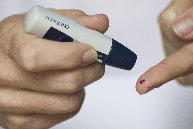
Diagram: An example of a BM stick used by patients with T1DM to measure their own blood glucose levels
Creative commons source by Reversing Your Diabetes Today [CC BY-SA 4.0 (https://creativecommons.org/licenses/by-sa/4.0)]
Type 2 Diabetes Mellitus (T2DM)
T2DM patients are typically older than those diagnosed with T1DM, and are usually overweight. T2DM is generally caused by insulin resistance (when cells respond poorly to insulin), which at first is compensated for by the pancreas increasing insulin production. However, over time pancreatic β-cell function decreases, and the patient suffers hyperglycaemia due to the inability of the pancreas to compensate for insulin resistance. By the time of diagnosis, β-cell function (the β-cells' ability to secrete insulin) has on average already fallen to less than half of normal. As it falls further, disorders of insulin resistance and secretion worsen, increasing the patient's hyperglycaemia.
Symptoms may be mild or absent, and when present they may develop gradually. Some patients have the classical triad of symptoms (polyuria, thirst, and weight loss) seen in T1DM. Other symptoms include lack of energy, persistent infections (particularly genital thrush, which is a yeast infection, or infections of the feet), minor skin damage that is slow to heal, and visual problems such as blurred vision.
T2DM can sometimes be managed by diet and exercise alone. However, NICE (updated February 2026) now recommends offering most adults with T2DM two drugs as initial treatment: metformin, which mainly reduces glucose production by the liver (gluconeogenesis) and increases glucose use by the tissues, and an SGLT-2 inhibitor (sodium-glucose co-transporter 2 inhibitor), which reduces glucose reabsorption in the kidney's proximal tubule so that more glucose is passed in the urine. SGLT-2 inhibitors are recommended as much for their heart and kidney benefits as for lowering glucose. If further drugs are needed, other classes can be added, including sulfonylureas, which stimulate β-cells to release insulin but can increase the risk of hypoglycaemia and weight gain.
In a person with symptoms of diabetes, the following venous plasma glucose criteria support the diagnosis. Without symptoms, an abnormal result requires confirmation as described below.
- A random venous plasma glucose concentration (taken at any time of day, regardless of meals) ≥11.1 mmol/L,
- OR a fasting plasma glucose concentration ≥7.0 mmol/L,
- OR plasma glucose concentration ≥11.1 mmol/L 2 hours after 75g anhydrous glucose in an oral glucose tolerance test.
Without symptoms, a single abnormal glucose result does not establish diabetes. Confirm with at least one further venous plasma glucose result in the diabetic range on another day, using a fasting, random or two-hour post-glucose-load sample. Glycosuria and a finger-prick glucose meter reading alone cannot establish the diagnosis.
Complications of Diabetes Mellitus
Persistent hyperglycaemia is associated, in some tissues, with the abnormal metabolism of glucose into products that may be harmful to cells. This is because, in some tissues, such as peripheral nerves, the eyes and the kidneys, glucose uptake into cells does not require insulin. Instead, it depends on the extracellular glucose concentration. When a patient is experiencing hyperglycaemia, the intracellular concentration of glucose in these tissues increases, and glucose is converted to sorbitol in the following reaction, catalysed by the enzyme aldose reductase:
Glucose + NADPH + H+ -> Sorbitol + NADP+
An increase in the above reaction depletes cellular NADPH, weakening the antioxidant defences of these cells (their protection against chemical damage, which depends on NADPH). This allows abnormal disulphide bonds (sulphur-to-sulphur links) to form in cellular proteins, altering their structure and function. In addition, the increased accumulation of sorbitol (a sugar alcohol) causes osmotic damage to cells.
Persistent hyperglycaemia is also associated with the increased glycation of plasma proteins (glucose attaching to proteins). The extent of glycation depends on the glucose concentration and the half-life of the protein. Glycation changes the net charge and 3D structure of the protein, therefore affecting the protein's function. Glucose can react with the terminal valine (the amino acid at the end of a haemoglobin chain) on haemoglobin to form glycated haemoglobin (HbA1c). Because HbA1c reflects blood glucose control over the previous 8 to 12 weeks, it is used as an indicator of how well diabetes is controlled and how effective treatment has been. NICE guidance gives HbA1c in mmol/mol, with the older percentage in brackets: for example, 48 mmol/mol (6.5%).
People who have had diabetes for a long time can develop a number of macrovascular (large blood vessel) and microvascular (small blood vessel) complications, made worse in individuals whose blood glucose is poorly controlled.
Macrovascular complications include increased risk of stroke, increased risk of myocardial infarction and poor circulation to the periphery, particularly to the feet (peripheral arterial disease).
There are a wide range of microvascular complications, which are explained below:
- Diabetic Eye Disease - this is usually caused by diabetic retinopathy, in which damage to blood vessels in the retina (the light-sensitive layer at the back of the eye) caused by hyperglycaemia can lead to blindness. The damaged blood vessels may leak and form protein exudate (deposits of leaked material) on the retina, or they can rupture and cause bleeding into the eye. Retinopathy does not usually cause symptoms at first, so in England people with diabetes aged 12 or over are invited for diabetic eye screening every 1 or 2 years. Finding and treating it early reduces the risk of sight loss.
- Diabetic Kidney Disease (Diabetic Nephropathy) - hyperglycaemia damages the renal glomeruli (the kidney's filtering blood vessels). An early sign of diabetic kidney disease is proteinuria (protein in the urine). In people with diabetes this is checked with a urine albumin:creatinine ratio (ACR) test, and a confirmed ACR of 3 mg/mmol or more is regarded as clinically important. Separately, glucose in the urine provides a substrate for bacteria, so urinary tract infections can be more common.
- Diabetic Neuropathy - hyperglycaemia damages peripheral nerves in a number of ways, which can cause a change in or complete loss of sensation.
- Diabetic Foot Disease - poor blood supply, nerve damage (loss of sensation) and an increased risk of infection together greatly increase the risk of damage to the feet, such as ulcers, in people with diabetes. NICE recommends assessing the foot risk of adults with diabetes when diabetes is diagnosed and at least once a year after that.

Diagram: Table outlining the main comparisons between type 1 and type 2 diabetes
SimpleMed original by Dr. Jenny Hubball
Metabolic syndrome is defined as a cluster of risk factors for cardiovascular disease and type 2 diabetes, including insulin resistance, dyslipidaemia, glucose intolerance and hypertension, associated with central adiposity. These features may share a common cause in insulin resistance, as they are commonly found together in patients with T2DM. Metabolic syndrome is seen commonly in the obese, and is related to an increase in obesity-related morbidity. The major factors that predispose the population to metabolic syndrome are obesity and a sedentary lifestyle, and these cause insulin resistance, which is associated with a dyslipidaemic profile (↑VLDL, ↑triglycerides, ↓HDL) that is highly atherogenic (it promotes atherosclerosis). VLDL stands for very low-density lipoprotein. Insulin resistance is also associated with an increased risk of hypertension. The WHO (1999) criteria for metabolic syndrome require insulin resistance or abnormal glucose regulation, plus at least two of the other features listed below:
- Glucose (required): type 2 diabetes, impaired fasting glucose (fasting plasma glucose ≥6.1mmol/L), impaired glucose tolerance (plasma glucose ≥7.8mmol/L 2 hours after a glucose load) or other evidence of insulin resistance
- Central obesity, with a waist:hip ratio of >0.9 in men and >0.85 in women, and/or a BMI >30kg/m2
- BP ≥140/90mmHg, or on treatment for high blood pressure
- Triglycerides ≥1.7mmol/L and/or HDL cholesterol <0.9mmol/L in men and <1.0mmol/L in women
- Microalbuminuria (small amounts of the protein albumin in the urine)
Other organisations use different definitions. For example, the International Diabetes Federation (IDF) definition requires central obesity plus at least two other features, and uses different thresholds: fasting glucose ≥5.6mmol/L (or known type 2 diabetes), HDL cholesterol <1.03mmol/L in men and <1.29mmol/L in women, and BP ≥130/85mmHg. Always check which definition is being used.
Metabolic syndrome first became common in Western developed countries, but with the spread of the Western lifestyle it is now a global problem. In one large Finnish and Swedish study using the WHO definition, it was present in 78% of women and 84% of men with type 2 diabetes.
When diagnosing metabolic syndrome, note that BMI does not allow for large muscle bulk. Central fat around the abdomen, not just total body fat, is associated with insulin resistance, fatty liver and the other features of metabolic syndrome. This means that BMI is not an ideal measurement of central fat. NICE therefore recommends measuring waist-to-height ratio as well as BMI in adults with a BMI below 35kg/m2. A ratio of 0.5 to 0.59 indicates increased health risks, and 0.6 or more indicates further increased risks.
Edited by: Dr. Thomas Burnell
Reviewed by: Dr. Marcus Judge
- 8937