

Next Lesson - Properties of Gas Exchange
Core
In this article, the process of ventilation and the mechanisms involved in inspiration and expiration will be discussed, alongside pathologies that can affect a patient’s ability to ventilate their lungs.
Ventilation is the physical movement of air in and out of the lungs. It can easily be confused with the process of respiration, which is the gaseous exchange of oxygen and carbon dioxide across a membrane either at a cellular level or in the lungs.
Quiet inspiration and quiet expiration occur when a person is unaware of their breathing. There is no extra effort to breathe, just the subconscious drive to breathe. Sensors in the brainstem (central chemoreceptors) detect the carbon dioxide level in the blood and adjust breathing to keep it normal.
Quiet Inspiration
At the beginning of quiet inspiration, the external intercostal muscles (the muscles between the ribs) contract causing the thoracic cavity to move outwards, whilst the diaphragm flattens. Both of these movements are active and increase the space in the thoracic cavity. As the volume increases in the thoracic space, the pressure drops below atmospheric pressure causing air to be drawn into the lungs through the mouth and nose.
Quiet Expiration
The diaphragm and external intercostal muscle cease contracting as the elastic recoil of the lungs leads to the thoracic cavity and lungs returning to their original position. The volume of the thoracic cavity decreases therefore the intra-thoracic pressure increases causing air to be pushed out of the lungs. The process of air moving out of the lungs is passive as the muscles relax.
A very small amount of pleural fluid exists between the visceral and parietal pleura (the layer covering the lung and the layer lining the chest wall) and forms a seal between the lung and the thoracic wall. The outer surface of the lungs and the inner surface of the thoracic cavity are held together by the surface tension between the pleural surfaces.
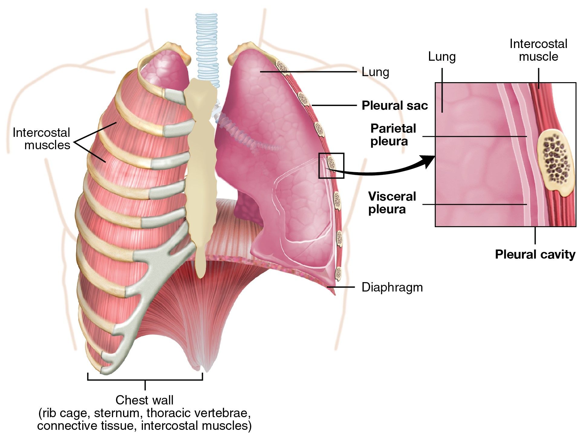
Diagram - The pleura of the lungs
Creative commons source by OpenStax College [CC BY-SA 4.0 (https://creativecommons.org/licenses/by-sa/4.0)]
The pleural seal allows the lungs to expand as the thoracic cavity expands.
The pressure in the pleural space is negative at rest (below atmospheric pressure). This is because the parietal pleura is pulled out by the chest wall, and the visceral pleura is pulled in by the elastic recoil of the lung. The pleural pressure becomes more negative during inspiration due to the expansion of the chest wall, and this increased negative pressure helps the lungs to expand with the chest wall.
The respiratory expiratory level is the point at the end of expiration, just before inspiration begins. The respiratory muscles are relaxed at this point. The lungs are exposed to two equal but opposing forces. The first is the elasticity of the lung, which favours a small lung volume. The second is the force exerted by the rib cage, which favours a large thoracic volume. These are balanced at the respiratory expiratory level.
During quiet inspiration, the main muscle involved in expanding the volume of the chest is the diaphragm, with help from the external intercostal muscles. In contrast, quiet expiration is a passive process and relies on the elastic recoil of the lungs. The respiratory expiratory level described above is also called the resting expiratory level (REL). It is the lung volume at which the elastic recoil forces of the thorax (chest wall) and the lungs counter each other.
When a patient is forcing inspiration, they will use their accessory muscles (extra muscles that help when breathing is forced) of inspiration. These include sternocleidomastoid, scalene, serratus anterior and pectoralis major muscles. They lift the ribs and sternum (breastbone) to enlarge the chest. Pectoralis major helps when the upper limb is fixed, and serratus anterior when the shoulder blades (scapulae) are fixed. The accessory muscles used during forced expiration are the internal intercostal muscles and abdominal muscles.
Spirometry is a type of pulmonary function test and assesses a patient’s lung volume in a dynamic way, meaning it can track inspiration and expiration. There are lots of different terms used when describing a patient’s spirometry reading and some of these terms can sound very similar, meaning it is important to understand each term. Simple spirometry can measure tidal volume, inspiratory reserve volume and expiratory reserve volume. It cannot measure residual volume, so it cannot measure functional residual capacity or total lung capacity either. These need other tests, such as helium dilution.
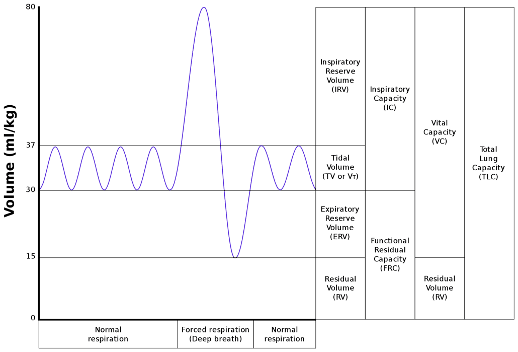
Diagram - A spirometry trace with lung volumes
Creative commons source by Vihsadas, modified by Rscottweekly [CC BY-SA 4.0 (https://creativecommons.org/licenses/by-sa/4.0)]
Tidal Volume - the volume of air that enters and leaves the lungs in each breath.
Inspiratory Reserve Volume - the volume of air that is the difference between the volume of quiet inspiration, and the maximum inspiratory volume possible. It occurs when a patient takes a normal breath in and then increases the volume of the lungs to the maximum capacity possible.
Expiratory Reserve Volume - the volume of air that is the difference between the volume of quiet expiration, and the maximum expiratory volume possible. It occurs when a patient takes a normal breath out and then decreases the volume of the lungs to the minimum capacity possible.
Residual Volume - after forced expiration, the lungs are not completely emptied as air must exist in the lungs for them to stay expanded. The remaining air in the lungs is known as the residual volume.
Lung Capacity - a lung capacity is the sum of two or more lung volumes. Unlike the lung volumes, which change with the pattern of breathing, lung capacities are fixed. They are defined between set points in the cycle of ventilation.
Inspiratory Capacity - from the end of quiet expiration to the maximum inspiration, and this can be calculated by the addition of inspiratory reserve volume and tidal volume.
Functional Residual Capacity - the volume of air in the lungs at the end of quiet expiration. This can be calculated by the addition of expiratory reserve volume and residual volume.
Vital Capacity - the maximal volume of air that can be expelled from a patient’s lung after maximal inspiration. This can be calculated by inspiratory capacity added to expiratory reserve volume OR inspiratory reserve volume plus tidal volume and expiratory reserve volume.
Total Lung Capacity - the volume of air present in the lungs after maximal inspiration. This can be calculated by the addition of vital capacity and residual volume.
Not all the air breathed in is available for gaseous exchange, because a proportion of the air fills the conducting airways (the airways that carry air to the alveoli but do not take part in gas exchange). This is an unavoidable ‘waste’ of air, usually about 150 mL. This is known as the anatomical dead space.
Damaged or poorly perfused alveoli are not able to perform gas exchange so ventilation to these alveoli is wasted. This volume of air is defined as the alveolar dead space.
The physiological dead space is the sum of the anatomical dead space and alveolar dead space.
During the process of inspiration, energy is used to overcome the resistance present in the airways and to stretch the lungs.
Compliance is the ability of the lungs to expand and stretch. Most of the effort of ventilation is used to stretch out the lungs because the elastic tissue in the lungs wants to recoil.
Surface tension is an important contributor to compliance. The alveolar surface is lined by fluid which generates surface tension, which decreases the compliance of the alveoli, therefore making it harder for the lungs to expand.
Surfactant is a lipoprotein secreted by type II pneumocytes (the cells in the alveolar wall that make surfactant). The molecule has hydrophilic (water-loving) and hydrophobic (water-repelling) regions. The hydrophilic region lies in the thin fluid lining the alveolus, whilst the hydrophobic tails face the air. The main lipid component of surfactant helps to decrease the surface tension of the alveoli.
The lipoprotein molecule is more effective on smaller alveoli because the surfactant molecules are closer together in comparison to larger alveoli. Where the surfactant molecules are closer together, on smaller alveoli, the surface tension is lower and the compliance is higher, meaning these alveoli are easier to inflate. It is important to remember that as the alveoli expand, the surfactant molecules become further apart meaning the surface tension increases.
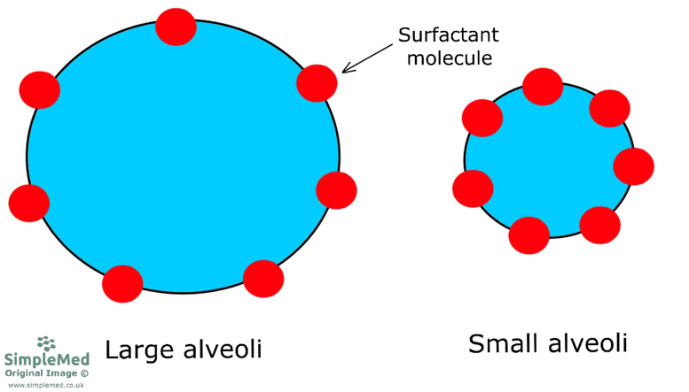
Diagram - Example of how surfactant molecules are further apart in large alveoli
SimpleMed original by Dr. Peter Parkinson
Surfactant aids the expansion of the alveoli, and also acts to stabilise the lungs through the Law of Laplace; defined as Pressure (P) = 2 x surface tension (T) / radius (r).
Alveolar size varies. The law of Laplace says that pressure is proportional to surface tension and inversely proportional to radius. So if the surface tension was the same in all alveoli, the smaller alveoli would contain higher pressures. Imagine two unequal sized alveoli connected by an airway, with no surfactant in the lungs. The smaller alveolus has the smaller radius, so its pressure is higher. Air would flow from the smaller alveolus into the larger one, and the smaller alveolus would collapse into the larger alveolus. This severely decreases the surface area available for gaseous exchange, as the combined surface area of thousands of smaller alveoli is larger compared to the combined surface area of fewer larger alveoli.
Because surfactant molecules are closer together on small alveoli compared to larger alveoli, the surface tension is less for small alveoli. As the alveolus expands (radius increases), the surfactant molecules become further apart, and the surface tension increases. This allows the small and large alveoli to expand until both large and small alveoli have equal pressure. This stops the small alveoli from collapsing into the larger alveoli, which stabilises the lungs.
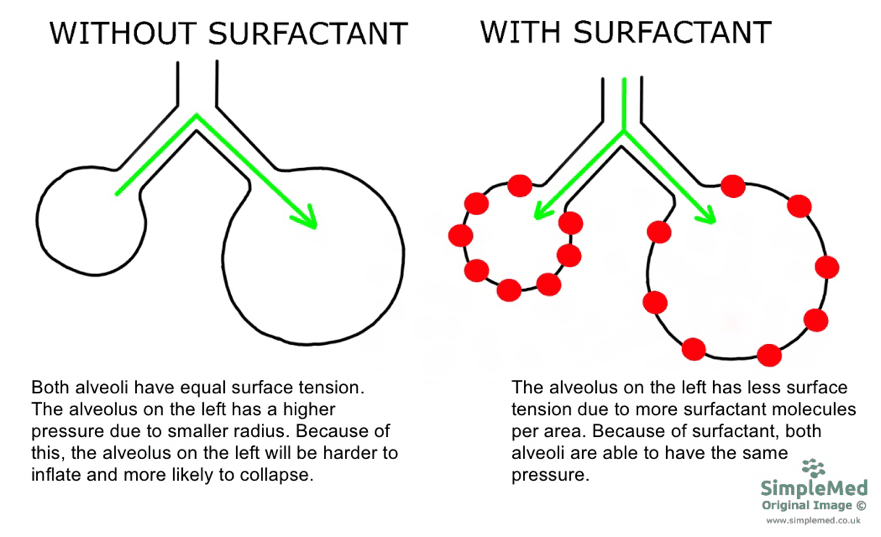
Diagram - The effect of surfactant on stabilising the lungs
SimpleMed original by Dr. Peter Parkinson
Surfactant production starts in a fetus at around 24 to 26 weeks of pregnancy, and many babies born before about 34 weeks do not yet have enough. This makes it very difficult to breathe and the baby’s lungs can collapse. This condition is called neonatal Respiratory Distress Syndrome (RDS), to tell it apart from acute respiratory distress syndrome in adults. In the UK, a mother who may give birth between 24 and 34 weeks of pregnancy is offered corticosteroids (steroid drugs) before the birth, which reduce the risk and severity of RDS (NICE NG25). Preterm babies who need breathing support soon after birth are given continuous positive airway pressure (CPAP), a constant gentle pressure that keeps the airways open, rather than invasive ventilation (a breathing tube attached to a ventilator) where clinically appropriate. Replacement surfactant is given to preterm babies who need invasive ventilation (NICE NG124).
Energy is required to force air through the airways of the respiratory system during inspiration and expiration. The most important factors are the radius of the airway and whether the flow of air is turbulent (chaotic) or laminar (smooth).
Resistance is inversely proportional to the fourth power of the radius of the airway (when flow is laminar), therefore as the radius decreases, the resistance increases steeply. Halving the radius increases the resistance 16-fold. Individually, small airways have higher resistance as radius decreases. However, because there are many bronchioles and alveolar ducts arranged in parallel, their total resistance to airflow is low compared with larger airways. The highest total resistance is in the trachea and the larger bronchi.
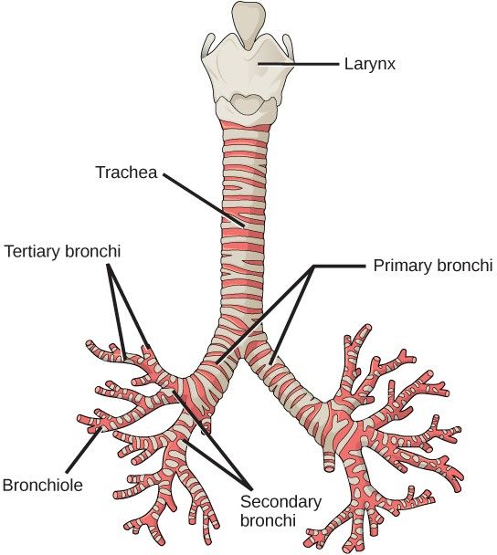
Diagram - The respiratory tract structure from the trachea to bronchiole
Creative commons source by CNX OpenStax [CC BY-SA 4.0 (https://creativecommons.org/licenses/by-sa/4.0)]
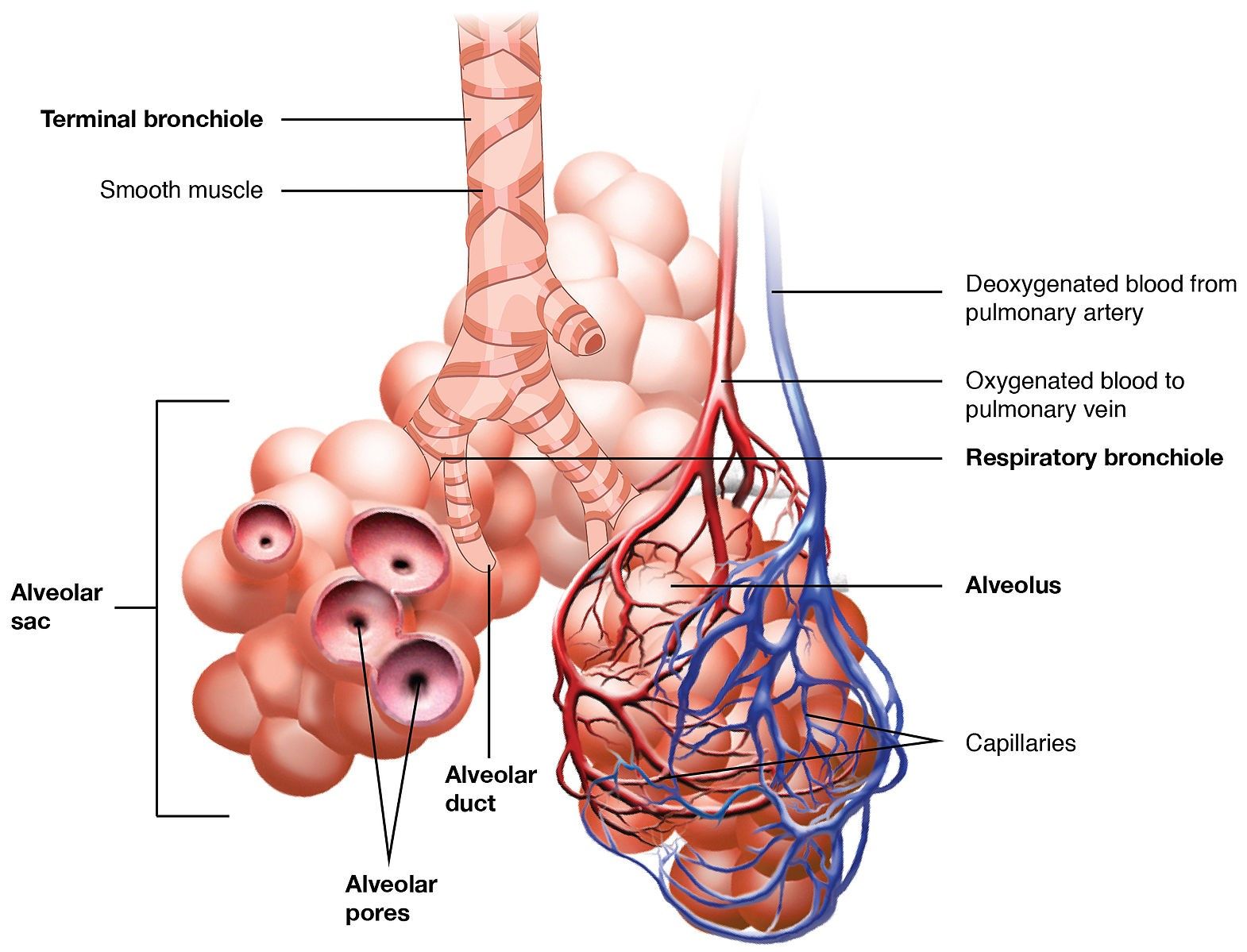
Diagram - The respiratory tract structure from the bronchioles to the alveoli
Creative commons source by OpenStax College [CC BY-SA 4.0 (https://creativecommons.org/licenses/by-sa/4.0)]
The resistance to airflow does change during inspiration and expiration depending on the radius of the airways. During inspiration, the thoracic cavity expands and the pressure around the small airways falls. This allows the small airways to widen, which increases the radius of the airways and decreases the resistance. During expiration, the opposite is seen: the intrathoracic pressure increases, the radius decreases and the resistance to airflow increases. When a person forcibly expires, the small airways are narrowed, increasing the resistance further. This leads to air being trapped in the alveoli and small airways, contributing to the residual volume. The residual volume is the volume of air that remains in the lungs after maximal expiration.
There is a relationship between resistance, pressure gradient and airflow. This is described by Ohm’s law (a rule from electricity that also applies to airflow), which involves the equation:
Airflow = pressure gradient / resistance.
This equation describes that an increase in resistance must be accompanied by an increase in pressure gradient to maintain the same airflow to the alveoli. This means that a larger pressure gradient between the inside and the outside of the lungs is needed to move air when the resistance is high. The diaphragm and the intercostal muscles then need to work harder to allow the lungs to expand and contract.
Disorders of Lung Mechanics and Ventilation
The following disorders we are going to discuss can be defined as having altered lung mechanics and ventilation.
Emphysema is defined as the chronic, progressive destruction of the alveolar wall without fibrosis (scarring) causing the permanent enlargement of air spaces distal to (beyond) the terminal bronchiole, the smallest airway that carries air without taking part in gas exchange. It is one form of chronic obstructive pulmonary disease (COPD), a group of lung conditions that also includes chronic bronchitis (long-term inflammation of the airways).
The main cause of emphysema is smoking; the inhaled smoke causes the breakdown of the alveolar wall and elastin fibres. Another cause of emphysema is alpha-1 antitrypsin deficiency. Alpha-1 antitrypsin is an antiproteinase (a protein that blocks enzymes that break down other proteins), and without enough of it the enzymes destroy the alveolar walls. This particular cause of emphysema is inherited, and a patient will present at a younger age compared to if the emphysema was caused by smoking.
In emphysema, the lack of elastic tissue means the lungs are easier to expand (increased compliance) but will not recoil as easily, which leaves the lungs hyperinflated (holding too much air). There is also a reduced surface area, meaning gas exchange is reduced. Both of these things lead to a ‘barrel chest’ and will reduce the radial traction (the pull of the surrounding lung tissue that holds the small airways open) on the small airways, causing an obstructive pattern on spirometry (air cannot be breathed out quickly, so the amount breathed out in the first second is low compared with the total amount).
Patients with emphysema will have a reduced exercise tolerance and shortness of breath. This is partly secondary to the reduced surface area available for oxygen to pass into the blood. Hyperinflation (too much air held in the lungs) also contributes to the breathlessness and to the reduced exercise tolerance.
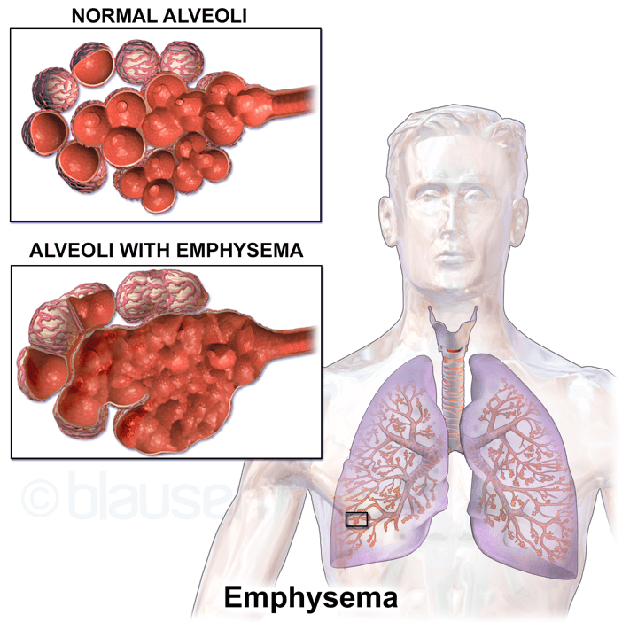
Image - The effects of emphysema on the structure of the alveoli
Creative commons source by Blausen Medical Communications, Inc. [CC BY-SA 4.0 (https://creativecommons.org/licenses/by-sa/4.0)]
Asthma is a chronic inflammatory condition of the respiratory airways that is caused by a mixture of genetic predisposition and environmental factors such as allergens and air pollution.
The chronic inflammation presents with smooth muscle contraction and mucosal oedema, leading to narrowing of the airways, particularly in the bronchi and bronchioles. On a spirometry tracing, asthma can show an obstructive pattern similar to COPD, but the airway narrowing is largely reversible. In adults, NICE NG245 uses a rise in the forced expiratory volume in one second (FEV1) of 12% or more, and 200 ml or more, after a bronchodilator as one test to diagnose asthma. Spirometry results are more likely to be normal in people who already take inhaled corticosteroids. Short-acting beta-2 receptor agonists such as salbutamol provide rapid bronchodilation (widening of the airways). Corticosteroids (steroid drugs) reduce airway inflammation over time, and inhaled corticosteroids are also used in some people with COPD. In current UK guidance (NICE NG245, 2024), a short-acting beta-2 receptor agonist is not prescribed without an inhaled corticosteroid. People aged 12 and over with newly diagnosed asthma are offered a low-dose inhaled corticosteroid and formoterol (another bronchodilator) combination inhaler, taken as needed for symptoms.
The interstitial space is found between the capillary basement membrane (the thin supporting layer under the lining of the capillary) and the alveolar cells. The space contains collagen fibres, elastin fibres and fibroblasts (synthesise collagen and the extracellular matrix). It is only in diseased states when the space becomes apparent by containing fibrous tissue, fluid or cells.
Interstitial Lung Disease (ILD) can develop idiopathically (with no known cause) or have a cause, for example:
- Drugs such as nitrofurantoin (urinary tract infections) and amiodarone (cardiac arrhythmias, which are abnormal heart rhythms)
- Occupational exposure to asbestos or silica
- Infection: ILD can develop after an infection (postinfectious ILD)
The pathophysiology of ILD (how the disease develops) is unclear, however, what is believed is that the alveolar epithelial cells become damaged due to an irritant (asbestos for example). The body increases the production of fibroblasts as a way to repair the damage by laying down scar tissue in the interstitial space. The increased amount of fibrotic tissue surrounding the remaining alveoli makes it harder for the alveoli to expand, therefore the compliance of the lungs decreases. The collagen is not very stretchy but tends to recoil meaning the elastic recoil of the lungs increases and the lungs become smaller as a result. The increased amount of collagen and extracellular matrix leads to an increased diffusion distance for gases moving from the alveolar spaces into the blood. This has a greater effect on the diffusion of oxygen than carbon dioxide, because carbon dioxide is more soluble.
When examining a patient with ILD, chest expansion will be reduced, and respiratory rate will be increased to supply enough oxygen to the body, which leads to a reduced exercise tolerance.
A pneumothorax is the presence of air in the pleural space caused by a disruption to the pleural membrane. It can develop spontaneously (without an injury), or following injury to the chest wall (for example a fractured rib), or when air-filled cysts found on lung tissue rupture.
At rest, the pressure within the pleural space is more negative than atmospheric pressure. Once air is able to enter the pleural space, air will usually flow down a pressure gradient until the pressure within the pleural space is equal to atmospheric pressure. This causes the lung tissue to collapse inward, as there is nothing keeping it attached to the chest wall. In a tension pneumothorax, the pressure in the pleural space keeps rising above atmospheric pressure. The raised pressure compresses the heart, the great vessels (the large vessels entering and leaving the heart) and the trachea, which reduces the blood returning to the heart and the cardiac output. This is life-threatening and needs immediate treatment, called decompression (releasing the trapped air).
As a pneumothorax can happen suddenly, it can present with acute, sharp pleuritic pain (pain that is worse when breathing in) and breathlessness.
Atelectasis refers to the complete or partial collapse of a whole lung, a lobe (one main section of a lung) or a smaller segment of lung. It has several causes. One is an obstruction (like a tumour, mucus plug or foreign object) in the airways, preventing the alveoli from ventilating. The air distal to (beyond) the obstruction is reabsorbed, collapsing that part of the lung. Others include surfactant deficiency, and compression of the lung from outside, for example by a pneumothorax or a mass in the chest.
Surgery can also lead to atelectasis. The anaesthesia, pain relief used, intubation (placing a breathing tube into the airway) and sedatives can lead to the patient’s breathing becoming shallower, meaning the alveoli may not inflate adequately and causing them to collapse.
Edited by: Dr. Maddie Swannackand Dr. Marcus Judge
- 11205



