

Next Lesson - Conditions of the Elbow
Core
Want more information on anatomy before you begin looking at conditions? Head to our articles on Muscles of the Upper Arm, Muscles of the Forearm and Bones of the Upper Limb for more information!
Supracondylar Fracture of the Distal Humerus
Supracondylar fractures of the distal humerus are usually caused by a fall onto an outstretched hand (FOOSH) with a hyperextended elbow. They occur most commonly in children and make up around 75% of ‘elbow’ injuries. The distal fragment is usually displaced posteriorly. This condition presents with pain, deformity and reduced function of the elbow joint.
Complications of a supracondylar fracture of the distal humerus include:
Damage to ulnar, radial or median nerves (all described later in this article) - the median nerve (often the anterior interosseous branch) is most commonly damaged.
Gunstock deformity results from fracture malunion causing cubitus varus: the carrying angle is reduced or reversed, and the forearm deviates medially towards the body when the elbow is extended.
Volkmann’s ischaemic contracture - where damage to the brachial artery causes compartment syndrome, which leads to infarction resulting in fibrotic repair. This complication presents with elbow flexion, forearm pronation, wrist flexion, metacarpal-phalangeal joint extension and interphalangeal joint flexion.
This condition is diagnosed with an X-ray. The treatment involves a cast or splint to promote natural healing for a less severe fracture, or surgery (usually a closed reduction) for a more severe fracture.

Diagram - An X-Ray of a normal humerus. The black line (the anterior humeral line) should normally pass through the middle third of the capitulum (highlighted green), but if it doesn't, it indicates that there is a supracondylar humerus fracture
Creative commons source by Tracy Kilborn, Halvani Moodley, Stewart Mears and Mikael Häggström [CC BY-SA 4.0 (https://creativecommons.org/licenses/by-sa/4.0)]
A high injury to the radial nerve usually occurs due to a mid-shaft humeral fracture. This is because the radial nerve runs in the ‘radial groove’ of the humerus, so can be damaged by this mechanism. It presents with:
- Wrist drop due to loss of wrist extension.
- Flexion at the metacarpal-phalangeal joints.
- Paraesthesia of the dorsal aspect of the 1st, 2nd and 3rd digits (excluding the fingertips).
Elbow extension is spared in the majority of high radial injuries, due to the branch that innervates triceps brachii being given off proximal to the site of injury.
Treatment typically involves restriction of movement with a splint, so that the nerve has time to heal, or it may require surgery. Complete recovery can take anywhere from a few weeks to six months, or recovery may not be possible at all.
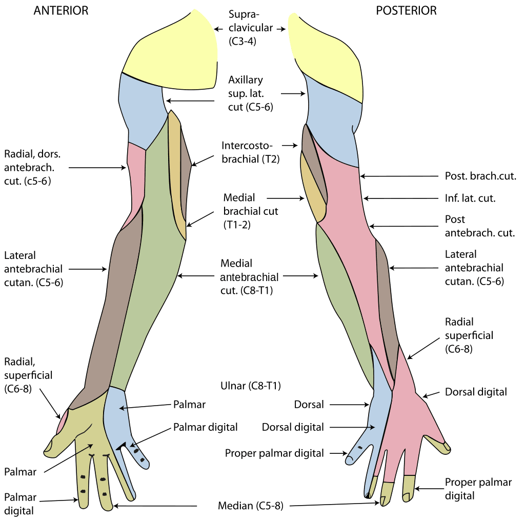
Diagram - The cutaneous innervation of the upper limbs. The radial nerve innervation can be seen in pink
Creative commons source by Henry Gray (1918) Anatomy of the Human Body, modified by Fred the Oyster [CC BY-SA 4.0 (https://creativecommons.org/licenses/by-sa/4.0)]
A high injury to the ulnar nerve usually occurs due to a medial epicondyle fracture. It presents with:
- A ‘high ulnar claw’ when asked to extend all the fingers:
- Hyperextension of the 4th and 5th digits at the metacarpal-phalangeal joints.
- Flexionof the 4th and 5th digits at the proximal interphalangeal joints but not at the distal interphalangeal joints because the ulnar-innervated part of flexor digitorum profundus supplying these fingers is paralysed in a complete high lesion.
- Paraesthesia of the 4th and 5th digits and medial border of the hand.
Management of a high ulnar nerve injury depends on whether the nerve is intact or divided. Splinting and observation may be appropriate for an intact injured nerve; a divided nerve needs specialist assessment for repair. Recovery may take many months, and the final outcome can take two years or longer to become clear.
Clawing may be less pronounced in a high ulnar lesion than in a low lesion because a high lesion also weakens the ulnar-innervated part of flexor digitorum profundus supplying the ring and little fingers. Reduced distal finger flexion makes the claw less marked: this is the ulnar paradox. The appearance varies with the extent of the lesion.

Image - A healthy hand imitating ulnar clawing. The photograph does not document a nerve lesion or establish whether a lesion is high or low.
Creative commons source by Mcstrother [CC BY 3.0]; resized for this article
A high injury to the median nerve usually occurs due to a supracondylar humeral fracture. It presents with:
- Loss of forearm pronation
- The traditional textbook ‘Hand of Benediction’ description on attempting a fist is not a reliable clinical sign:
- Index interphalangeal flexion may be impaired, but index metacarpophalangeal flexion can remain through the ulnar-innervated interossei. Middle-finger flexion may also remain through variable innervation and connections between the deep flexor tendons. Absence of the traditional posture does not exclude a high median nerve lesion.
- Flexion of the 4th and 5th digits is preserved.
- Extension and adduction of the 1st digit.
- Paraesthesia of the palmar aspect of the lateral hand and 1st, 2nd and 3rd digits.
Management depends on the mechanism and extent of nerve injury. Splinting and observation may be appropriate for an intact injured nerve, while a divided nerve requires specialist assessment for repair. Recovery is variable and may take many months; the final outcome can take two years or longer to become clear.
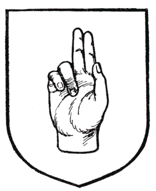
Image - A heraldic drawing of the traditional hand of benediction posture. This is not a reliable depiction of the clinical appearance of a high median nerve lesion.
Illustration source from A Complete Guide to Heraldry by Arthur Charles Fox-Davies [public domain]
Edited by: Dr. Thomas Burnell and Dr. Maddie Swannack
Reviewed by: Dr. Marcus Judge
- 6536



