

Next Lesson - Action Potentials
Core
A membrane potential is the difference in electrical potential (the voltage) across a plasma membrane, expressed as the potential inside the cell relative to the extracellular solution (the fluid outside the cell). It is measured in millivolts (mV).
All cells have a membrane potential, although the resting membrane potential and the ability to change it (excitability: how readily a cell can produce electrical signals called action potentials) vary between different cell types. Membrane potentials are the basis of electrical signalling in excitable cells such as neurones and muscle cells, and in other cells they help to control processes such as cell volume and the cell cycle (the cycle of cell growth and division).
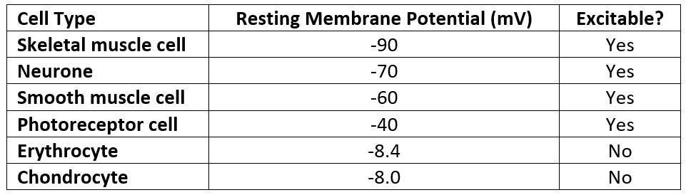
Table - The resting membrane potentials of different cell types
SimpleMed original by Dr. Joshua Bray
The establishment of a membrane potential requires two key criteria:
- Ionic concentration gradients (resulting in an asymmetrical distribution of ions)
- Selective permeability to particular ions (the membrane lets some ions cross more easily than others, via ion channels) - mainly potassium (K+), sodium (Na+) and chloride (Cl-)
The movement of ions results in a separation of charge across the membrane: this is the membrane potential.
The Na+/K+ pump (a protein that uses energy to move sodium out of the cell and potassium in) is slightly electrogenic: it moves three Na+ out for every two K+ in, which adds a small negative component to the resting membrane potential. However, its main role is indirect: it sets up and maintains the ionic gradients required to generate the membrane potential, and without it the membrane potential would gradually run down.

Diagram - The ionic concentration gradients responsible for the membrane potential
Creative commons source by smonsays [CC BY-SA 4.0 (https://creativecommons.org/licenses/by-sa/4.0)]
The Resting Membrane Potential
There are two gradients which determine the net movement of ions:
- Chemical gradient - Equivalent to the concentration gradient.
- Electrical gradient - The tendency of positive ions to move to areas of negative charge and negative ions to move to areas of positive charge.
Assume that the membrane is selectively permeable to one ion only - for example, K+. If the concentration of K+ is greater inside the cell compared to outside the cell, then K+ will begin to move out of the cell. However, as K+ leaves the cell, the cell will become less positive and the extracellular solution will become more positive - this causes the opposing electrical gradient to increase. When the chemical and electrical gradients become equal and opposite, they cancel each other out so that there is no net movement of K+. However, a negative charge remains across the membrane. In this simplified case, that potential is the equilibrium potential for K+ (defined below). In a real cell, the resting membrane potential is strongly influenced by K+ but other ions also cross the membrane, so it is less negative than the equilibrium potential for K+ (in a neurone, about -70 mV compared with -95 mV).
The membrane potential when the membrane is selectively permeable to only one type of ion is known as the equilibrium potential (E) for that ion. We can calculate an ion’s equilibrium potential using the Nernst equation. The Nernst equation for K+ is as follows: EK = (RT/zF) ln([K+]out/[K+]in), where:
- R is the gas constant (8.314 J mol-1 K-1)
- T is temperature in kelvin (K)
- z is the charge on the ion (+1 for K+, -1 for Cl-)
- F is the Faraday constant, the charge carried by one mole of singly charged ions (96500 C mol-1)
- [K+]out and [K+]in are the concentrations of K+ outside and inside the cell respectively.
To make calculations easier, we can work out the value of RT/F (T = 37°C, which is 310 K) and convert the natural log (ln) to log10 (the base-10 logarithm, as on a calculator). This gives a constant of about 61 mV, so: EK = (61 mV / z) × log10([K+]out / [K+]in).
The Nernst equation can be used for any ion (both positive and negative ions).
If a membrane is selectively permeable to K+ alone, its membrane potential will be at EK. However, in reality a membrane is never permeable to K+ alone, as real cells are permeable to other ions too.
The equilibrium potentials of K+, Na+ and Cl- at physiological (normal body) temperature and concentrations are as follows:
- EK = -95 mV
- ENa = +60 mV
- ECl = -70 mV to -90 mV
Changing the Membrane Potential
The membrane potential can be changed by altering the membrane’s permeability to different ions - i.e. opening and closing ion channels.
If the membrane becomes more permeable to an ion, the membrane potential will shift towards the equilibrium potential for that ion.
The value of the membrane potential can change in two directions:
- Depolarisation - The membrane potential becomes less negative
- Hyperpolarisation - The membrane potential becomes more negative
Opening Na+ or calcium (Ca2+) channels usually results in depolarisation, whereas opening K+ or Cl- channels usually results in hyperpolarisation. The actual effect depends on where the ion’s equilibrium potential lies relative to the current membrane potential. The membrane potential moves towards the equilibrium potential of the ion, so the cell depolarises if that equilibrium potential is above the current membrane potential (less negative, or positive), and hyperpolarises if it is below it (more negative).

Diagram - Graph showing the changes in membrane potential by altering Na+ and K+ flow (a simplified graph: the baseline is drawn at 0 mV, but a real resting membrane potential is negative)
SimpleMed original by Dr. Joshua Bray
A synapse is a connection between two excitable cells. At a chemical synapse, a neurotransmitter (a chemical messenger) is released from the pre-synaptic cell (the cell sending the signal). It diffuses across the synaptic cleft (the tiny gap between the two cells) and binds to receptors on the post-synaptic membrane (the membrane of the cell receiving the signal). This allows the conversion of an electrical signal (action potential) to a chemical signal (neurotransmitter), which generates a new electrical signal in the post-synaptic cell.
Synaptic transmission can be classed as fast or slow:
- Fast transmission - Involves ligand-gated ion channels (channels that open when a ligand, such as a neurotransmitter, binds to them; e.g. nicotinic acetylcholine receptors, named after nicotine, which also activates them).
- Slow transmission - Involves G-protein coupled receptors (receptors that work through a G protein inside the cell; e.g. muscarinic acetylcholine receptors, named after muscarine, a mushroom toxin that also activates them), which activate ion channels via direct and indirect signalling pathways (the indirect route uses second messengers, which are chemical signals made inside the cell).
Synapses can also be excitatory or inhibitory:
- Excitatory synapses cause membrane depolarisation, which makes the post-synaptic cell more likely to fire an action potential - Transmitters commonly include glutamate and, depending on receptor type, dopamine and acetylcholine
- Inhibitory synapses cause membrane hyperpolarisation, which makes the post-synaptic cell less likely to fire an action potential - Transmitters commonly include gamma-aminobutyric acid (GABA) and glycine
The more depolarised the post-synaptic cell becomes, the more likely it is to reach the threshold for action potential generation.

Diagram - Graphs showing the excitatory and inhibitory post-synaptic potentials
SimpleMed original by Dr. Joshua Bray
Reviewed by: Dr. Thomas Burnell and Dr. Marcus Judge
- 12696

