

Next Lesson - Thrombosis and Embolism
Core
Haemostasis (not to be confused with homeostasis) comes from haemo (pertaining to blood) and stasis (to halt) and thus literally translates to halting blood. It is an essential process in the moments after vessel injury that prevents small wounds from becoming fatal.
The Three Steps of Haemostasis
- Vessel wall damage
- The severed or damaged artery contracts. This contraction isn’t enough to stop the bleeding but does assist in reducing the pressure ‘downstream’ of the site of injury and control the bleeding. Contraction is less pronounced in veins due to their thinner smooth muscle layer; however, bleeding is usually less severe because venous blood flow is under lower pressure.
- A primary haemostatic plug of activated platelets will form at the site of injury. This plug will stick to the injured vessel and the tissue outside of it. Once more, this won’t stop the bleeding completely but will help to control the bleeding. The primary plug will form seconds to minutes following the vessel severing.
- A secondary haemostatic plug forms as fibrin reinforces the platelet plug. Platelet activation and coagulation overlap after vessel injury; there is no universal 30-minute delay before fibrin reinforcement. Thrombin generates fibrin and activated factor XIII cross-links it to strengthen the clot.
Below are the aspects of haemostasis and clotting in more detail.
Platelets are cell fragments which have no nucleus and are involved in the process of blood clotting. There are approximately 150-400 x109 cells per litre of blood. Platelets are formed in the bone marrow by the process of budding from the cytoplasm of megakaryocytes.
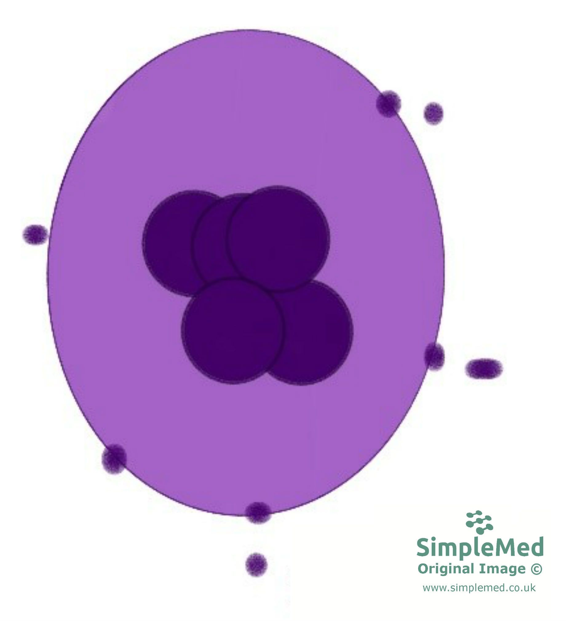
Image - Platelets (represented by the small dark purple circles) budding from a megakaryocyte
SimpleMed original by Benjamin Norris
Platelets can be activated in numerous ways:
- Contact with collagen surfaces - collagen is found in the blood vessel wall below the endothelium and is therefore exposed when the blood vessel is damaged and can activate platelets.
- ADP - released by activated platelets and red blood cells. This causes an amplified platelet response.
- Thromboxane A2 - a powerful platelet aggregator that is released by activated platelets in order to amplify the platelet response.
- Thrombin - activated in the clotting cascade and recruits more platelets.
Platelets adhere to the injured vessel wall with the help of von Willebrand factor (vWF). Activated platelets aggregate through fibrinogen bridges between their surface receptors. The clotting cascade produces a fibrin mesh that reinforces this platelet plug. Activated platelets also release ADP and thromboxane A2, which recruit and activate further platelets.
The drug aspirin irreversibly deactivates cyclooxygenase, an enzyme responsible for thromboxane A2 production. Therefore, long-term low-dose aspirin is used as secondary prevention against myocardial infarction and stroke (i.e. aspirin makes it more difficult for blood clots to form).
For blood to clot, fibrin is needed which are threads of protein that can cross-link to form a fibrin mesh. This mesh catches platelets and holds the blood clot together to strengthen the platelet plug. The formation of fibrin is achieved via the clotting cascade, which is shown in the diagram below.

Diagram - The clotting cascade. The intrinsic and extrinsic pathways lead to thrombin generation. Thrombin converts fibrinogen to fibrin, which reinforces the platelet plug; activated factor XIII cross-links the fibrin strands to strengthen the clot.
SimpleMed original by Benjamin Norris
Coagulation can occur via two pathways which produce the same result. The two pathways use some different clotting factors shown in the Venn diagram. This means that timings of these pathways can be measured when investigating coagulation disorders to narrow down the clotting factors affected. The two pathways and their measurement are explained below:
- Intrinsic pathway (measured by APTT) - Triggered by the exposure of collagen after a blood vessel is damaged.
- Extrinsic pathway (measured by PT) - Triggered by the release of thromboplastin from damaged endothelial cells. It can be released in response to breaking of the blood vessel wall, hypoxia, sepsis, malignancy and inflammation.
PT can also be expressed by the international normalised ratio (INR) which compares the PT to a normal value in the form of a ratio. For example, an INR of 1 is normal, and blood with an INR of 3 will clot more slowly, for example if a patient is on warfarin.
There is an additional measurement which has historically been used to investigate clotting by measuring how long the patient bleeds for after a break to the skin. A longer bleeding time (BT) suggests a problem with platelet aggregation, although BT is now largely obsolete and has been replaced by laboratory platelet function tests.
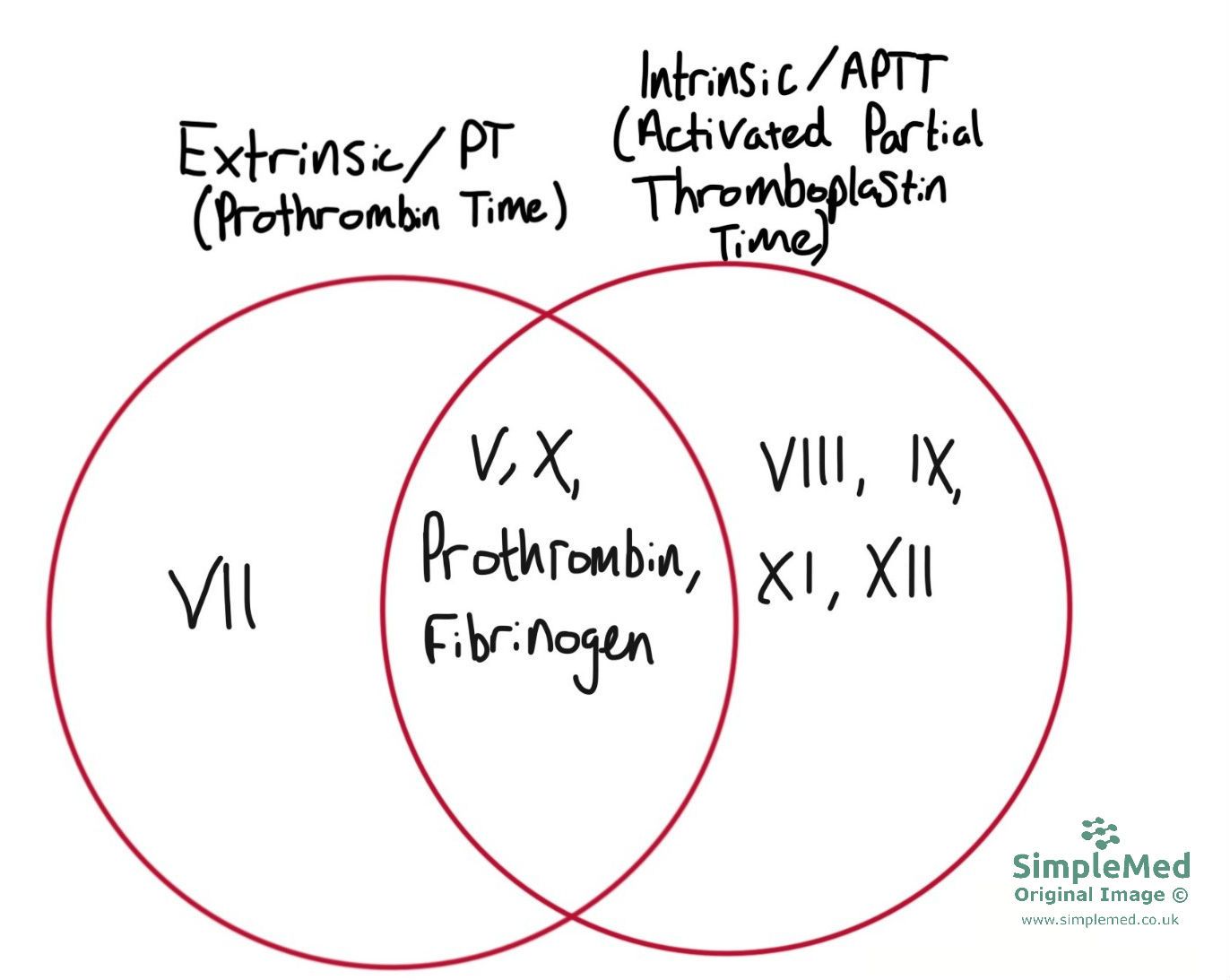
Diagram - A Venn diagram illustrating the factors present in the extrinsic and intrinsic pathways
SimpleMed original by Benjamin Norris
These factors include dilution of clotting factors by blood flow and natural anticoagulants. The main natural anticoagulants in the blood are antithrombin III, protein S and protein C. Natural anticoagulants don’t destroy clots (that deed is performed by the enzyme plasmin in fibrinolysis); rather they oppose the formation of fibrin in the first place to control the clotting cascade.
As platelets within the clot contract, they pull on fibrin via actin-myosin filaments. As a result of this, the edges of the wound to which the clot has adhered are drawn together to a certain extent. This is wound contraction, a part of the healing process of the blood vessel.
Plasminogen is an inactive circulating precursor synthesised mainly by the liver. Endothelial cells release tissue plasminogen activator (tPA), which converts plasminogen to plasmin; fibrin enhances this activation at the clot. Plasmin breaks down fibrin into soluble fibrin degradation products. tPA is an activator, not an intermediate into which plasminogen is converted.
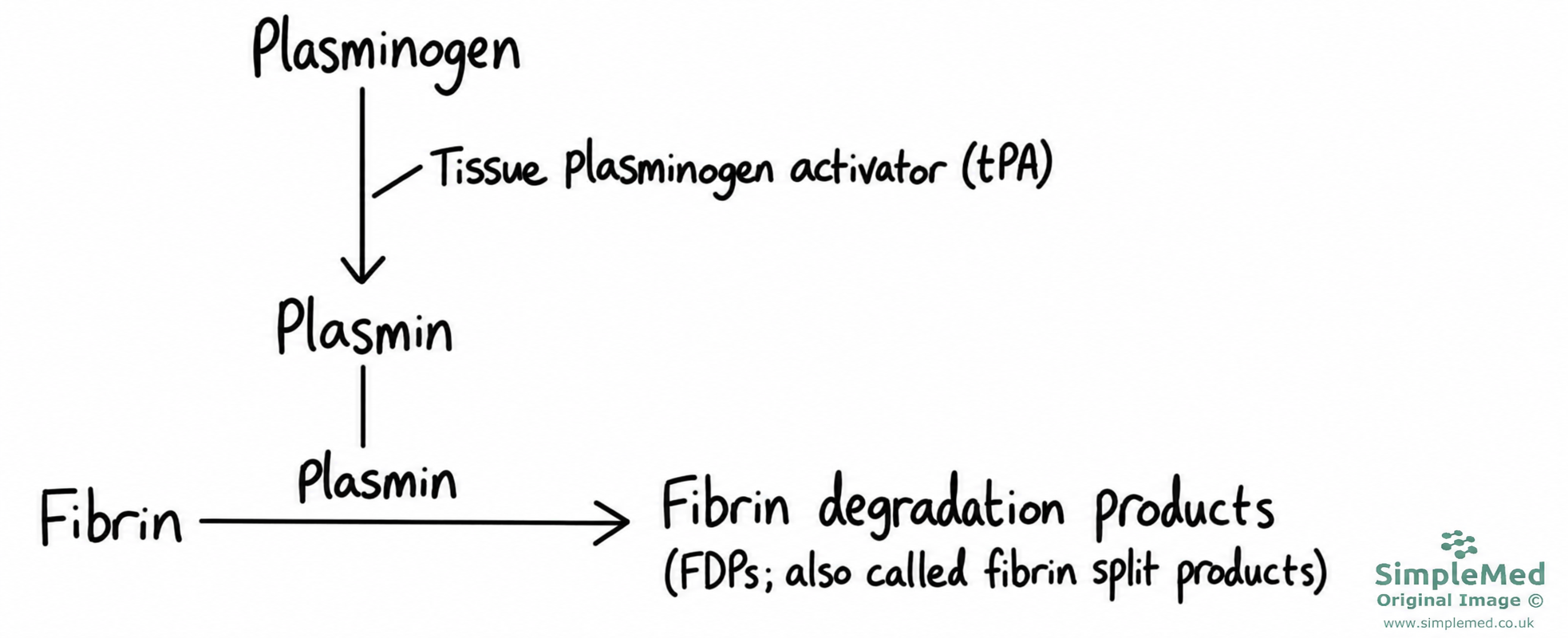
Diagram - tPA activates plasminogen to form plasmin. The same plasmin then breaks down fibrin into fibrin degradation products (also called fibrin split products).
Adapted from the SimpleMed original by Benjamin Norris. Updated in 2026 to show tPA as an activator rather than an intermediate.
tPA can be used therapeutically in dissolving thrombi and thromboembolisms, however, a side effect of this can be undesired bleeding. An additional fibrinolytic agent which can be used therapeutically is streptokinase, however this is obtained from streptococci, meaning that it is antigenic and can only be used once.
Disseminated Intravascular Coagulation (DIC)
Disseminated intravascular coagulation is a pathological activation of the coagulation cascade. In DIC, clotting activators enter the systemic circulation and many microthrombi (small pathological clots in the vessels) are formed throughout the circulation. During this process many coagulation factors and platelets are consumed.
When enough of these clotting factors have been used, haemorrhage can begin to occur as the body has no more platelets or clotting factors to form clots when they're actually needed. The affected patient is at risk of bleeding, neurological impairment, gangrene, renal failure, respiratory distress and a wide variety of other complications. Red blood cells are often damaged in DIC as they are sheared by flowing past the microthrombi, and between this and haemorrhage the patient can develop anaemia.
Triggers for DIC can include malignancy, massive trauma (e.g. burns), infections, massive haemorrhage, and transfusion. Although in the short term DIC can be managed by transfusing the patient with clotting factors and platelets, in order to truly treat the condition, the underlying cause must be treated first.
There are two main types of haemophilia, both of which are congenital disorders of clotting factors. These two types are haemophilia A and haemophilia B (also known as Christmas disease). Platelets are unaffected in haemophilia, however, as there are inadequate amounts of clotting factors, less fibrin is produced.
Both varieties of haemophilia are X-linked recessive and are therefore much more common in males than in females. Haemophilia A and haemophilia B are clinically indistinguishable from one another, with both having normal bleeding time and PT but prolonged APTT (factor VIII and IX are used in the intrinsic pathway causing the APTT to be prolonged, but neither factor is involved in the extrinsic pathway so PT is normal).
Haemophilia A is either a deficiency of the amount of factor VIII, or reduced factor VIII activity It is the most common hereditary disorder associated with serious bleeding.
Individuals with mild haemophilia A may experience prolonged bleeding after major trauma or surgery, and after venepuncture. In more severe cases, patients can present with spontaneous bleeding into joints (haemarthrosis), intramuscular haemorrhage and, in severe cases, intracranial haemorrhage.
Mild haemophilia A is seen with 5-40% of normal factor VIII activity, moderate disease with 1-5%, and the severe form with under 1% of factor VIII.
Patients with haemophilia A can be managed through infusion of recombinant factor VIII.
Haemophilia B is a deficiency in factor IX, and presents with similar symptoms to haemophilia A, but with an increased risk of bruising and experiencing mucosal bleeds. It can be treated through an infusion of recombinant factor IX.
Von Willebrand disease is the most common inherited bleeding disorder and can range in nature from asymptomatic to severe. Von Willebrand factor is either deficient or abnormal in this condition.
The function of this protein includes protecting factor VIII from premature destruction and assisting in the formation of the platelet plug. Both bleeding time and APTT can be affected by this condition.
Features include bleeding following minor trauma or surgery, epistaxis and menorrhagia. Bleeding into muscles and joints is rare.
Management depends on the type and severity of von Willebrand disease. Many patients respond to desmopressin, while others require concentrates containing von Willebrand factor (with factor VIII).
Thrombocytopenia means a platelet count below the normal range, commonly below 150 × 109/L in adults. Bleeding risk depends on the severity and cause. In isolated thrombocytopenia, PT and APTT may remain normal because they assess plasma coagulation pathways rather than platelet number. A prolonged bleeding time is a traditional teaching association, but this test is rarely used in current clinical practice.
Patients with thrombocytopenia can present with petechiae, which are small red or purple spots under the skin; these occur due to spontaneous bleeding from capillaries.
Thrombocytopenia has a variety of causes such as autoimmune thrombocytopenic purpura, DIC, liver failure or drugs such as heparin.
Thrombophilia can be due to congenital or acquired defects of haemostasis that lead to an increased chance of thrombosis (pathological blood clotting).
Examples of thrombophilias include factor V Leiden thrombophilia, antithrombin deficiency, protein S or protein C deficiency, and antiphospholipid syndrome.
Edited by: Dr. Bethany Turner
Reviewed by: Dr. Marcus Judge
- 13322
