

Next Lesson - Regeneration and Repair of Tissues
Core
There are several different ways chronic inflammation arises. These include:
- Taking over from acute inflammation.
- Develop alongside acute inflammation with severe or persistent irritation.
- De novo - developing with no period of acute inflammation preceding it, for example, in autoimmune conditions. This is one way chronic inflammation can arise
These cells are derived from blood monocytes. Their functions include:
- Phagocytosis and destruction of bacteria.
- Antigen presentation and therefore initiation of an immune response.
- Synthesis of cytokines, complement components, blood clotting factors and proteases.
- Control of other cells by cytokine release.
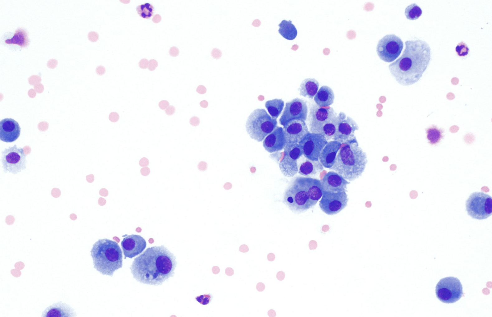
Image - Macrophages under a light microscope
Creative commons source by Librepath [CC BY-SA 4.0 (https://creativecommons.org/licenses/by-sa/4.0)]
There are two types of lymphocytes: B lymphocytes and T lymphocytes. The different types have different functions:
- B cells: produce and secrete antibodies by differentiating into plasma cells.
- T cells: kill virus-infected cells and sometimes tumour cells, and mediate the immune response.
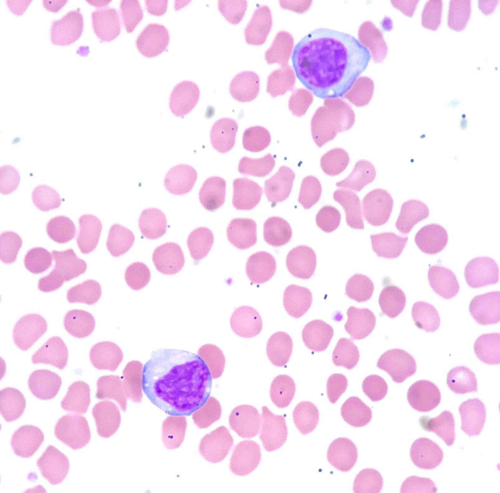
Image - Histological sample showing two lymphocytes
Creative commons source by Ed Uthman [CC BY-SA 4.0 (https://creativecommons.org/licenses/by-sa/4.0)]
These cells are seen in large numbers with atopic sensitivity reactions such as asthma. They also attack large parasites such as worms and may be seen in association with some types of cancer, for example Hodgkin’s lymphoma.

Image - Histological sample of an eosinophil
Creative commons source by Dr Graham Beards [CC BY-SA 4.0 (https://creativecommons.org/licenses/by-sa/4.0)]
These cells respond to chemotactic stimuli to move to the site where they are needed. They play a role in wound healing and scar production by producing collagen, elastin and glycosaminoglycans. They can also differentiate into myofibroblasts which contain actin to be able to contract and close wounds.
In certain circumstances macrophages are unable to phagocytose, for example when there are foreign bodies or certain bacteria present. When this occurs, the macrophages fuse together to form giant cells that isolate the offending substance.
There are different types of giant cell and the type that is present can aid diagnosis:
Langhans Giant Cells - nuclei are arranged at the periphery, often in a horseshoe pattern. They occur in granulomatous inflammation, including tuberculosis (TB), but their morphology alone does not establish TB. Persistent stimuli such as infection with Mycobacterium tuberculosis can promote macrophage fusion and giant-cell formation. The cause must be established from the clinical context and further investigations.
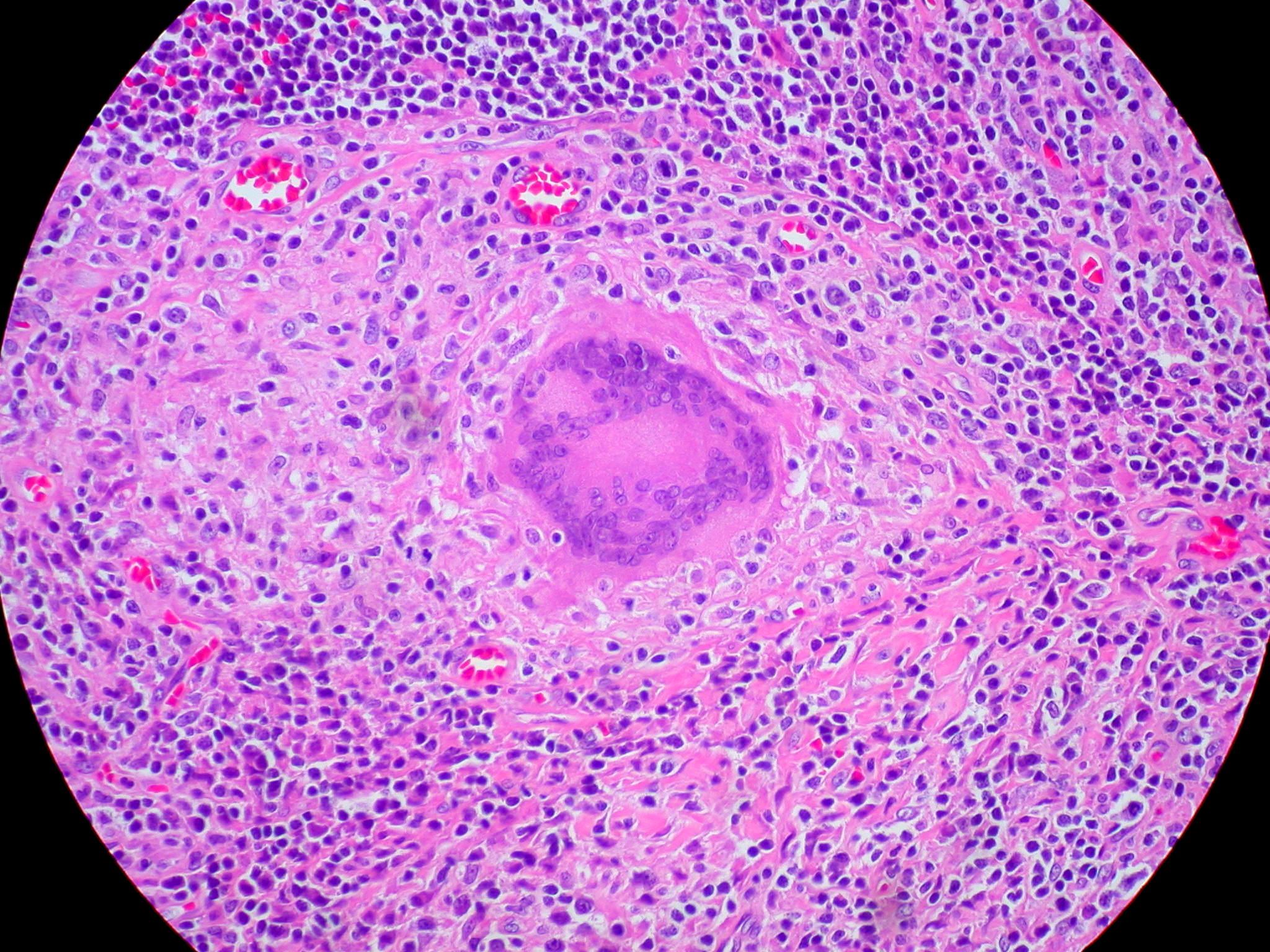
Image - Histological Sample Showing a Langhans Type Giant Cell
Creative commons source by Ed Uthman [CC BY-SA 4.0 (https://creativecommons.org/licenses/by-sa/4.0)]
Foreign Body Type - nuclei are arranged randomly in the giant cell. They occur when there is a foreign body that macrophages can’t phagocytose.
Touton Giant Cells - nuclei form a ring or wreath around central cytoplasm, with foamy, lipid-rich cytoplasm outside the ring. They are seen in areas with high lipid content, such as fat necrosis and xanthomas. These giant cells have a foamy appearance due to the phagocytosis of lipid.
Effects of Chronic Inflammation
Fibrosis
This occurs when fibroblasts are stimulated to produce collagen. This process is initially helpful as it produces a fibrous scar to replace lost tissue and wall off the infected area.
However, fibrosis can become harmful when it replaces normal parenchymal tissue of an organ, leading to impaired function of the organ. An example of this is when the liver undergoes cirrhosis. Long-term damage to the liver causes fibrosis and over time this scarring leads to contraction and shrinking of the liver which impairs portal blood flow. This causes portal hypertension which leads to ascites.
Stimulation of Inappropriate Immune Responses
Inflammation is used by the immune system as a way of destroying its targets. However, it can occasionally cause inflammation at an inappropriate site. This occurs in autoimmune conditions such as rheumatoid arthritis. In rheumatoid arthritis the immune system attacks the soft tissues in the joints causing chronic inflammation. This leads to swelling and joint pain, and if untreated can cause joint destruction.
Granulomas
Sometimes chronic inflammation can be accompanied by the formation of granulomas.
A granuloma is formed when there are particles which are difficult to break down but need to be isolated and contained. These particles can include foreign bodies such as splinters, or bacteria such as Mycobacterium tuberculosis. Granulomas are a collection of epithelioid macrophages. These are macrophages which have been modified due to increased activity so look like epithelium. Giant cells can also be found in and around granulomas.
There are two general types of granuloma:
- Foreign Body Granulomas
- Develops around material which isn't antigenic e.g. surgical thread.
- Contains macrophages, foreign body giant cells, epithelioid macrophages and fibroblasts.
- Immune Type Granulomas
- Develop around insoluble particles with antigenic materials.
- Undergo central necrosis, especially in granulomas associated with tuberculosis.
- Present in infections such as tuberculosis, syphilis, leprosy and fungal infections.
Granulomas can also be present in conditions without antigenic material such as sarcoidosis, granulomatosis with polyangiitis (GPA) and Crohn's disease.
Edited by: Dr. Bethany Turner
Reviewed by: Dr. Josh Sturgeon
- 11363


