

All done here! Check out our other subjects!
Core
The axilla can be imagined as a pyramid, with the apex starting behind the lateral border of the first rib.
The borders are as follows:
- Apex - cervico-axillary canal, bounded by the lateral border of the first rib, the clavicle, and the superior border of the scapula
- Lateral wall - intertubercular groove of the humerus
- Anterior wall - pectoralis major (and pectoralis minor)
- Posterior wall - subscapularis, teres major, latissimus dorsi
- Medial wall - thoracic wall and serratus anterior
The axilla contains the following key neurovasculature:
- Axillary artery
- Axillary vein
- Axillary lymph nodes
- Brachial plexus
Knowing the contents helps locate the axillary lymph nodes. These nodes may contain metastases from malignancy, including breast cancer, or enlarge reactively in response to infection. Infection-related node enlargement is not metastasis.
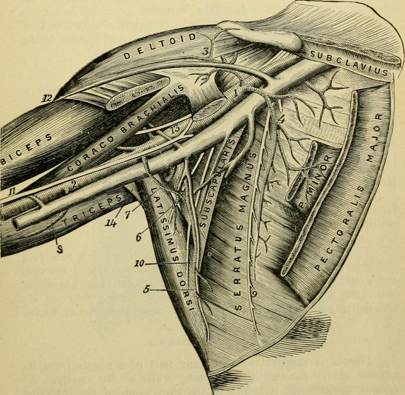
Diagram - Illustration showing the contents of the axilla.
Public Domain Image [Public domain]
This triangular region is found on the anterior side of the elbow.
The borders are as follows:
- Lateral border - medial border of brachioradialis
- Medial border - lateral border of pronator teres
- Superior border - an imaginary line between the condyles of the humerus
The contents are as follows (moving lateral to medial):
- Radial nerve
- Biceps tendon
- Brachial artery
- Median nerve
A way to remember the order of these structures is 'Really Need Beer To Be At My Nicest'.
Knowledge of the placement of contents within the cubital fossa is incredibly important. For example, the brachial artery can be felt by palpating medial to the biceps tendon - useful when taking blood pressure or testing peripheral pulses. The superficial fascia of the roof contains the median cubital vein, a common site for venepuncture. It lies superficial to the bicipital aponeurosis and is separate from the deep contents of the fossa.
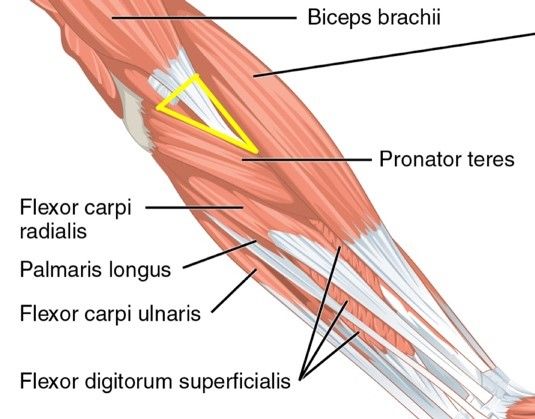
Diagram - The highlighted triangle locates the region of the cubital fossa
Creative commons source by CFCF [CC BY-SA 4.0 (https://creativecommons.org/licenses/by-sa/4.0)]
This is a small tunnel/passage located on the anterior wrist. Tendons and the median nerve pass through it to access from the anterior forearm to the palm of the hand.
There are two borders forming the tunnel:
- Carpal arch - formed laterally by the scaphoid and trapezium; medially by the hamate and pisiform.
- Flexor retinaculum - forms the roof of the carpal tunnel. It is a thick connective tissue that connects the medial and lateral edges of the arch.
The contents of the tunnel are as follows:
- Four tendons of flexor digitorum profundus (deep)
- Four tendons of flexor digitorum superficialis (superficial)
- Tendon of flexor pollicis longus
- Median nerve
Understanding the carpal tunnel is critical when understanding carpal tunnel syndrome and damage caused by compression of the median nerve.
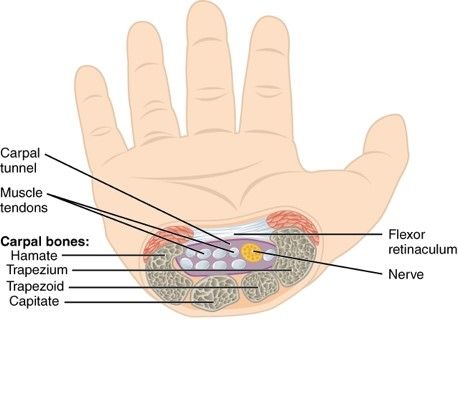
Diagram - Transverse section illustrating the carpal tunnel and its contents
Creative commons source by OpenStax College [CC BY 3.0 (https://creativecommons.org/licenses/by/3.0)]
This is a small depression located on the posterior aspect of the hand, just proximal to the base of the thumb.
Borders forming the anatomical snuffbox:
- Medial border - tendon of extensor pollicis longus
- Lateral border - tendons of abductor pollicis longus and extensor pollicis brevis
- Proximal border - styloid process of the radius
- Roof - skin
- Base - Scaphoid and trapezium
Contents include:
- Radial artery
- Radial nerve (superficial branch)
- Cephalic vein
Understanding the contents and boundaries of the anatomical snuffbox are important! For example, localised pain to this region could indicate a fracture of the scaphoid, an injury that must be diagnosed quickly to avoid avascular necrosis of the bone.
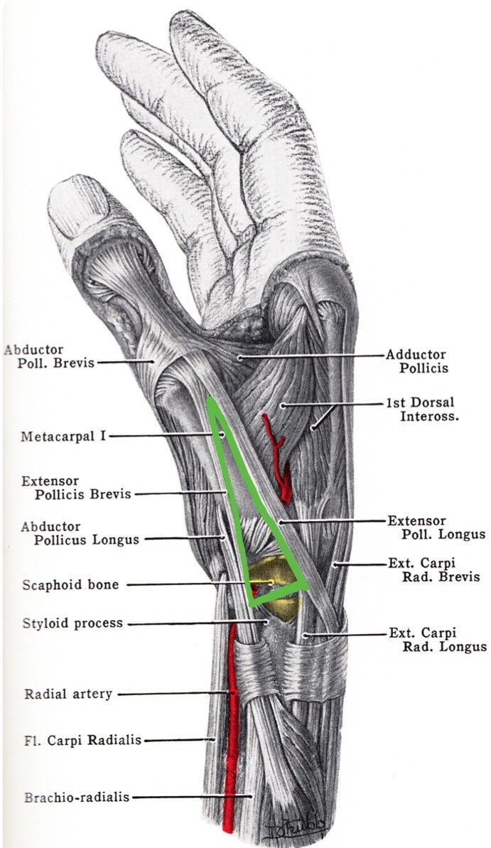
Diagram - The green triangle highlights the location of the radial fossa
Public Domain Image by Grant, John Charles Boileau [Public domain]
Guyon's canal, or the ulnar canal, carries the ulnar nerve and artery into the hand. It occupies the proximal hypothenar region and extends from the proximal border of the pisiform towards the hook of hamate. The hamate hook is a lateral landmark of the canal, rather than a point proximal to the whole passage.
Borders of Guyon's canal include:
- Roof - palmar carpal ligament
- Floor - flexor retinaculum and hypothenar muscles
- Medial border - pisiform bone
- Lateral border - hook of hamate bone
It is a key anatomical feature due to Guyon's Canal Syndrome, or 'Handlebar' palsy, which is an ulnar neuropathy at the wrist caused by compression of the ulnar nerve in Guyon's canal.
For Pathologies and Conditions of the Upper Limb, check out our articles on Conditions of the Shoulder and Clavicle, Arm, Elbow and Forearm and Wrist.
Reviewed by: Dr. Marcus Judge
Edited by: Dr. Maddie Swannack
- 8390


