

By Dr. Ethan Kavanagh and Dr. Ben Appleby
Next Lesson - The Infection Model
Core
An Infection can be defined as the invasion of a host’s body tissues by disease-causing agents, followed by multiplication of these agents and the subsequent reaction of these agents with the host’s defences.
An infectious disease is thus a disease that is caused by such an infection. Anytime you see the term “agent”, it is just referring to a micro-organism (also known as a microbe) that causes an infection.
There are five generic stages for how a microorganism, (in our example a bacterium) could cause an infection within a human:
An infectious disease is thus a disease that is caused by such an infection. Anytime you see the term “agent”, it is just referring to a micro-organism (also known as a microbe) that causes an infection.
There are five generic stages for how a microorganism, (in our example a bacterium) could cause an infection within a human:
1. Exposure
2. Adherence
3. Invasion
4. Multiplication
5. Dissemination
Let’s go through each step:
1. Exposure
For a microbe to infect a host it must first come in contact with the host.
This is known as the transmission of the microorganism and it is split into two broad categories:
Vertical transmission: this is transmission of an infection from a mother to their fetus/baby during pregnancy or childbirth. Another term for this would be "mother-to-child transmission". The main viruses that are transmitted in this manner include cytomegalovirus (CMV) and human immunodeficiency virus (HIV), but many other infections can also be transmitted via this method. Transmission may occur during pregnancy across the placenta, during delivery if the agent is swallowed by the child in the birth canal and, postnatally via the mother’s breast milk (similar to the mechanism of passive immunity via breast milk in the neonate)
The second type, horizontal transmission, encompasses any form of transmission which is not from parent to child. To try to further structure this, there are three sub-categories of horizontal transmission; contact, inhalation and ingestion.
1. Contact- involves direct contact between the source and the host. This can be either direct or indirect.
- Direct contact transmission occurs when there is physical contact between an infected person and a susceptible person. For example- Most sexually transmitted infections (STIs) are transmitted in this way.
- Indirect contact transmission occurs when there is no direct human-to-human contact, but the susceptible patient comes into contact with a contaminated surface or object (a fomite). Transmission can also occur via vectors, which are living organisms that carry and transmit infection, such as mosquitoes, flies, mites, fleas and ticks. In indirect contact transmission, the susceptible person comes into contact with a contaminated object (fomite), such as a door handle that has been sneezed on, or with a vector, such as an Anopheles mosquito that has fed on an infected human.
2. Inhalation - involves the entry of infective microorganisms through the respiratory tract. This can be broadly split into airborne and droplet transmission. In airborne transmission, very small particles generated by coughing, sneezing or breathing can remain suspended in the air for prolonged periods. Droplet spread however has a much larger particle which when coughed or sneezed out will fly for a short distance before dropping to the floor or a surface which it may then contaminate.
3. Ingestion - involves the entry of infective organisms through the gastrointestinal system. It mainly consists of faeco-oral transmission which is usually when food or water contaminated with faeces is ingested. Although it is rare for individuals to knowingly directly ingest faeces, this may happen with microscopic amounts of faeces in cases of poor hand hygiene. A few examples of how this happens would be: drinking water contaminated with sewage, eating shellfish/raw fruit and vegetables that have been harvested or washed in contaminated water, sexual activity, or poorly managed/chlorinated swimming pools.
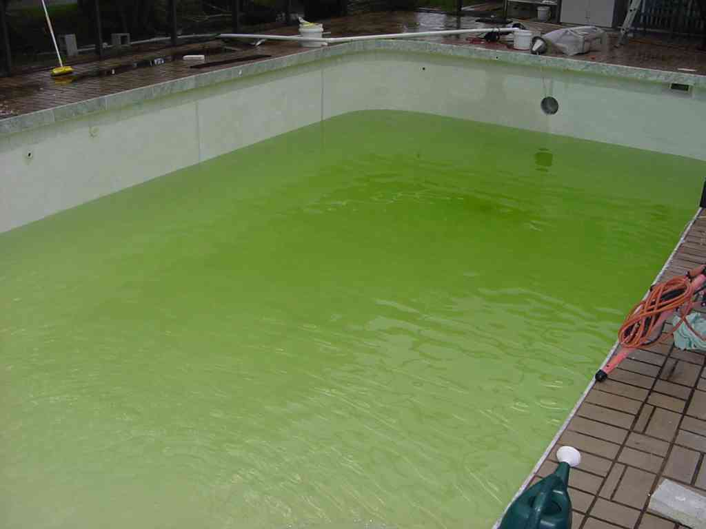
Image: High quality swimming pool management - anyone for a swim?
Creative commons source by Richard J Kinch, 2001, http://www.truetex.com/. [CC BY-SA 3.0 (http://creativecommons.org/licenses/by-sa/3.0/)]
2. Adherence
Simply put adherence is the binding of a pathogen to a host cell. This is done using an adhesion protein, or ligand, on the bacterial cell wall or viral capsule surface, which then binds to a receptor (a carbohydrate structure bound to the lipid membrane) on a host cell.
Fimbriae are also believed to be involved in attaching some bacteria to a solid surface or another cell (there is a structural difference between pili and fimbriae but the terms are mostly used interchangeably) - this is one example of a virulence factor, a specific adaptation of a pathogen (organism capable of infection) that allows it to better infect cells, replicate and survive.
3. Invasion
The way that viruses and bacteria invade the host are unique to the pathogen. Our article on HIV (and how viruses invade) covers viral cellular entry, so this introduction will focus on how bacteria invade the body.
There are two types of cells within the body that bacteria can bind to and invade: phagocytic and non-phagocytic:
Phagocytic cell invasion
Phagocytosis is a normal process for macrophages (macro - "large", phage "eater" - these cells consume waste and pathogens throughout the body) and is a process some bacteria can actually withstand and use to invade a macrophage (using virulence factors, such as strengthened cell walls and efflux pump systems). Tuberculosis (TB) is one such common example. TB is a serious lung infection caused by Mycobacterium tuberculosis which is inhaled into the lungs and ingested by alveolar (within the lungs) macrophages. The M. tuberculosis bacterium is very hardy due to the structure of its cell wall so can not be easily cleared by the immune system. As an alternative the immune system cordons off the bacteria as a granuloma. TB can be reactivated if the patient becomes immunosuppressed due to failure of the granuloma ‘cordon’, leading to active infection in the lungs. TB can be life threatening and spread to other organs. Famous celebrities that died from TB include George Orwell, Emily Bronte and the pianist, Frederic Chopin.
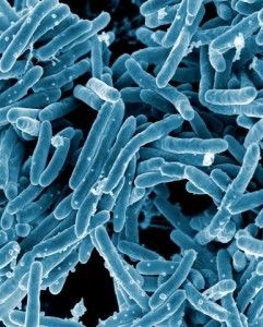
Image: Mycobacterium tuberculosis is a pathogenic mycobacterium and is the causative agent of Tuberculosis.
Creative commons source by NIAID [CC BY 2.0 (https://creativecommons.org/licenses/by/2.0)]
Non-Phagocytic cell invasion
Bacteria that invade non-phagocytic cells work slightly differently by something called the zipper method. Using Yersinia pseudotuberculosis as an example, the bacterium will bind to a receptor that is involved in binding to other cells, which tricks the cell into forming a cell junction with it. The bacterium is relatively small compared to the host cell so the host cell tries to spread over the adhesive surface of the bacterium, endocytosing it (receptor-mediated endocytosis).
The other way bacteria infect nonphagocytic cells is by the trigger method. In short the bacterium injects molecules into the host cell which activates the Rho family of GTPases, that changes the shape of the actin cytoskeleton so it can fold over the bacterium and invaginate it.
4. Multiplication
Multiplication of bacteria is via the mechanism of binary fission. The process starts with replication of the main strand of circular DNA of the bacteria into two copies. The next step is the replication of any plasmids; small circular strands of DNA which contain any ‘luxury genes’ ie those not crucial in the basic functioning of the bacteria. Multiple copies of these plasmids may be made. The final step is when the main strand copies move to opposite poles of the cell, the bacteria pinches in the middle and finally divides into two.
In viruses, the host cell's multiplication machinery is taken over and used as a "factory" for further viruses to be produced based on the RNA/DNA of the initial virus which infected the cell. The copies are then released from the cell to infect further cells.
5. Dissemination
Dissemination involves the spreading of the bacteria/ viral load from one part of the body to another. For example, infection with TB, this is from the lungs to a secondary site (normally another organ) through the blood and lymphatics. Viruses released from body cells will circulate around the body looking for cell-surface receptors that they can bind to, in order to enter the target cell and begin multiplication again.
Commensal bacteria are part of the normal flora of the body and are generally harmless, however commensals can lead to pathology either by overgrowth, invasion or being destroyed.
An example of destruction is the case of vaginal douching. Lactobacilli are a genus of bacteria which reside within and are vital in maintaining an acidic vaginal pH (typically around 3.8 to 4.5). To carry out this function the Lactobacilli convert glycogen to lactic acid reducing the pH. A common problem is that when the vagina is cleaned by douching the Lactobacili can be washed out. Therefore, as lactic acid is no longer produced the pH in the vagina can rise and allow bacterial infections to take hold such as G. vaginalis causing bacterial vaginosis.
An example of invasion is bowel perforation leading to peritonitis. The large bowel is populated with commensal so if perforated these commensals can escape into the usually sterile peritoneal cavity. This will lead to peritonitis which is a life threatening condition and a medical emergency.
An example of commensal overgrowth causing pathology is that of C. difficile (C. diff) infection. Although C. diff can be contracted via ingestion of its spores, it is also present in the gut of some members of the population causing little to no issues. Problems arise however if a patient is prescribed a course of antibiotics. Some antibiotics will reduce the gut flora population whilst leaving C. diff unharmed. This means that C. diff can opportunistically multiply and come to dominate the large bowel. This infection can then lead to complications such as Pseudomembranous colitis on top of its normal symptoms of diarrhoea and vomiting.
The microbiome is the community of microorganisms, their genes and their local chemical environment at a body site. Its composition differs between the bowel, skin, mouth and vagina because each site supplies different nutrients, oxygen tensions, surfaces and host secretions. A stable microbiome is not simply passive. Gut organisms ferment otherwise indigestible carbohydrate to short-chain fatty acids, including butyrate, which can fuel colonocytes and support the epithelial barrier. Microbial products also help immune tissues learn to tolerate ordinary residents while remaining able to respond to invasion. Some organisms contribute to vitamin and bile-acid metabolism, although the importance of any one function varies by site and community.
Colonisation resistance is the protection that established organisms provide against an incoming pathogen. They occupy attachment sites, consume limiting nutrients, alter local pH and oxygen conditions, and may release bacteriocins or other inhibitory products. They also support mucus, epithelial junctions and a measured basal immune response. A pathogen must overcome several of these barriers before it can multiply successfully.
This protection is ecological rather than absolute. Broad-spectrum antibiotics can remove susceptible members of the community and leave nutrients or niches available to resistant organisms, helping explain the opportunistic expansion of Clostridioides difficile described below. Normal residents can also cause disease if they enter a normally sterile compartment or host defences fail. For the cell structures, oxygen classifications, spores and growth phases that determine how bacterial populations survive and expand, see Bacterial Structure, Classification and Growth.
Virulence is the ability of a micro-organism to infect a host (at all steps of the process), and virulence factors are what help the micro-organism do just that. General examples of these include toxins, haemolysins (proteins and lipids that cause lysis of erythrocytes (blood cells) by destroying their cell membrane) and proteases - but toxins are the easiest to understand.
Toxins are subdivided into endotoxins and exotoxins:
Endotoxins consist of the lipopolysaccharide part of the cell wall of gram negative bacteria. Endotoxins cause an inflammatory response. If the response is severe and unregulated the patient may become septic or develop other systemic symptoms. These endotoxins do not produce a strong, protective acquired immune response, as they are only weakly immunogenic. Read about Innate and Adaptive immune responses.
Exotoxins are toxins secreted by bacteria that can destroy body cells and affect normal cellular metabolism.
So now you know generally how a micro-organism has infected a host - how can you tell if it has happened?
Infection History
As with any condition it is crucial that a history is taken. If an infective cause is suspected it is important to focus on some of these specific questions including
- Potential exposures a patient may have had which includes: travel history for the last 6-12 months, the calendar time (eg Norovirus as the winter vomiting bug), contact with any animals
- Relative time from exposure to presentation to indicate incubation time of the infection
- Vaccination history (don’t forget some people opt not to have their children vaccinated so it is important not to make assumptions)
- Symptoms of the presenting complaint (severity, duration, rash, radiation, muscular problems, numbness, vomiting, appetite etc)
It is then important to explore patient symptoms further. One useful mnemonic for identifying more information about a particular symptom is SQITARS.
Site of symptom/pain
Quality - how would they describe it? For example, a pain could be stabbing or crushing
Intensity of the symptom
Time since onset
Aggravating factors
Relieving factors
Secondary features/other symptoms
Examination
It would then be important to conduct an examination of the patient, making sure to look at anything relevant the patient mentioned in their history. This could include things like rashes or muscle stiffness. Where rashes are mentioned, it is important to check where exactly they are and their appearance. The Herpes zoster virus that causes shingles is diagnosed from its vesicular appearance and the fact that it is usually limited to a single dermatome on the body.
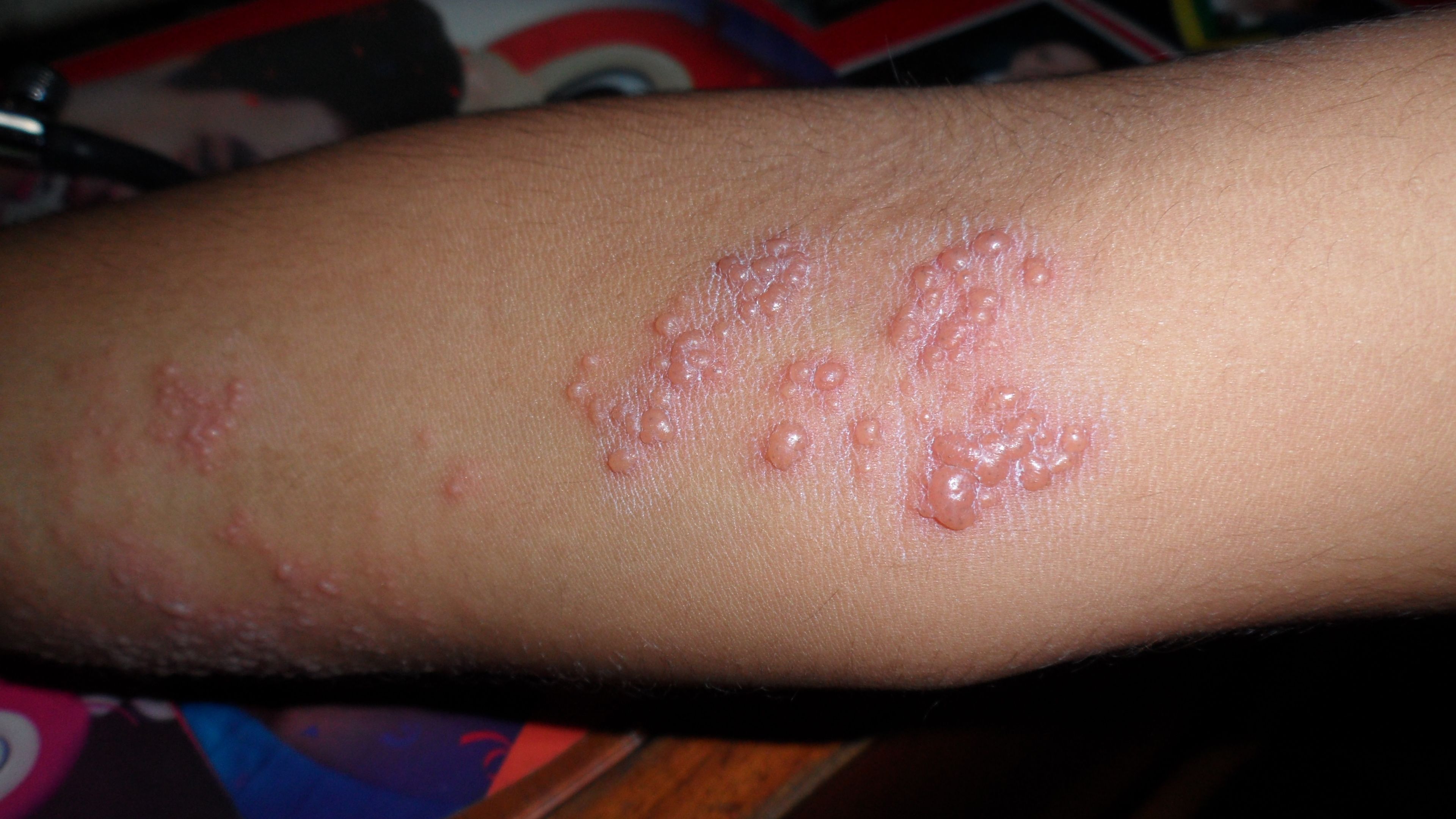
Image: Picture of Herpes zoster affecting the C7 dermatomal region of a patient’s elbow.
Creative commons source by Asvmdrn [CC BY-SA 3.0 (https://creativecommons.org/licenses/by-sa/3.0)]
Meanwhile the rash associated with Neisseria Meningitidis is a non blanching (does not disappear when a tumbler/clear glass is rolled over it) petechial rash. Small differences in examination findings can lead to large changes in diagnosis.
Investigations
There are a variety of investigations that may be carried out to help determine the cause of an infection. Some of them are listed below:
- Full blood count - covered in Haematology
- C-reactive protein (a sign of systemic inflammation, produced by the liver)
- LFTs (liver function tests - sign of liver damage)
- U's and E's (urea and electrolytes - a sign of multiple issues, including kidney damage/failure)
- Imaging (X-ray, ultrasound, MRI)
- Blood cultures (to grow bacteria if it is present and potentially test it for antibiotic susceptibility)
- Antigen detection (of the micro-organism)
- Antibody detection (understand the patient’s response - particularly relevant in Hepatitis B)
- Nucleic acid and viral nucleic acid amplification testing (used to detect specific viruses or bacteria)
Edited by: Dr. Marcus Judgeand Dr. Ben Appleby
- 12797


.jpg)
