

Next Lesson - Basics of Bone Anatomy
Core
This article describes the structure of bones at the level of cells and tissues. More information can be found on the anatomical relationships of bones in the rest of our Musculoskeletal Section.
The skeleton in an adult human is made up of 206 bones and facilitates the movement of the body and the protection of important internal organs.
See our article ‘Basics of Bone Anatomy’ which includes information about the types of bones and their functions.
There are three main types of cell that make up bone tissue.
Osteoblasts are derived from mesenchymal stem cells (stem cells from connective tissue) and are responsible for the laying down of bone. This is done by secreting osteoid (a collagen-rich matrix), which is then mineralised with hydroxyapatite (the mineral component of bone). Osteoblasts lay down bone matrix in layers (lamellae). In compact bone, these layers form osteons (see below). Once the osteoblast has surrounded itself with bone matrix, it becomes trapped in its lacuna and becomes an osteocyte.
Osteocytes are star-shaped cells that cannot divide. Osteocytes are formed from osteoblasts that have been trapped in the bony matrix that they have secreted and exist in a space called a lacuna. Once trapped, the cell is not inactive. Osteocytes monitor the minerals and proteins in the matrix to regulate bone mass, and they sense mechanical strain on the bone. Their thin extensions stretch out, connecting to other osteocytes through canaliculi (tiny channels).
Osteoclasts are the type of bone cell responsible for the breakdown of bone tissue through the release of acid (to dissolve the hydroxyapatite crystals) and enzymes such as cathepsin K (which digest collagen). They have an important role in remodelling of bone and regulation of calcium levels in the blood, but also play a key part in the pathophysiology (how a disease develops) of osteoporosis. The osteoclasts originate from haemopoietic stem cells (blood-forming stem cells), rather than mesenchymal stem cells which produce osteoblasts.
There are two types of bone tissue namely compact (cortical) bone and spongy (trabecular) bone.
The main building block of compact bone is an osteon, shown in the image below. It is a collection of osteocytes trapped between concentric layers of bone matrix (lamellae). The individual osteocytes communicate with each other through microscopic canals called canaliculi. An osteon has a central canal called a Haversian canal, which contains blood vessels and nerves to supply the bone.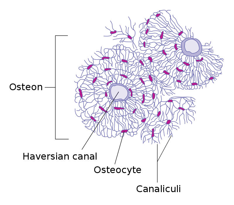
Image: this diagram shows the structure of an osteon; note the Haversian canal and the lamellae laid down by osteoblasts.
Source: https://en.wikipedia.org/wiki/Osteocyte#/media/File:Transverse_section_of_bone_en.svg
Compact bone consists of closely packed osteons. An osteon is made up of a central canal called the Haversian canal, surrounded by concentric rings of matrix called lamellae. These Haversian canals lie parallel to the axis of the long bone.
Between the lamellae are osteocytes, mature bone cells which have been surrounded by bony tissue and now exist in lacunae. The lacunae are connected by canaliculi.
Smaller canals known as perforating or Volkmann’s canals connect the Haversian canals to provide passageways through the hard matrix. They run at 90 degrees to the Haversian canals, across the width of the long bone.
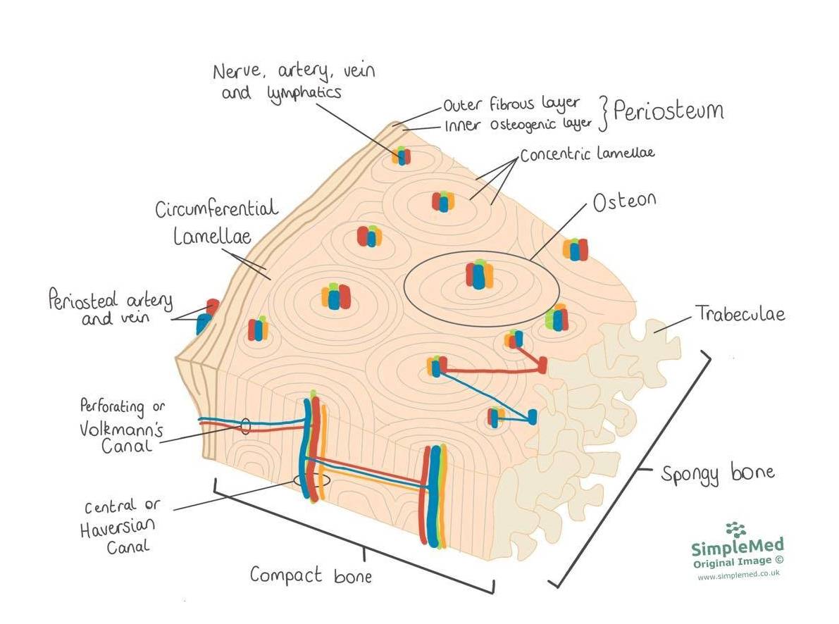
Image: this diagram shows the structure of a long bone. Key features include the periosteum, the lamellae of the osteons and the trabeculae of the spongy bone centre.
SimpleMed original by Dr. Maddie Swannack
Spongy or cancellous bone is lighter and less dense than compact bone. It consists of a lattice of fine columns and plates called trabeculae, with irregular spaces between them that contain red bone marrow (responsible for haematopoiesis, the making of blood cells). While it may seem that the trabeculae are arranged randomly, in fact they are organised to provide maximum support. They are aligned along the lines of mechanical stress in the bone and can realign if the direction of the stress changes.
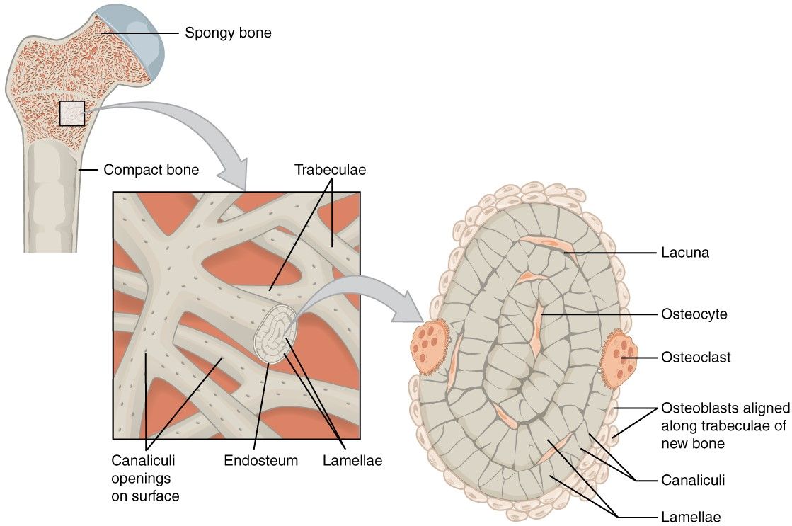
Image: this diagram shows the structure of spongy bone, highlighting the trabeculae and marrow spaces within cancellous bone.
Source: https://commons.wikimedia.org/wiki/File:606_Spongy_Bone_esp.jpg
In the body there are two mechanisms through which bones are created.
The first, endochondral ossification, involves converting a cartilage template into bone. The second, intramembranous ossification, involves the production of bone from stem cells without a cartilage template. Endochondral ossification is responsible for the lengthening of long bones through growth at epiphyseal growth plates (areas of growing cartilage near the ends of long bones).
Endochondral ossification produces long bones from a cartilaginous template.
The stages are shown in the following diagram:
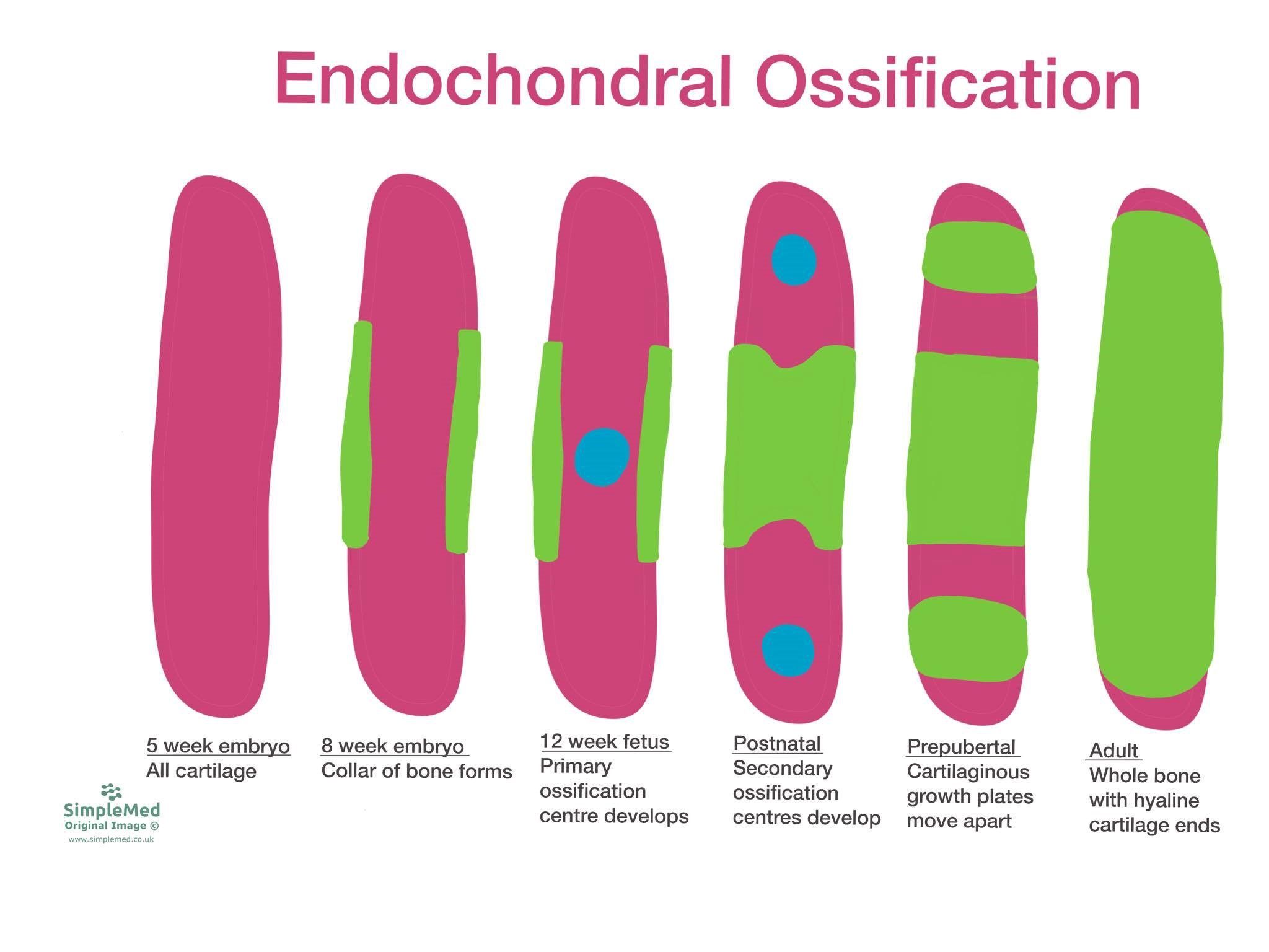
Image: this diagram shows the development of a long bone from cartilaginous template through endochondral ossification. In this diagram, pink represents cartilage, green represents bone and blue represents an ossification centre (a site where bone formation starts).
SimpleMed original by Dr. Maddie Swannack
Endochondral ossification begins at around 5 weeks gestation (weeks of pregnancy), when the long bones have a complete cartilaginous template formed. Around 3 weeks later, a collar of bone begins to form around the middle of the bone.
At 12 weeks gestation, the primary ossification centre of the bone develops. From this point, the development of bone spreads out from the primary ossification centre until the middle of the bone is complete.
Secondary ossification centres develop in the ends of the bone (the epiphyses). Many of them appear after birth, at different ages through childhood. These centres then grow into bone themselves. Cartilage is left in two places. One is articular cartilage, which covers the ends of the bones at the joints. The other is a band of cartilage between the widened part of the shaft (the metaphysis) and the end of the bone (the epiphysis), which allows the bone to grow in length. These bands are called epiphyseal growth plates.
When the bone has reached its full length, the epiphyseal growth plates fuse (harden into solid bone) and leave only the ends capped with cartilage. This happens at different ages in different bones. For example, the growth plates of the elbow fuse between about 10 and 18 years.
The stages of intramembranous ossification are as follows:
- Mesenchymal stem cells form an ossification centre and differentiate into osteoblasts, which begin to lay down osteoid (unmineralised bone matrix, which later mineralises to form bone).
- Rudimentary bone forms as bony spicules (tiny needle-like pieces of bone), surrounded by functional osteoblasts and containing osteocytes.
- These bony spicules are remodelled into trabeculae, which eventually are replaced by the lamellae of compact bone.
Osteoporosis is a disease that involves a mismatch between osteoblast and osteoclast action, with bone breakdown being much faster than bone creation. This causes a decrease in bone mineral density, leading to an increased risk of fractures.
Fractures are the main symptom of osteoporosis, with common sites of fracture being vertebral bodies, femoral neck, radius and ulna. These fractures can occur with a comparatively small amount of force due to the decreased density of bone. Complications of such fractures can include chronic pain and decreased mobility.
Risk factors for osteoporosis include:
- Menopause - oestrogen is a protective factor for osteoporosis, meaning that decreased levels of oestrogen experienced after menopause increase the risk of osteoporosis.
- Chronic kidney disease - the diseased kidneys are unable to properly regulate levels of minerals in the body, which makes laying down bone more difficult.
- Medications such as oral corticosteroids - steroids mainly reduce bone formation, increase bone breakdown early on and also change how the kidneys and gut handle calcium. The risk rises with higher doses and longer use.
- Smoking, alcohol (3 or more units daily) and inactivity (immobility) - all three are listed as risk factors for osteoporosis and fragility fracture.
- Increasing age - fracture risk rises with age.
- A previous fragility fracture, or a parent who has had a hip fracture.
A healthy balanced diet throughout life and reducing use of medications that increase the rate of bone loss is key to preventing osteoporosis.
Prevention of fractures in a patient who has established osteoporosis can be done through improving diet, increasing exercise, stopping smoking, reducing alcohol and preventing falls.
Bisphosphonates (drugs that slow bone breakdown by osteoclasts) are used in patients with osteoporosis to reduce the risk of future fractures. In the UK, alendronate or risedronate taken by mouth once a week is usually offered first. A DXA scan (a low-dose X-ray scan that measures bone density) is used to assess people at higher risk of fracture. Other drugs are used if an oral bisphosphonate is unsuitable.
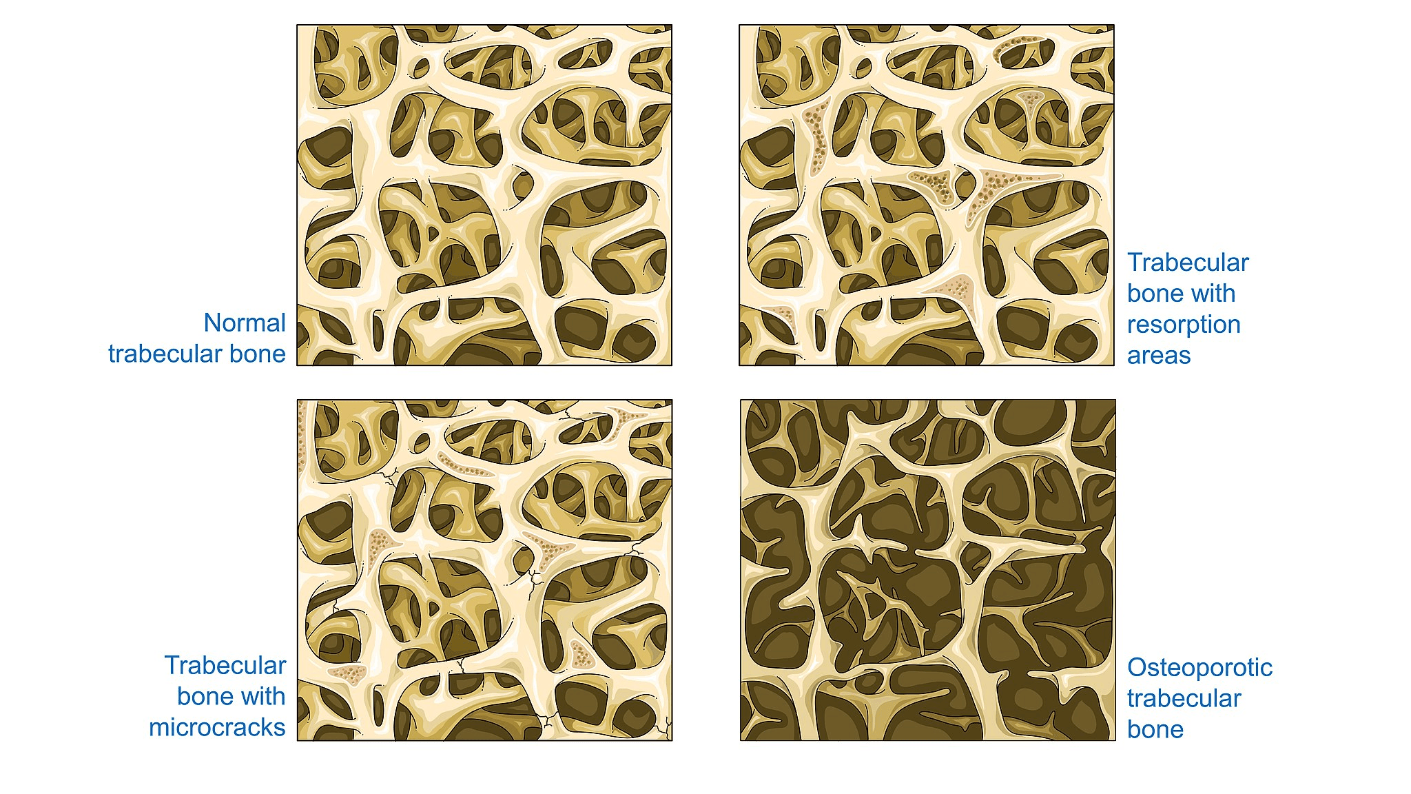
Image: this diagram shows the stages of bone condition leading to osteoporosis.
Source: Laboratoires Servier / CC BY-SA (https://creativecommons.org/licenses/by-sa/3.0)
A fracture is a broken bone: the continuity of the bone is interrupted. Fractures require significant force in the young, but less force in the elderly due to the higher incidence of osteoporosis in this age group.
One important consideration when assessing the severity of a fracture is whether the fracture is open or closed. An open fracture is one where a wound in the skin leads down to the broken bone. A fragment of bone may stick out through the skin, or a blow may break the skin at the time of the fracture. This is clinically important as a break in the skin provides a route into the bone for infection. Open fractures need urgent hospital treatment, including intravenous antibiotics given immediately and surgery to remove damaged tissue from the wound (wound excision), fix the bone and cover the wound.
Another consideration is whether a fracture is complete (all the way through the bone) or partial, also called incomplete (only part of the way through the bone, as in a greenstick fracture).
Common types of fracture include:
- Transverse - a fracture with a horizontal fracture line, straight across the bone.
- Spiral - a fracture that spirals around the bone, caused by a twisting force.
- Comminuted - a fracture that involves many pieces of bone.
- Impacted - a fracture in which one fragment of bone is driven into the other, usually as a result of compression (a squashing force).
- Greenstick - an incomplete fracture occurring mostly in children, because the bones of children are softer and more pliable. A bending force breaks the outer layer of bone (cortex) on the stretched side only. The cortex on the other side stays intact and the bone bends.
- Oblique - a fracture that crosses the bone in a diagonal direction.
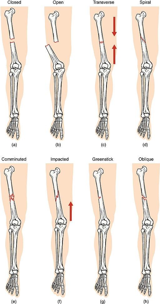
Image: this diagram shows the different types of fractures.
Source: OpenStax College / CC BY (https://creativecommons.org/licenses/by/4.0)
There are two types of fracture healing, direct (primary) and indirect (secondary), depending on how stable the fracture site is.
Direct (primary) healing occurs when two ends of bone are fixed together surgically with absolute stability (no movement at the fracture), so the fracture site is held under compression. Osteoclasts tunnel across the fracture line in structures called cutting cones, and osteoblasts then fill the tunnels with new bone. This heals the bone without the need for callus formation.
Indirect (secondary) healing is the most common type. It occurs when there is some movement at the fracture site, as in most fractures treated without surgery and in some operations, such as intramedullary nailing (fixing a metal rod inside the bone). This type of healing follows the stages below, which are important to know to understand the management of fractures. The stages of repair are as follows:
- Bleeding from damaged blood vessels within the bone forms a haematoma (a collection of clotted blood), which causes swelling.
- New blood vessels permeate the haematoma, forming a pro-callus of granulation tissue.
- Fibroblasts produce collagen and chondroblasts (cartilage-making cells) produce cartilage.
- This collagen and cartilage forms the soft callus, which makes the fracture site more stable.
- Trabeculae form through endochondral ossification, producing the hard callus.
- The hard callus is remodelled. Osteoclasts break down parts of the hard callus and osteoblasts lay down lamellar bone (mature bone made of organised layers) in its place.
It is important that the bone is protected from further damage while it heals. Many fractures are held still (immobilised) in a cast or splint, but not all: for example, NICE advises immediate weight-bearing, as tolerated, for non-surgical ankle fractures that involve only one malleolus (a bony bump at the ankle). In any plaster or support it is also important to allow space for swelling to form and recede without restriction. A splint (a support that does not go all the way round the limb) is often used first for a fresh injury, and a full cast may replace it once the swelling has gone down. A cast applied straight away may be cut (valved) to allow for swelling and then repaired at the first follow-up appointment. A cast may also need to be replaced if it gets too big as the swelling goes down. Patients should contact their doctor straight away if pain increases and the cast feels too tight, or if there is numbness, tingling, burning, excessive swelling below the cast or loss of movement of the fingers or toes (this needs urgent assessment).
Remembering the stages of fracture repair can be very difficult. A mnemonic can be helpful, and one that I use is:
Herds of Haematoma
Giraffes Granulation tissue (and angiogenesis)
Stampede Soft callus
Hiroshima for Hard callus
Lovely Lamellar bone formation
Radiation Remodelling
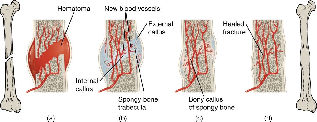
Image: this diagram shows a simplified version of the stages of fracture repair. In this image, the label of external callus represents the soft callus as at this point it only contains soft tissue.
Source: OpenStax College / CC BY (https://creativecommons.org/licenses/by/3.0)
Edited by: Dr. Ben Appleby
Reviewed by: Dr. Marcus Judge
- 11800


