

By Dr. Thomas Burnell and Dr. Bethany Turner
Next Lesson - Congenital Heart Defects
Core
The cardiovascular system begins development at around the 10th day after fertilisation. The heart is the first functional organ to develop, with the heart beat able to be heard on sonography by around the 6th week.
Formation of the Primitive Heart Tube
At the start of its development, the cardiovascular system exists as two regions near the cranial end of the embryo. These regions are called cardiogenic fields and they are derived from the mesoderm layer of the embryo. The cells that will go on to form the cardiovascular system reside here.
The cardiogenic fields consist of blood islands which are primitive tissues, and mark the beginning of blood, vessel and heart development. These areas develop further and fuse to form two tubes, which are called endocardial tubes, one on each side of the embryo.
In the fourth week of development the embryo begins to fold, which will put the heart tissue in the correct position to form the primitive heart tube surrounded by the pericardial sac. The embryo folds in two directions:
- Cephalo-caudal folding brings the cardiogenic fields from the cranial end towards the centre of the embryo to sit in thoracic region where the heart will be.
- Lateral folding fuses the two lateral sides of the embryo. This brings the two cardiogenic fields into the midline so they can fuse and form the primitive heart tube.
This primitive heart tube is formed at around day 25 of gestation. It is divided into six parts:
- Aortic sac
- Form the arteries of the aortic arch
- Truncus arteriosus
- Involved in the formation of the pulmonary trunk and aorta (outflow from the heart)
- Bulbus cordis
- Involved in the formation of the pulmonary trunk and aorta (outflow from the heart)
- Primitive ventricle
- Forms the ventricles
- Primitive atrium
- Forms the atria
- Sinus venosus
- Forms the smooth part of the right atrium and the coronary sinus
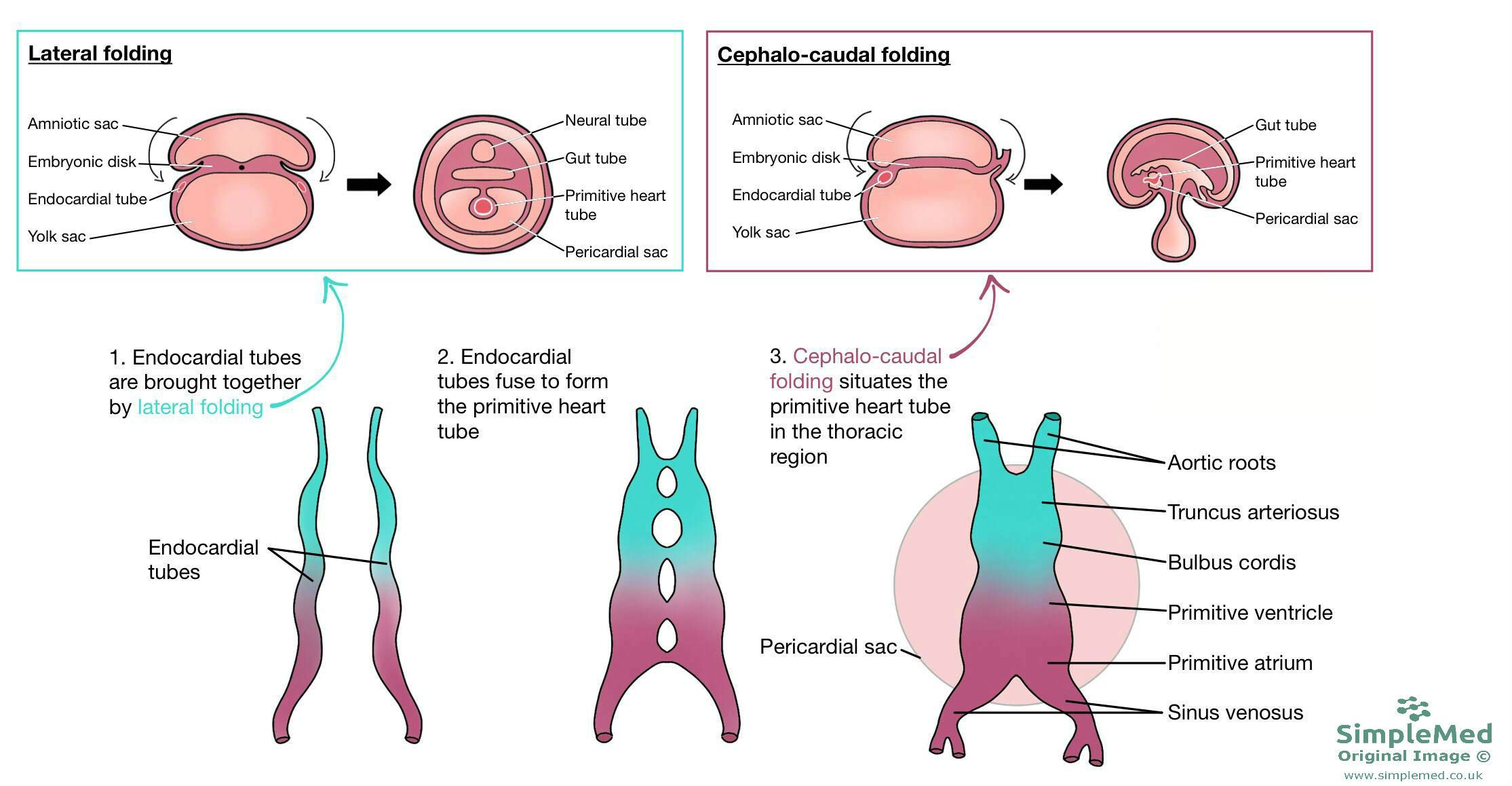
Diagram - Shows how the embryo folds, and endocardial tubes fuse to form the primitive heart tube in the pericardial cavity. The heart tube sits in the thoracic region of the embryo
SimpleMed original by Dr. Bethany Turner
The newly formed heart tube is surrounded by the pericardial sac. As the heart tube grows and elongates, differential growth produces looping.
During looping, the tube changes shape. The primitive ventricle moves ventrally and to the right, while the primitive atrium moves dorsally and to the left. This puts the inflow portion of the heart (veins and atria) behind the outflow portion (ventricles and arteries) - the same shape and orientation as mature hearts.
Breakdown of the dorsal mesocardium, which initially suspends the heart tube, creates the transverse pericardial sinus. In the mature heart, this passage lies behind the aorta and pulmonary trunk and in front of the superior vena cava; looping positions the chambers but is not itself the cause of this passage.
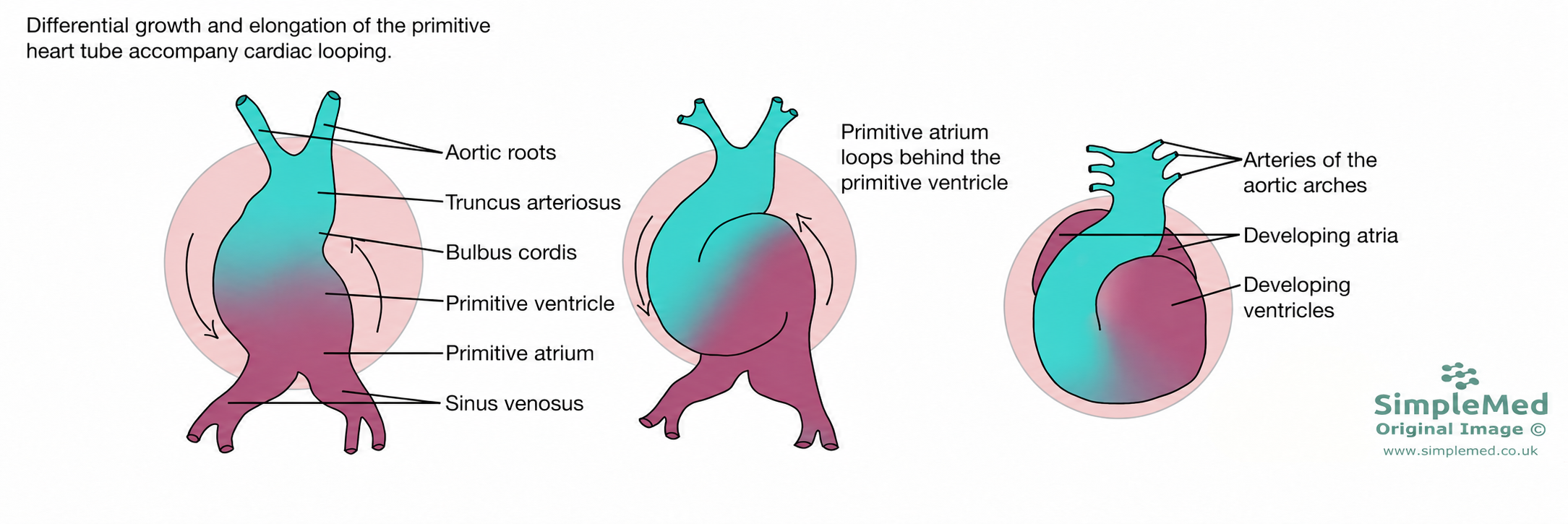
Diagram - The changing position of the heart tube during looping, with the primitive atrium moving behind the primitive ventricle. The dorsal mesocardium and its breakdown, which forms the transverse pericardial sinus, are not shown
SimpleMed original by Dr. Bethany Turner
Development of the Right Atrium
The right atrium develops from most of the primitive atrium, and part of the sinus venosus.
The sinus venosus receives blood from the right and left sinus horns, bringing blood into the primitive heart tube. The right sinus horn is partially absorbed by the primitive atrium as it grows. This forms the smooth-walled sinus venarum of the right atrium, rather than the superior and inferior vena cava. The left sinus horn becomes the coronary sinus which drains venous blood from the coronary vessels into the right atrium.
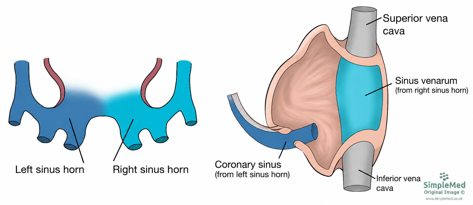
Diagram - The blue represents the sinus venosus. The light blue portion represents the right sinus horn, which is incorporated into the right atrium to form its smooth-walled sinus venarum; it does not form the superior and inferior vena cava. The dark blue portion represents the left sinus horn which becomes the coronary sinus
SimpleMed original by Dr. Bethany Turner
Development of the Left Atrium
The left atrium develops from a small part of the primitive atrium, and the proximal portions of the pulmonary veins.
The pulmonary veins begin as a single vein entering the left atrium. This vein is formed from four branches which converge to form one vein draining into the developing left atrium. As the left atrium grows, it absorbs the single pulmonary vein, absorbing all the way to the four branches. This means that when the left atrium has finished growing, it is receiving blood from four pulmonary veins, as seen in mature hearts.
The part of the mature left atrium that came from the primitive atrium is trabeculated. This forms the left auricle in the mature heart, which is a little pouch in the wall of the left atrium. The part derived from the pulmonary veins (which makes up most of the future left atrium) is smooth. The auricle is an important feature of the mature heart because with atrial fibrillation (a condition where the atria do not contract efficiently) blood can collect here. This stasis can go on to cause blood clots, which can travel to the brain causing stroke or other organs causing infarction.
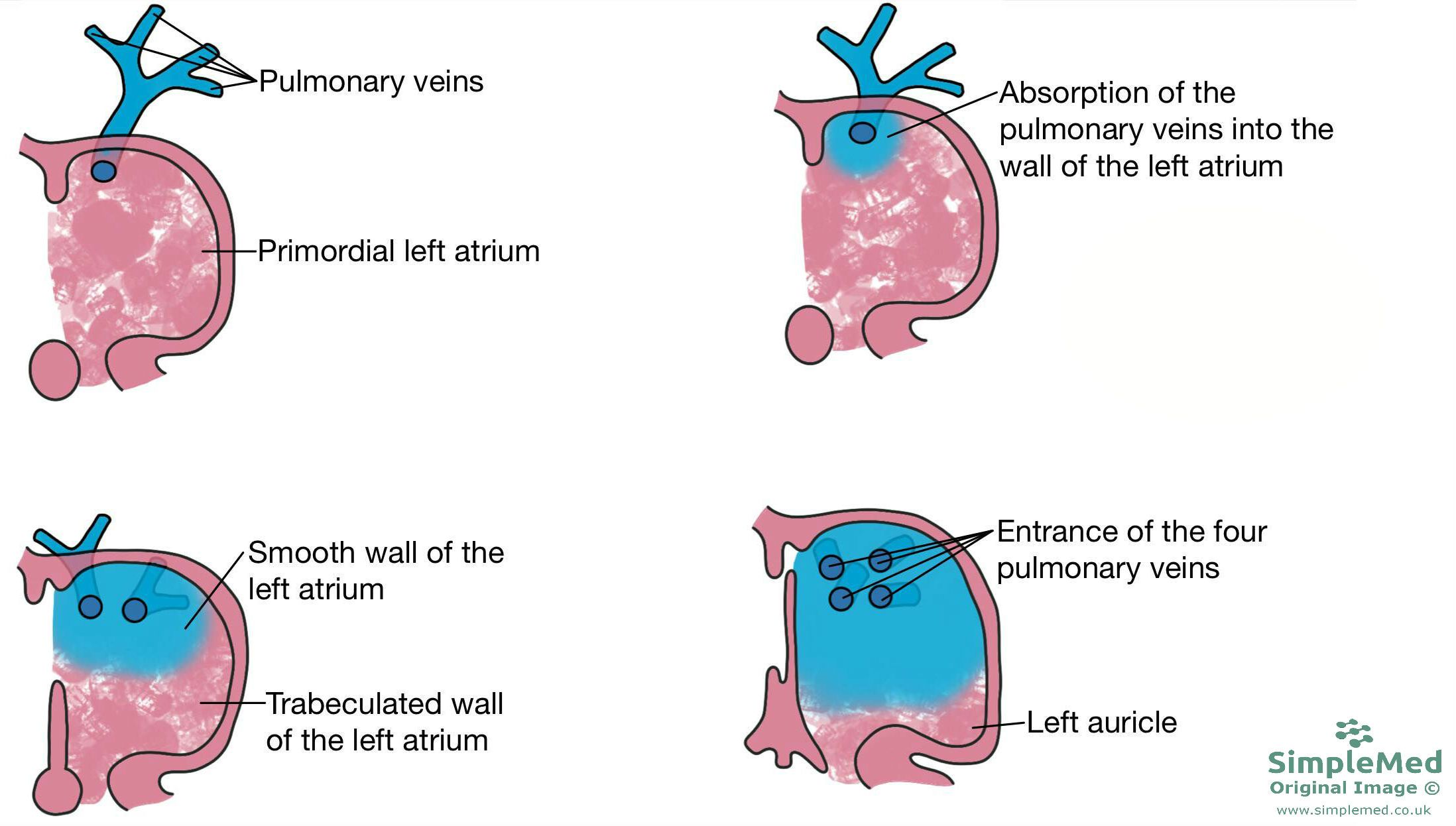
Diagram - The proximal parts of the pulmonary veins become absorbed into the wall of the left atrium. The contribution of the pulmonary veins is shown in blue, and the primitive left atrium is shown in pink
SimpleMed original by Dr. Bethany Turner
The lining of the dorsal and ventral aspects of the developing heart grow small bumps called endocardial cushions. These grow to meet in the middle of the heart and are key in the separation of the left and right sides of the heart. They act almost as a target for the septa that develop as each septum will grow towards the cushions.
The atrial septum is formed from two septa which grow in a specific order:
- A septum of tissue grows down from the top of the developing atria towards the endocardial cushions. This is the septum primum, and as it grows down, the communicating hole between the atria is called the ostium primum.
- Just before the septum primum meets the endocardial cushions and the ostium primum is closed, a hole forms in the middle of the septum. This is the ostium secundum.
- The septum primum meets the endocardial cushions, and the ostium secundum allows blood to continue to move from the right atrium to the left.
- Another septum grows down from the top of the atria, called the septum secundum.
- As the septum secundum grows down, it leaves another hole just below the ostium secundum.
These two septa, and two holes together form the foramen ovale - a right to left shunt allowing blood to flow from the right atrium into the left. This shunt will cease after birth and the septa functionally close, as discussed later. The ostium secundum and foramen ovale are staggered in their orientation and this is key in allowing for shunt closure.
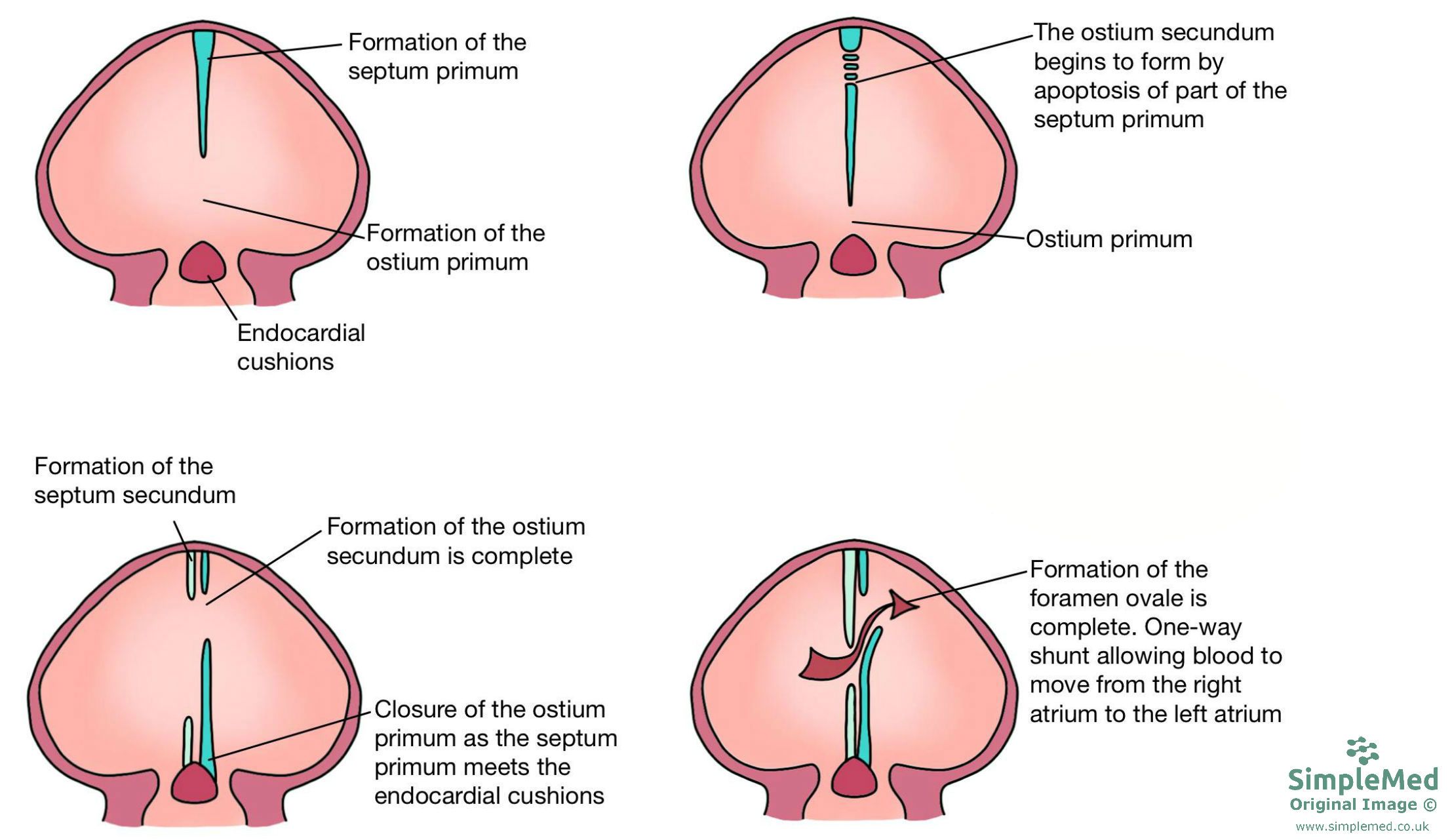
Diagram - The formation of the septum. Also shown is the expansion of the atria by the contributions of the veins
SimpleMed original by Dr. Bethany Turner
The formation of the ventricular septum takes place in two steps.
First, a muscular portion of heart tissue grows upwards from the floor of the primitive ventricle towards the endocardial cushions. It doesn’t quite reach the cushions, forming the primary interventricular foramen.
The remaining communication closes at the junction of the muscular septum, developing outlet septal structures and atrioventricular cushion tissue. The membranous portion derives from cushion tissue at this junction as the outflow tract aligns with the ventricles; closure is not simply downward growth from the cushions. A defect in the formation of the membranous portion is a common reason for ventricular septal defects, as discussed in the Congenital Heart Defects article.
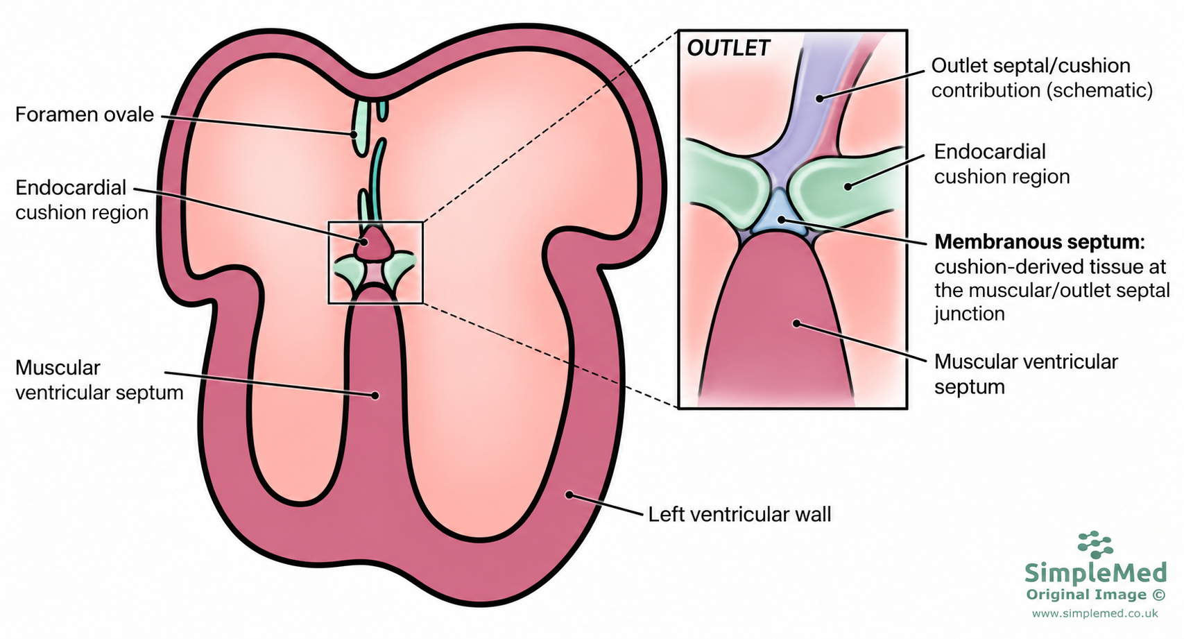
Diagram - The formation of the interventricular septum. The muscular portion grows upwards from the base of the ventricles. The membranous portion forms from cushion tissue at the junction with the muscular and outlet septal structures as outflow alignment is completed. Also shown in the diagram is the septum separating the ventricles
SimpleMed original by Dr. Bethany Turner
Septation of the Outflow Tract
The bulbus cordis and truncus arteriosus form one tube allowing outflow from the heart. This tube needs to be split in order to form the aorta and pulmonary trunk.
The outflow tract develops paired endocardial cushions containing mesenchymal cells derived from the endocardium, with an additional contribution from migrating neural crest cells. These cushions extend in a spiral arrangement through the proximal and intermediate outflow tract; they are not made solely of neural crest cells.
The outflow cushions grow towards one another and fuse to form a spiral septum through the proximal and intermediate outflow tract. Together with the distal aorticopulmonary partition and subsequent remodelling, this separates and aligns the aorta and pulmonary trunk with their respective ventricles.
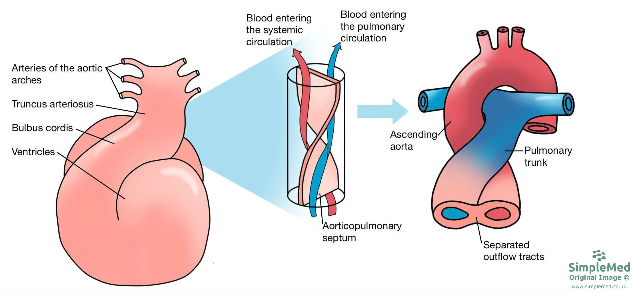
Diagram - The formation of the aorticopulmonary septum. It shows the development of the pulmonary artery and the arch of the aorta with its branching vessels
SimpleMed original by Dr. Bethany Turner
Formation of the Great Vessels
Vessels connecting the aortic sac to the dorsal aortae develop as aortic arches. The five pairs are conventionally numbered I, II, III, IV and VI. These arches, along with the truncus arteriosus, contribute to the formation of large arteries:
- Truncus arteriosus: divided by the aorticopulmonary septum to contribute to the pulmonary trunk and the proximal ascending aorta.
- Sixth arch: the proximal portions contribute to the right and left pulmonary arteries. The distal left portion persists as the ductus arteriosus, connecting the pulmonary circulation to the descending aorta, while the distal right portion regresses. After birth, closure of the ductus removes this arterial connection.
- The sixth arch is also known as the pulmonary arch.
- Fourth arch: on the left forms a segment of the arch of the aorta, between the left common carotid and left subclavian arteries; on the right it forms the proximal part of the right subclavian artery, with the remaining length derived from other embryonic vessels.
- Third arch: becomes the common carotids and the first part of the internal carotids.
- Second and first arches: largely regress, but leave contributions including the maxillary arteries from the first arch and transient stapedial arteries from the second.
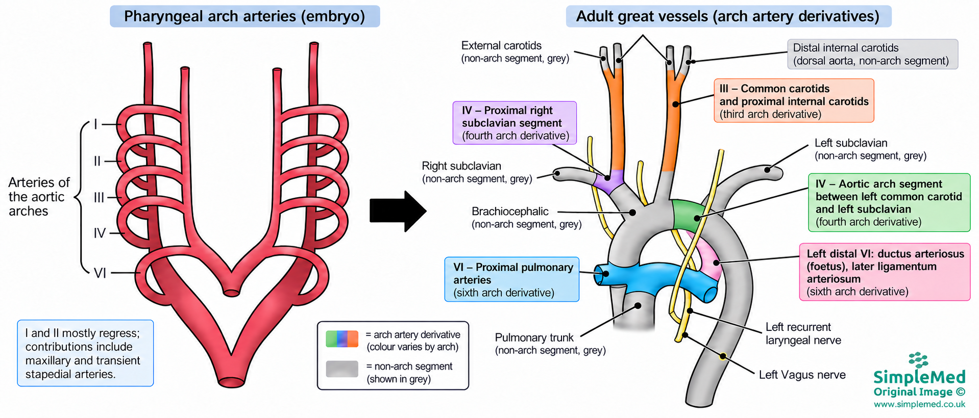
Diagram - Shows the development of the vessels from aortic arches, to the final arrangement of the vessels. Also shows the final position of the right and left vagus nerves and recurrent laryngeal nerves. The left recurrent laryngeal nerve loops under the arch of the aorta while the right recurrent laryngeal nerve loops under the right subclavian artery
SimpleMed original by Dr. Bethany Turner
The fetal circulation works very differently to adult circulation. The following steps outline the flow of blood from the placenta to the systemic circulation of the fetus:
- Oxygenated blood is carried from the placenta into the fetal circulation via the umbilical vein.
- The oxygenated blood enters the inferior vena cava and mixes with deoxygenated blood. It bypasses the developing liver via the ductus venosus.
- The liver is very metabolically active, so bypassing it means that oxygenated blood can maintain its oxygen saturation for when it reaches the heart to be pumped around the body (most importantly, the brain)
- The blood enters the right atrium and passes into the left atrium via the foramen ovale, therefore bypassing the pulmonary circulation. The blood is able to shunt from the right to left side of the heart because pressure in the right side of the heart is higher than the left in the fetus.
- The baby is not breathing and instead the fetus' blood is being oxygenated by the mother's blood. Therefore blood doesn’t have to go to the alveoli for oxygenation, so the pulmonary circulation can be bypassed.
- The blood is pumped from the left ventricle into the aorta.
- Blood that doesn’t pass through the foramen ovale, and instead is pumped into the pulmonary trunk from the right ventricle, enters the systemic circulation at the arch of the aorta via the ductus arteriosus. This system exists so that the right ventricle still has some blood to pump against. In the developing heart, the rule of 'use it or lose it' applies, so if the right ventricle has no blood passing through it, it will be underdeveloped in the mature heart. This is relevant in congenital heart defects such as tricuspid atresia.
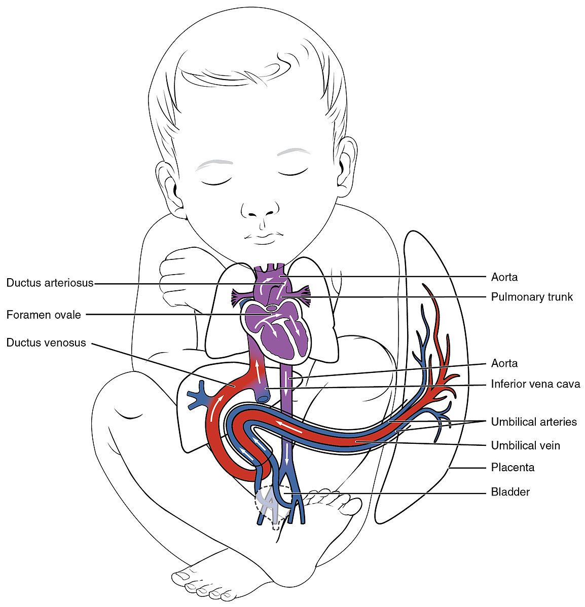
Diagram - The fetal circulation. Blood comes in through the umbilical vein, around the fetal body, and back to the placenta via the two umbilical arteries
Creative commons source by OpenStax college [CC BY-SA 4.0 (https://creativecommons.org/licenses/by-sa/4.0)]
Changes to the Circulation After Birth
- When the baby takes its first breath, the pO2 increases. This change causes the ductus arteriosus to close.
- Ductus arteriosus becomes the ligamentum arteriosum.
- More blood now flows through the pulmonary circulation as blood in the pulmonary trunk cannot leave via the ductus arteriosus anymore. This causes increased venous return to the left atrium, leading to an increase in left atrial pressure. The pressure in the left atrium exceeds that of the right, which causes the foramen ovale to close.
- Foramen ovale becomes the fossa ovalis.
- When the umbilical cord is cut, there is no longer blood flowing through the umbilical vein, causing the ductus venosus to collapse.
- Ductus venosus becomes the ligamentum venosum.
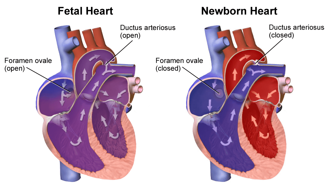
Diagram - The changes that have occurred after the baby has been born. The changes shown are the ligamentum arteriosum (closed ductus arteriosus) and fossa ovalis (closed foramen ovale)
Creative commons source by BruceBlaus [CC BY-SA 4.0 (https://creativecommons.org/licenses/by-sa/4.0)]
Edited by: Dr. Ben Appleby
Reviewed by: Dr. Marcus Judge
- 40596


