

Next Lesson - The Cell Cycle, Checkpoints and Apoptosis
Core
Deoxyribonucleic acid or DNA is a molecule, which is found in the nucleus of every cell in the human body. It has a double helix structure and it carries the genetic instructions for cell reproduction, growth, development and function.
The width of the DNA chain is 2nm, but the total length of DNA in each cell is around 2m (no you didn’t read that wrong, 2 meters in every cell!) therefore DNA needs to packaged to fit inside each cell. DNA wraps around an octamer of histone proteins: two copies each of H2A, H2B, H3 and H4. About 147 base pairs of DNA wrap around this octamer to form the core of a nucleosome. Nucleosomes are packaged into a thread which under a transmission electron microscope has the classical appearance of ‘beads on a string’. Nucleosomes form higher-order chromatin structures whose packing varies within cells. The classical solenoid model describes a 30 nm fibre, but this is not a universal arrangement of chromatin in living cells. Chromatin becomes highly condensed into the chromosomes visible during mitosis and meiosis.
Chromatin is a complex of DNA and histones with the basic structural unit being a nucleosome. In the nucleus of the cell, there is heterochromatin and euchromatin:
- Heterochromatin is relatively densely packed chromatin. Its DNA is generally less accessible to transcription machinery, so transcription is usually lower than in euchromatin.
- Euchromatin is relatively less condensed chromatin. Its DNA is generally more accessible to transcription machinery, favouring gene expression. Both forms contain nucleosomes; neither is defined by a single fixed higher-order structure.
Under an electron microscope, heterochromatin appears dark due to its condensed nature and euchromatin is much lighter or not visible.
A gene is a region of DNA that encodes a functional product: a protein or a functional RNA. Each gene has a specific location on a specific chromosome known as a locus. There are around 25,000 genes in humans spread across 23 pairs of chromosomes - 22 autosomes and a pair of sex chromosomes.
Both DNA and Ribonucleic Acid (RNA) are nucleic acids (polymer of nucleotides(be mindful of spelling)).
- NucleoSide = base + sugar
- NucleoTide = base + sugar + phosphate
The pentose sugar in DNA is 2-deoxyribose whereas the pentose sugar in RNA is ribose.
As you can see in the diagram, 2-deoxyribose has only a hydrogen atom on the second carbon (therefore no extra oxygen hence deoxyribose), whereas ribose has a hydroxyl group on second carbon instead.
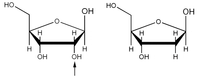
Diagram - The difference in structure between Ribose (pictured left) and Deoxyribose (pictured right)
Creative commons source by Pixie [CC BY 3.0 (https://creativecommons.org/licenses/by/3.0)]
The bases in DNA and RNA are known as nitrogenous bases and can be classed as either a purine or a pyrimidine. Purines are double-ring structures while pyrimidines are single ring structures. The purines are the same in both DNA and RNA, however, the pyrimidines used in DNA and RNA are different:
- The purines are Adenine and Guanine.
- The pyrimidines are Cytosine, Thymine and Uracil
- N.B. In DNA Thymine is used in pairs with Adenine, whereas in RNA Uracil pairs with Adenine. (for pairing rules see below)
DNA’s primary structure is that of a polymer of nucleotides covalently linked by phosphodiester bonds. These covalent bonds form the supporting ‘phosphate backbone’. The secondary structure of DNA is a right handed double helix with the chains being complementary and anti-parallel to each other. The two chains are bound to each other with hydrogen bonds between the paired purine/ pyrimidine bases.
The terms 5 prime (5’) and 3 prime (3’) refer to the distinct ends of the chain distinguished by the 5’ phosphate group and 3’ hydroxyl group.
There are major and minor grooves formed from the double helix structure of DNA and the difference between the grooves is the width. As the width of the major groove is wider, the bases are more accessible, therefore proteins that bind to DNA (such as transcription factors) are able to bind.
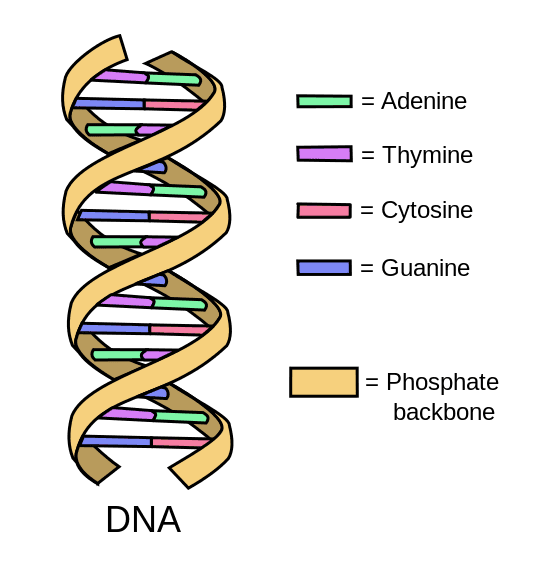
Diagram - The DNA double helix with the base pairings
Creative commons source by Forluvoft [CC BY 3.0 (https://creativecommons.org/licenses/by/3.0)]
The bases on each chain form a hydrogen bond with a specific counterpart on the complementary chain to form a base pair. A purine will always bond with a pyrimidine and vice versa.
In DNA:
- Guanine (purine) forms three hydrogen bonds when it is paired with cytosine (pyrimidine).
- Adenine (purine) forms two hydrogen bonds when it is paired with thymine.
In RNA this is slightly different (pay close attention to how adenosine pairs):
- Guanine (purine) forms three hydrogen bonds when it is paired with cytosine (pyrimidine).
- Adenine (purine) forms two hydrogen bonds when it is paired with uracil.
A way of remembering which base pairs with which can be remembered using animals.
For DNA- GCAT- As G pairs with C and A with T. Imagine a cat with a long tail which curls round to make it look in the shape of a g.
For RNA- GCAU- As G pairs with C and A with U. Imagine a letter G with black and white spots similar to a cow.
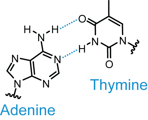
Diagram - The hydrogen bonds between thymine and adenine
Creative commons source by Roadnottaken [CC BY 3.0 (https://creativecommons.org/licenses/by/3.0)]
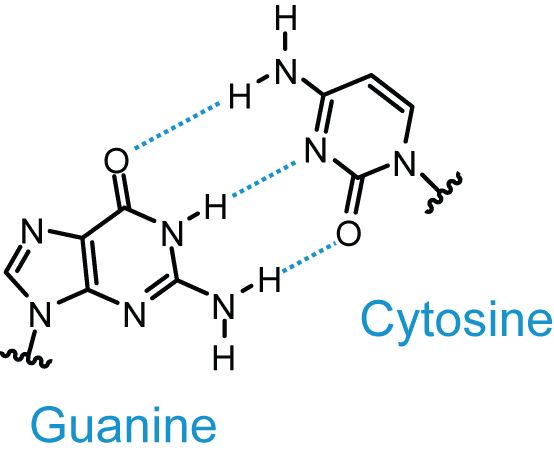
Diagram - The hydrogen bonds between guanine and cytosine
Creative commons source by Roadnottaken [CC BY 3.0 (https://creativecommons.org/licenses/by/3.0)]
Commonly in microbiology, the nitrogenous bases are shortened to the first letter of their name, for example adenine is shorted to A. As well as this, when depicting a DNA sequence, you begin at the 5’ end on the left to the 3’ end on the right.
Replication of DNA occurs in the Synthesis phase (S phase) of the cell cycle (between Growth phase 1 and 2). The reaction is catalysed by the enzyme DNA polymerase. DNA replication is semi conservative in nature and this process is described below.
The existing DNA strands can be described by the direction in which DNA polymerase moves along them. In DNA replication, each original strand acts as a template for the synthesis of a new complementary strand.
DNA polymerase reads the template strand in the 3’ to 5’ direction and synthesises a new strand in the 5’ to 3’ direction. The new strand is complementary to its template.
There are three stages to DNA replication: initiation, elongation and termination.
- DNA Helicase breaks the hydrogen bonds between the strands to unwind them.
- Primase (type of RNA polymerase) catalyses the synthesis of a short RNA segment (primer) that is complementary to single stranded DNA.
- DNA polymerase cannot begin DNA replication without an initial primer. After elongation, the RNA primer is removed and replaced with DNA by other enzymes before the strand is sealed.

Diagram - The first part of DNA replication showing a replication fork
SimpleMed original by Dr. Peter Parkinson
- On the template running 3’ to 5’ towards the advancing replication fork, DNA polymerase synthesises the new leading strand continuously in the 5’ to 3’ direction.
- On the oppositely oriented template, the new lagging strand is synthesised discontinuously. Each fragment begins from a new primer and is extended in the 5’ to 3’ direction, away from the advancing fork.
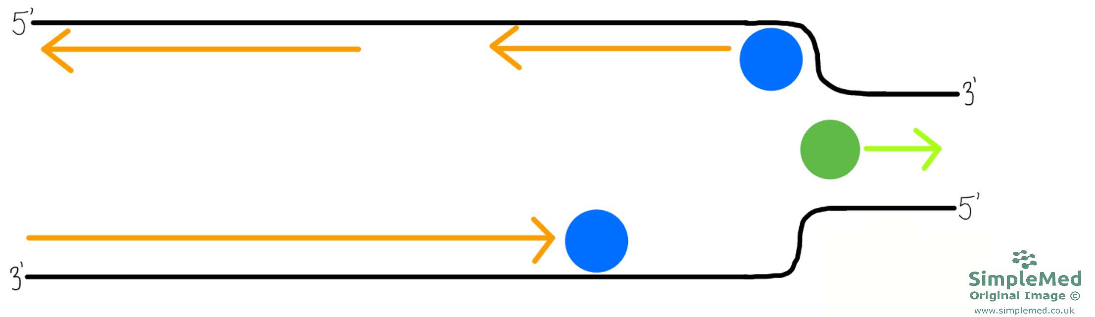
Diagram - DNA replication continued
SimpleMed original by Dr. Peter Parkinson
- The short segments of newly synthesised lagging-strand DNA are called Okazaki fragments. They belong to the daughter strand, not the original template.
- The terms Lagging strand and Leading strand describe the newly synthesised daughter strands at a replication fork. The lagging strand is made as Okazaki fragments, whereas the leading strand is made continuously; both are synthesised 5’ to 3’.
- These Okazaki fragments on the lagging strands have to be joined up to form a single chain. This is the job of DNA ligase which joins the fragments together.
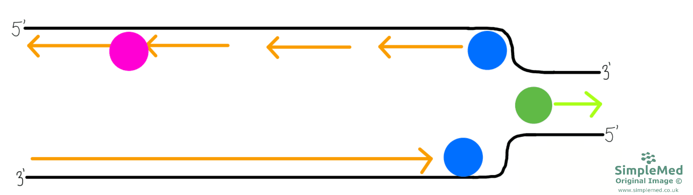
Diagram - DNA replication continued
SimpleMed original by Dr. Peter Parkinson
- As more than one helicase acts on the DNA when replication is required, there are multiple replication forks (sites of replication). These forks with DNA polymerase at the head will of course eventually meet.
- The two remaining Okazaki fragments and the sections at which two forks have met are all joined together by DNA ligase to form two new continuous DNA molecules.
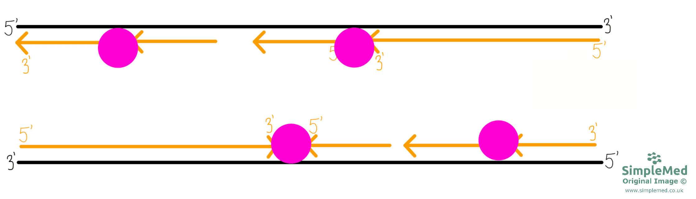
Diagram - DNA replication continued
SimpleMed original by Dr. Peter Parkinson
- After joining of the fragments, there are two new DNA strands contributing to two complete DNA molecules, each with one strand of original DNA and one strand of new DNA. This is why DNA replication is called semi-conservative replication (as half of the original strands are conserved).
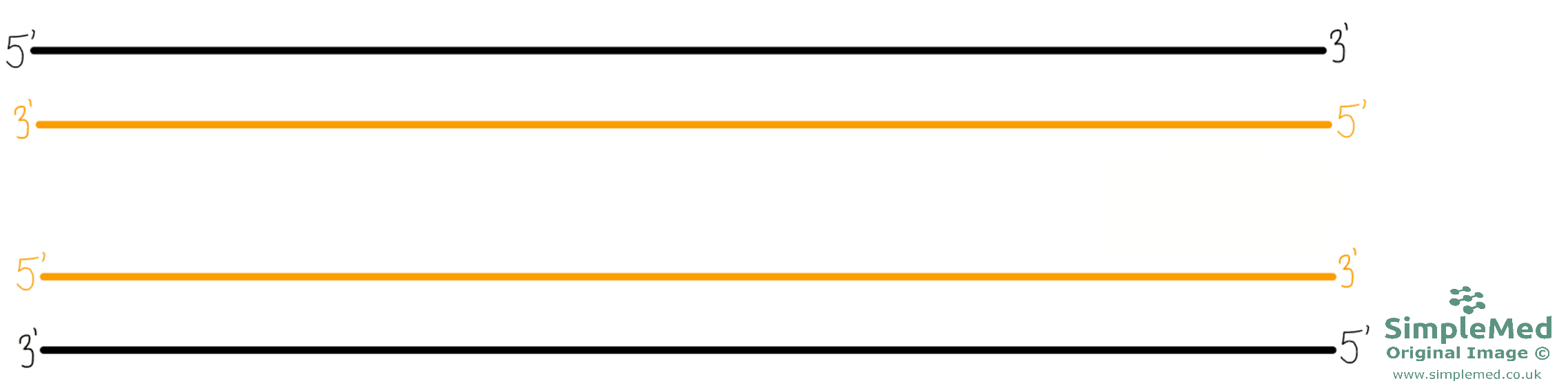
Diagram - The end product of DNA replication
SimpleMed original by Dr. Peter Parkinson
DNA replication is a complex process, and the knowledge covered here can be supported by the article in our Genetics series on Mitosis and Meiosis, found here.
Edited by:Dr. Ben Appleby
Reviewed by: Dr. Thomas Burnell
- 10946



