

Next Lesson - Gastrointestinal Motility and the Enteric Nervous System
Core
The hindgut gives rise to the distal 1/3 of the transverse colon, the descending colon and the sigmoid colon, and these are sections of the large intestine that lie in the left half of the abdominal cavity.
The transverse colon is an intraperitoneal structure and has its own mesentery, the transverse mesocolon.
The descending colon is a retroperitoneal structure, but it lies anterior to the left kidney. Its arterial supply is from the left colic artery which branches off the inferior mesenteric artery (IMA) and its venous drainage is into the left colic vein then into the inferior mesenteric vein (IMV). Lymphatic drainage is into the inferior mesenteric lymph nodes.
The sigmoid colon has its own mesentery (the sigmoid mesocolon) and is an ‘S’ shaped section of the large intestine proximal to the rectum. Its arterial supply is from the sigmoid arteries which branch off the IMA and its venous drainage is into the sigmoid veins then into the IMV. Lymphatic drainage is into the inferior mesenteric lymph nodes.
The colon is much wider and shorter than the small intestine, and it has crypts not villi. The external longitudinal muscle of the colon is incomplete - it is made up of teniae coli (bands of muscle) which cause the formation of haustra (sacculations) when they contract. These can be differentiated from the plicae circularis of the small intestine because the plicae circularis traverse the whole diameter of the lumen, and the haustra do not.
The main functions of the colon are final water and electrolyte absorption (facilitated by eNaC channels which move sodium from the lumen into the enterocytes), and formation and temporary storage of faeces prior to defaecation.

Diagram - The anatomy of the large intestine
Public Domain Source by Mikael Häggström and B jonas, edited by Dr. Laura Hansell [Public domain]
The rectum has a curved shape and sits anteriorly to the sacrum in the pelvis. It is distinguishable from the sigmoid colon because it has a continuous band of longitudinal muscle instead of the intermittent teniae coli like the colon.
Peritoneal covering varies with rectal level: the upper rectum is covered anteriorly and laterally, the middle rectum anteriorly, and the lower rectum has no peritoneal covering. These are descriptions of covering, not sharply divided vascular territories.
The superior rectal artery, the terminal continuation of the IMA, is the principal supply to the upper rectum and communicates with other rectal arteries. The superior rectal vein drains through the IMV towards the portal system. Lymphatic drainage is into the pararectal lymph nodes then into the inferior mesenteric nodes.
The middle rectal arteries arise from the internal iliac arterial system, often through its branches, and make a variable contribution to the middle and lower rectum. The middle rectal veins drain into the internal iliac veins. Lymphatic drainage is a combination of the upper and lower third’s drainage.
The inferior rectal arteries, branches of the internal pudendal arteries, mainly supply the distal anorectal region and communicate with the other rectal arteries. The inferior rectal veins drain through the internal pudendal veins into the internal iliac veins. Lymphatic drainage is into the internal iliac lymph nodes.
The rectal venous plexus connects the superior rectal veins of the portal system with the middle and inferior rectal veins of the systemic circulation. These communicating veins form a portosystemic anastomosis, rather than separate upper- and lower-third drainage compartments. In portal hypertension, for example from cirrhosis, these collateral channels can enlarge.
The function of the rectum is the temporary storage of faeces prior to defaecation. Stretching of the rectum is what stimulates the urge to open the bowels.
The anal canal is the distal end of the gastrointestinal tract - it begins at the terminal rectum and ends with the anus.
The canal is divided into two sections by a line known as the pectinate line or the dentate line. Above this line, the anal canal is developed from the hindgut, and below this line the anal canal is developed from the proctodeum, an embryological structure formed by the folding of the embryo. For more information, please see our article Development of the Gastrointestinal Tract (here).
Above the pectinate line, the anal canal’s arterial supply is the superior rectal artery which branches off the IMA and its venous drainage is into the superior rectal vein then into the IMV. This means that the superior portion of the anal canal drains into the portal system. Lymphatic drainage is into the internal iliac lymph nodes. The epithelium of the anal canal above the pectinate line is columnar (glandular) epithelium and it has a visceral nerve supply (S2,3 and 4) that allows for a sense of stretch.
Below the pectinate line, the anal canal’s arterial supply is the inferior rectal artery which branches off the internal pudendal artery and its venous drainage is into the inferior rectal vein then into the internal pudendal vein. This means that the inferior portion of the anal canal drains into the systemic circulation. Lymphatic drainage is into the superficial inguinal lymph nodes. The epithelium of the anal canal below the pectinate line is stratified squamous epithelium and it has a somatic nerve supply (S2,3 and 4) that allows for a sense of pressure, pain, and temperature.
The functions of the anal canal are faecal continence and defaecation, which are controlled by sphincters.
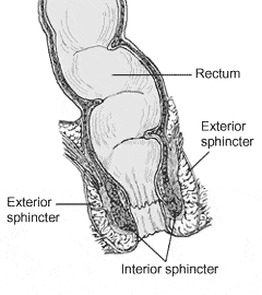
Diagram - The anatomy of the anal canal
Public Domain Source by U.S. Government National Institutes of Health [Public domain]
Connect this region to the integrated physiology: use Gastrointestinal Motility and the Enteric Nervous System for colonic mixing, mass movement and defaecation control; Gastrointestinal Secretion, Digestion and Absorption for water and electrolyte handling across the gut; and Liver, Biliary and Exocrine Pancreatic Physiology for the upstream bile-acid cycle that returns through the portal circulation.
Edited by: Dr. Maddie Swannack
Reviewed by: Dr. Thomas Burnell
- 1972


