

Next Lesson - Energy Storage
Core
A reactive oxygen species (ROS) is a reactive molecule that contains oxygen. Some ROS are free radicals with an unpaired electron, while others such as hydrogen peroxide (H2O2) are not. Not all free radicals are ROS, as some do not contain oxygen.
Oxidative stress occurs when there is an imbalance between reactive oxygen species and the cell defences, with too many reactive oxygen species. It is thought to play a part in the pathogenesis (the way a disease develops) of many diseases by damaging cells. Examples include cancer, chronic obstructive pulmonary disease (COPD, a long-term lung disease with narrowed airways), cardiovascular disease, multiple sclerosis (an autoimmune disease that damages the covering of nerves in the brain and spinal cord) and Parkinson’s disease (a brain disease that causes tremor and slow movement).
There are many types of damage that can occur because of reactive oxygen species. Each can stop a cell working properly, and severe damage can kill the cell. Three important types are:
- Base damage or sugar damage to DNA (damage to the bases, the letters A, C, G and T, or to the deoxyribose sugar in the DNA backbone, including strand breaks), leading to DNA mutation.
- Protein damage through the formation of inappropriate disulphide bonds (links between sulphur atoms in different parts of a protein, which change its shape).
- Disruption of lipid structures (including cell membranes) by creating a chain reaction of lipid radicals.
Nitric oxide (NO•) is a free radical, but it is classed as a reactive nitrogen species (RNS, reactive molecules containing nitrogen) rather than a ROS. It is useful in the body as it signals vasodilation (widening of blood vessels), angiogenesis (growth of new blood vessels) and neurotransmission (signalling between nerve cells). It also helps to defend against infection (see Respiratory Burst below). However, it is toxic at high levels, partly because it reacts with superoxide (a radical form of oxygen) to form peroxynitrite, a very damaging molecule. This is why nitric oxide levels need to be controlled.
Nitric oxide is formed through a reaction between arginine (an amino acid) and oxygen to produce nitric oxide, water and citrulline (a different amino acid). This is catalysed by the enzyme nitric oxide synthase (NOS).
The respiratory burst is a sudden release of reactive oxygen species from a phagocytic cell (a white blood cell, such as a neutrophil or macrophage, that engulfs microbes), designed to kill bacteria. First, an enzyme called NADPH oxidase (which uses NADPH, a molecule that supplies electrons) makes O2-• (superoxide, a free radical form of oxygen). Superoxide is converted to H2O2 (hydrogen peroxide). The enzyme myeloperoxidase then uses hydrogen peroxide and chloride ions to make HOCl (hypochlorous acid, bleach). The cell also releases peroxynitrite (ONOO-, a very reactive molecule), produced from nitric oxide and superoxide. Both HOCl and ONOO- are used to kill the bacteria.
Chronic Granulomatous Disease (CGD) is a form of immunodeficiency caused by a fault in NADPH oxidase, the enzyme that starts the respiratory burst. In CGD, phagocytes fail to produce this respiratory burst, so bacterial and fungal infections can become very severe, especially with catalase-positive organisms (microbes that make the enzyme catalase). The disease gets its name from the granulomas that form in organs to try to contain the infection. The formation of these granulomas damages the organs.
Cells have defences against the ROS that can cause damage.
The first of these is an enzyme called superoxide dismutase (SOD). It catalyses the reaction in which two molecules of superoxide (O2-•) and two hydrogen ions (H+) are converted to hydrogen peroxide (H2O2) and oxygen (O2). Removing superoxide is key because otherwise it reacts with nitric oxide to form peroxynitrite, which is much more damaging. Even though hydrogen peroxide is damaging to cells, it can undergo a reaction to become water and oxygen. This reaction is catalysed by the enzyme catalase. In this way a damaging molecule is converted into two far less harmful products, water and oxygen.

Diagram - The reactions of superoxide dismutase and catalase
SimpleMed original by Dr. Maddie Swannack
A second type of cellular defence is glutathione (GSH). The enzyme glutathione peroxidase uses GSH (a small antioxidant molecule) to convert H2O2 into H2O, as shown in the diagram below. This oxidises GSH to GSSG (glutathione disulphide, two glutathione molecules joined together). The enzyme glutathione reductase then regenerates GSH from GSSG, using NADPH (a molecule that supplies electrons) from the pentose phosphate pathway (a glucose-using pathway).

Diagram - The reaction pathway of glutathione
SimpleMed original by Dr. Maddie Swannack
The NADPH used for this comes from the pentose phosphate pathway, but the pathway requires the enzyme glucose-6-phosphate dehydrogenase (G6PD), which can be genetically deficient in some individuals. If there is a deficiency in G6PD, NADPH levels are too low to regenerate GSH. This matters most in red blood cells, because the pentose phosphate pathway is their only source of NADPH. These cells then cannot neutralise ROS, and inappropriate disulphide bonds form in their proteins. This causes Heinz bodies to form in the red blood cells. Heinz bodies are clumps of haemoglobin that have come out of solution because of the inappropriate disulphide bond formation. Affected people can develop haemolytic anaemia (anaemia caused by red blood cells breaking down early). This breakdown can be triggered by extra oxidative stress, such as an infection, eating fava beans or taking certain drugs (including some antimalarials).
A third way that cells defend themselves against free radicals is through free radical scavengers (molecules that mop up free radicals). These include vitamins C and E, and carotenoids (plant pigments such as beta-carotene, which the body can convert into vitamin A). Vitamin E donates a hydrogen atom (a proton and an electron) to the radical to neutralise it. Vitamin C regenerates vitamin E so that vitamin E can keep working.
Paracetamol metabolism also involves glutathione. When taken at therapeutic doses (the normal, recommended doses), most paracetamol is safely metabolised into glucuronide and sulphate conjugates (water-soluble products that can be excreted in the urine). A small fraction is converted by cytochrome P450 enzymes (a group of liver enzymes) into the toxic metabolite (breakdown product) NAPQI (N-acetyl-p-benzoquinone imine). Glutathione joins to NAPQI and makes it safe to excrete.
However, in an overdose, the safe pathways are saturated (their enzymes are working at full capacity), so more paracetamol is metabolised through the alternative pathway. This alternative pathway produces much more NAPQI. NAPQI uses up glutathione stores by conjugating with (joining to) GSH. There may also be insufficient NADPH to regenerate reduced glutathione. Once glutathione has run low, NAPQI builds up, binds to proteins in liver cells and causes oxidative damage, which can kill the cells.
Acetylcysteine is the antidote for a paracetamol overdose. It supplies cysteine (an amino acid), which the liver uses to make more glutathione. Glutathione can then join to NAPQI and prevent or reduce the damage to liver cells. Acetylcysteine works best when given within 8 hours of the overdose, but it can still reduce liver damage when given later.
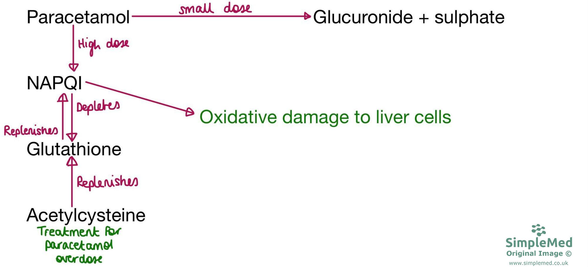
Diagram - The pathway of paracetamol metabolism
SimpleMed original by Dr. Maddie Swannack
Edited by: Dr. Ben Appleby
Reviewed by: Dr. Thomas Burnell and Dr. Marcus Judge
- 8279
