

SimpleMed - Learning Medicine, SimplifiedSimpleMed - Learning Medicine, Simplified
Hit enter to search or ESC to close

Get in Touch

Contact Us

Use our contact form.

The Larynx

Share:

By Dr. Joshua Bray

Next Lesson - Head and Neck Cancers

  Head & Neck


Contents

Contents

  1. Introduction to the Larynx
  2. Structure of the Larynx
  3. Laryngeal Muscles
  4. Actions of the Larynx
  5. Nerve Supply of the Larynx
  6. Conditions Affecting the Larynx
  7. Quiz
Abstract

Abstract

  • The larynx is part of the respiratory tract found in the anterior neck.
  • Functions of the larynx include airway protection, ventilation, coughing and phonation.
  • The larynx is divided into 3 sections: the supraglottis, glottis and infraglottis.
  • The larynx has 9 cartilages: 3 unpaired (epiglottis, thyroid, cricoid) and 6 paired (arytenoid, corniculate, cuneiform).
  • Laryngeal cartilages are connected by various membranes and ligaments.
  • The larynx has many muscles: extrinsic muscles move the entire larynx while intrinsic muscles act on the vocal cords.
  • The larynx is innervated by branches of the vagus nerve.
  • Many conditions can present with hoarseness of voice: these include acute laryngitis, croup, epiglottitis, laryngeal nodules, laryngeal cancer and recurrent laryngeal nerve palsy.
  • Epiglottitis is a medical emergency as it compromises airway patency.
Core

Core

Introduction to the Larynx

The larynx (‘voice box’) is a component of the respiratory tract found in the anterior neck. It lies at the level of the C3-6 vertebral bodies and connects the laryngopharynx to the trachea.

The larynx has several functions:

  • Airway Protection - this is arguably the most important role of the larynx, preventing food and fluid from entering the airways. When food/fluid enters the airway, this is known as aspiration.
  • Ventilation - as part of the respiratory tract, the larynx provides a channel for the movement of air in and out of the lungs.
  • Coughing - the larynx plays an important role in the cough reflex, which rapidly expels unwanted material from the respiratory tract.
  • Phonation - the production of sound.

The larynx lies anterior to the laryngopharynx. Food should not enter the larynx and is normally diverted into the laryngopharynx via recesses called the piriform fossae.

The larynx is divided into 3 sections: the supraglottis, glottis and infraglottis. The glottis (or rima glottidis) is the narrow area at the level of the vocal cords and divides the supraglottis and infraglottis.
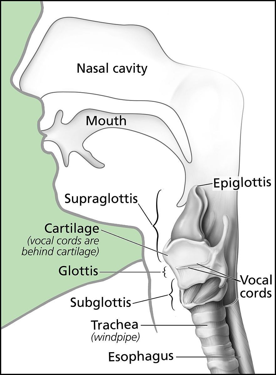
 

Anatomy of the Larynx SimpleMed

Diagram - Anatomy of the larynx

Public Domain Source by Alan Hoofring (Illustrator), edited by Dr. Joshua Bray [Public domain]


Structure of the Larynx

The larynx is formed by a framework of cartilages, membranes and ligaments which is predominantly lined by respiratory mucosa, with the true vocal cords covered by stratified squamous epithelium.

The laryngeal skeleton consists of 9 cartilages: 3 are single and 3 are paired.

The 3 unpaired cartilages are:

  • Epiglottis - the most superior of the laryngeal cartilages, the epiglottis is a heart-shaped cartilage which plays an important role in airway protection. Its elastic cartilage allows it to fold over the laryngeal inlet while swallowing to prevent material from entering the larynx. It lies just posterior to the root of the tongue and the space between the two is known as the vallecula.
  • Thyroid Cartilage - the largest of the cartilages, the thyroid cartilage consists of two laminae which extend anteriorly and join to form the laryngeal prominence (Adam’s apple).
  • Cricoid Cartilage - the most inferior cartilage, the cricoid cartilage is a complete ring of hyaline cartilage which encircles the airway and forms the inferior border of the larynx. It is shaped like a signet ring with its band facing anteriorly.

The 3 paired cartilages are:

  • Arytenoid Cartilages - these pyramid-shaped cartilages articulate with the superior aspect of the cricoid cartilage. These cartilages are important as they provide attachment for the aryepiglottic folds and are also visible on laryngoscopy.
  • Corniculate Cartilages - these smaller cartilages articulate with the apex of the arytenoid cartilages.
  • Cuneiform Cartilages - these cartilages sit within the aryepiglottic folds to provide reinforcement to the folds.

These cartilages are connected by numerous membranes and ligaments to unite the larynx as a single structure.

Three membranes connect the unpaired cartilages of the larynx to each other and to other nearby structures:

  • Thyrohyoid Membrane - connecting the hyoid bone to the thyroid cartilage.
  • Cricothyroid Membrane - connecting the thyroid cartilage to the cricoid cartilage. A cricothyroidotomy involves making a hole in this membrane in order to create an emergency airway.
  • Cricotracheal Membrane - connecting the cricoid cartilage to the 1st tracheal cartilage.


External Laryngeal Cartilages and Membranes SimpleMed

Diagram - External laryngeal cartilages and membranes

Creative commons source by Olek Remesz [CC BY-SA 4.0 (https://creativecommons.org/licenses/by-sa/4.0)]


The quadrangular membrane spans between the epiglottis and arytenoid cartilages. The free edges of this form important structures:

  • Aryepiglottic Folds - formed by the superior free edge of the quadrangular membrane, connecting the epiglottis to the arytenoid cartilages. These form an oval entrance to the larynx called the laryngeal inlet.
  • Vestibular Ligaments - formed by the inferior free edge of the quadrangular membrane, connecting the angle of the thyroid cartilage to the arytenoid cartilage. These are otherwise known as the false vocal cords.

The true vocal cord, otherwise known as the vocal ligament, is formed by the thickened superior free border of the conus elasticus (cricovocal membrane).


Cartilages of the Larynx SimpleMed

Diagram - View of the larynx and vocal cords on nasendoscopy

Creative commons source by OpenStax College [CC BY-SA 4.0 (https://creativecommons.org/licenses/by-sa/4.0)]


The larynx is predominantly lined with pseudostratified ciliated epithelium, except for the true vocal cord and the lingual surface of the epiglottis, which are lined by stratified squamous non-keratinised epithelium.


Laryngeal Muscles

The larynx has many muscles. This article will focus on some of the more important muscles.

In broad terms, there are 2 groups of laryngeal muscles: extrinsic and intrinsic. The extrinsic muscles attach the larynx (directly or indirectly) to other structures and act to move the entire larynx. The intrinsic muscles are found within the larynx itself and act primarily on the vocal cords.

Extrinsic Muscles:

  • Stylopharyngeus - this muscle of the pharynx acts to elevate the larynx.
  • Suprahyoids - this group of muscles act to elevate the hyoid bone and therefore elevate the larynx.
  • Infrahyoids - this group of muscles act to depress the hyoid bone and therefore depress the larynx.

Intrinsic Muscles:

Most of the intrinsic laryngeal muscles (e.g. lateral cricoarytenoid, transverse arytenoid) act to adduct the vocal cords. Other important muscles of note are:

  • Posterior Cricoarytenoids - this pair of muscles are the only muscles to abduct the vocal cords.
  • Cricothyroid - this pair of muscles increase tension in the vocal cords in order to produce higher pitched sounds.


Actions of the Larynx

The larynx executes different movements depending on the action which is being performed:

  • Ventilation - when taking a deep breath in or out, the vocal cords are fully abducted (via posterior cricoarytenoids) in order to create a wide airway.
  • Phonation - in order to create sounds, the vocal cords adduct to vibrate a thin column of air. The pitch of the sound created is altered by varying the length and tension of the vocal cords (via cricothyroid).
  • Coughing - in order to produce a cough, the vocal cords initially adduct in order to build up intrathoracic pressure. The cords then suddenly snap open which causes the air to be expelled at a high velocity.

During swallowing, it is imperative that food/drink does not enter the airway (aspiration). To avoid this, the larynx has several protective mechanisms:

  • The vocal cords adduct in order to close the glottis.
  • Contraction of the aryepiglottic muscles acts to narrow the laryngeal inlet and fold the epiglottis over the inlet.
  • The larynx is pulled upwards by contraction of stylopharyngeus and the suprahyoid muscles. This helps to close the laryngeal inlet.


Nerve Supply of the Larynx

The larynx is innervated by branches of the vagus nerve (CN X). These branches are the superior laryngeal nerve and the recurrent laryngeal nerve.

The recurrent laryngeal nerve has an important path, branching distally from the vagus nerve and then travelling back on itself towards the larynx:

  • Right Recurrent Laryngeal Nerve - loops under the right subclavian artery
  • Left Recurrent Laryngeal Nerve - loops under the aortic arch
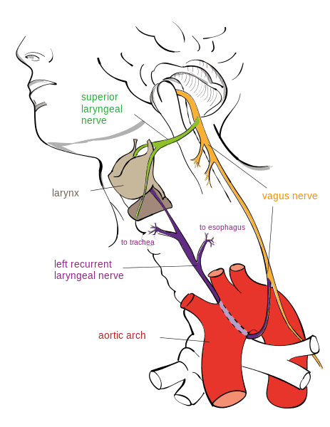
 

Course of the Left Recurrent Laryngeal Nerve SimpleMed

Diagram - Course of the left recurrent laryngeal nerve

Creative commons source by Jkwchui [CC BY-SA 4.0 (https://creativecommons.org/licenses/by-sa/4.0)]


In terms of sensory innervation, the supraglottis is supplied by the internal branch of the superior laryngeal nerve, while the infraglottis is supplied by the recurrent laryngeal nerve. Sensation at the level of the glottis is generally attributed to the internal laryngeal nerve.

As for motor supply, all intrinsic muscles of the larynx are innervated by the recurrent laryngeal nerve, except for cricothyroid which is innervated by the external branch of the superior laryngeal nerve.

Injury to the recurrent laryngeal nerve leads to paralysis of the ipsilateral vocal cord.

  • If the lesion is unilateral - this will lead to a hoarse voice and/or an ineffective cough reflex. These lesions are often well tolerated as the contralateral vocal cord compensates over time.
  • If the lesion is bilateral - this is a surgical emergency. Both vocal cords are paralysed and lie in a paramedian position, which compromises the airway. An emergency airway is needed.


Conditions Affecting the Larynx

Acute laryngitis is inflammation of the larynx. It is associated with upper respiratory tract infections and is most commonly viral, for example parainfluenza or influenza viruses. Symptoms include dry cough, hoarse voice and sore throat. If the inflammation extends to the trachea and bronchi, this is known as laryngotracheobronchitis, or croup. Croup is common in children and is characterised by stridor and a barking cough (which sounds like a seal).

Epiglottitis is inflammation of the epiglottis. It is a medical emergency as the swollen epiglottis can compromise the airway. It presents with dyspnoea, stridor, hoarseness of voice and often drooling. Patients require urgent senior assessment, with airway management in a controlled setting by experienced specialists if there are signs of airway compromise. The most common cause of epiglottitis is Haemophilus influenzae B (HiB) infection. For this reason, children are routinely vaccinated against HiB in the UK.

The airway can also become compromised during anaphylaxis, a severe allergic reaction where histamine release by mast cells results in laryngeal oedema. Securing a definitive airway is a priority in these patients.

Laryngeal nodules (vocal nodules) are benign growths on the vocal cords. They are generally due to voice overuse which causes chronic irritation of the vocal cords. Therefore, they are common in singers, cheerleaders, teachers, etc. Patients generally present with voice changes (e.g. hoarse, raspy, low-pitched voice) and/or pain in the throat. Initial treatment is vocal rest, however, if this does not work the nodules can be removed surgically.

Laryngeal cancer often presents with voice changes. Other symptoms include dysphagia, globus and weight loss. On rare occasions a neck lump may be palpable. The mainstay of treatment is surgery. For more information, see our article on Head and Neck Cancers.


Reviewed by: Dr. Thomas Burnell

Quiz

Quiz

Preview the The Larynx quiz

In this article

The larynx is part of the respiratory tract found in the anterior neck. Functions of the larynx include airway protection, ventilation, coughing and…

Free account

Keep reading for free

Create a free account to continue this article and save your revision progress.

Your account includes:

  • Thousands of free practice questions
  • Tools to track your revision progress
Sign up free Already have an account? Log in

No payment. No spam. Just free medical revision.

SimpleMed+

Want More?

Access more on SimpleMed+

Lock it in with practice questions, the full question bank and revision tools that track what you know.

Practice now See what's in SimpleMed+
7726
simplemedlonglogo.png
  • Home
  • Terms & Conditions
  • Privacy Policy
  • Contact

The medical information on this site is provided as an information resource only, and is not to be used or relied on for any diagnostic or treatment purposes. This information is intended for medical education, specifically to biological/medical learning inclined individuals, and does not create any doctor-patient relationship, and should not be used as a substitute for professional diagnosis and treatment.

©2026 Lili's Ark Limited, UK Company #15424953
S+

Search

  • Pre-Clinical
    • Cardiovascular System
      • 1. Anatomy of the Cardiovascular System
      • 2. The Cardiac Cycle
      • 3. Pulses and Pressures
      • 4. Control of Cardiac Output
      • 5. Cardiac Conduction and Contraction
      • 6. Embryology of the CVS
      • 7. Congenital Heart Defects
      • 8. Hypertension
      • 9. Electrocardiograms
      • 10. Electrocardiogram Pathology
      • 11. Cardiovascular Drugs
      • 12. Murmurs
      • 13. Special Circulations
      • 14. Heart Failure
      • 15. Artery and Vein Disease
      • 16. Acute Coronary Syndromes
      • 17. Haemodynamic Shock
    • Musculoskeletal System
      • The Human Body
        • 1. Anatomical Terms
        • 2. Structure of the Muscles
        • 3. Muscles in Action
        • 4. Connective Tissue
        • Cartilage: Structure, Growth and Repair
        • 5. Structure of Bones
        • 6. Basics of Bone Anatomy
        • 7. Joints
        • 8. The Spine
        • 9. Dermatomes and Myotomes
        • 10. The Brachial Plexus
        • 11. The Lumbar Plexus
        • 12. The Sacral Plexus
      • Clinical Conditions
        • 1. Conditions of the Foot
        • 2. Conditions of the Ankle
        • 3. Conditions of the Lower Leg
        • 4. Conditions of the Knee
        • 5. Conditions of the Thigh
        • 6. Conditions of the Hip
        • 7. Conditions of the Lower Back
        • 8. Conditions of the Back and Neck
        • 9. Conditions of the Shoulder and Clavicle
        • 10. Conditions of the Arm
        • 11. Conditions of the Elbow
        • 12. Conditions of the Forearm and Wrist
        • 13. Conditions of the Hand
      • Musculoskeletal Anatomy
        • 1. Muscles of the Gluteal Region
        • 2. Muscles of the Thigh
        • 3. Key Areas of the Lower Limb
        • 4. Bones of the Lower Limb
        • 5. Bones of the Foot
        • 6. Muscles of the Lower Leg
        • 7. Bones of the Upper Limb
        • 8. Muscles of the Shoulder
        • 9. Muscles of the Upper Arm
        • 10. Bones of the Hand
        • 11. Muscles of the Forearm
        • 12. Key Areas of the Upper Limb
    • Cell Physiology & Biology
      • 1. An Introduction to the Cell
      • 2. Introduction to DNA
      • 3. The Cell Cycle, Checkpoints and Apoptosis
      • 3. Structure of Proteins
      • 4. Protein Function in Oxygen Transport
      • 5. Enzymes and Regulation of Protein Function
      • 6. Molecular Techniques for Medics
      • 7. Post-translational Modification and Collagen Biosynthesis
      • 8. Protein Targeting
      • 9. Biological Membranes
      • 10. G-Protein Coupled Receptor
      • 12. Enzyme-linked and Intracellular Receptor Signalling
      • 11. Membrane Transport and Intracellular Calcium Regulation
      • 12. Membrane Potentials
      • 13. Action Potentials
      • 14. Autonomic Nervous System Introduction
    • Metabolism
      • 1. Nutrition, Diet and Body Weight
      • 2. Energy Reactions in Cells
      • 3. Dietary Energy Production
      • 4. Vitamins, Cofactors, Minerals and Trace Elements
      • 4. Glycolysis
      • 5. Oxygen-dependent Pathway
      • 6. Lipid Metabolism and Transport
      • 7. Protein and amino acid metabolism
      • 9. Purine and Pyrimidine Metabolism
      • 10. Inborn Errors of Metabolism and Newborn Screening
      • 8. Alcohol Metabolism
      • 9. Oxidative Stress
      • 10. Energy Storage
      • 11. Temperature Regulation
      • 12. Metabolic and Endocrine Regulation
    • Endocrinology
      • 1. An Introduction to Endocrinology
      • 2. Control of Appetite
      • 3. The Adrenal Glands and Adrenal Disorders
      • 4. HPA and Growth Hormone
      • 5. Pituitary Disorders
      • 6. The Thyroid Gland
      • 7. The Endocrine Pancreas
      • 8. Diabetes Mellitus
      • 9. Calcium Metabolism
    • Haematology
      • 1. Haematopoiesis and the Spleen
      • 2. Full Blood Count
      • 3. Blood Groups, Crossmatching and Transfusion Reactions
      • 3. Anaemia
      • 4. Vitamin B12 and Folate
      • 5. Iron Metabolism
      • 6. Haemoglobinopathies
      • 7. When Haematopoiesis goes wrong
      • 8. Haematology in Systemic Disease
    • Genetics
      • 1. Mitosis and Meiosis
      • 2. Genotype, Phenotype and Inheritance
      • 3. Gene Expression
      • 4. Epigenetics, Genomic Imprinting and Mitochondrial Inheritance
      • 4. Introduction to Gene Mutation
      • 5. DNA Repair and Cancer
      • 7. Genetic Testing and Counselling
    • Infection
      • 1. An Introduction To Infection
      • 2. The Infection Model
      • 3. Bacterial Structure, Classification and Growth
      • 3. Biofilms
      • 4. Innate Immunity
      • 5. Adaptive Immunity
      • 6. Gram Staining and Infection Investigations
      • 8. Staphylococci and Other Gram-positive Bacteria
      • 9. Streptococci
      • 10. Gram-negative Bacteria: Structure, Entry Routes and Endotoxin
      • 11. The Organisms Gram Staining Misses
      • 7. Sepsis
      • 8. Antibiotics
      • 14. Principles of Medical Virology
      • 10. Hepatitis
      • 11. Human Immunodeficiency Virus
      • 17. Fungi and Parasites: The Eukaryotic Pathogens
      • 12. The Immunocompromised Host
      • 13. Travel-related Infections
    • Pathology
      • 1. An Introduction to Pathology
      • 2. How to Read a Histology Slide
      • 2. The Basics of Pathology
      • 3. Epithelium, Skin and Exocrine Glands
      • 4. Acute Inflammation
      • 5. Chronic Inflammation
      • 6. Regeneration and Repair of Tissues
      • 7. The Lymphatic System
      • 8. Haemostasis and Clotting
      • 9. Thrombosis and Embolism
      • 10. Atherosclerosis
      • 11. Cellular Adaptations
      • 12. Introduction to Neoplasia
      • 13. Pathogenesis of Neoplasms
      • 14. Behaviour of Neoplasms
    • Population & Social Science
      • 1. Introduction to Population Science
      • 2. Describing Data and Testing Hypotheses
      • 2. Epidemiological Studies
      • 3. Models of Health
      • 4. Beliefs of the Public
      • 5. Health Economics
      • 6. Evidence-Based Practice
      • 7. Patient Safety and Quality Control
      • 8. Summary and Glossary
    • Patient-Centred Care
      • 1. Lifestyle Medicine
      • 2. Childhood Immunisations
      • 3. Sepsis in Paediatrics
      • 4. Child Safeguarding
      • 5. Ageing
      • 6. Falls
      • 7. Men's Health
      • 8. Palliative Care
    • Reproductive System
      • 1. Anatomy of the Man
      • 2. Anatomy of the Woman
      • 3. The Pelvic Floor
      • 4. Development of the Reproductive System
      • 5. Puberty
      • 6. The Menstrual Cycle and Menstrual Abnormalities
      • 7. Gametogenesis
      • 8. Sex, Infertility and Contraception
      • 9. Physiology of the Mother
      • 10. Diseases of the Mother in Pregnancy
      • 11. Physiology of the Fetus
      • 12. The Placenta
      • 13. Labour and Delivery
      • 14. The Postpartum Period
      • 15. Infections of the Reproductive Tract
      • 16. Pelvic Inflammatory Disease
      • 17. The Menopause
      • 18. Malignancies of the Reproductive Tract
      • 19. Introduction to the Breast
      • 20. Benign Conditions of the Breast
      • 21. Malignant Breast Disease
    • Respiratory System
      • 1. Anatomy of the Respiratory System
      • 2. Development of the Respiratory System
      • 3. Ventilation and Lung Mechanics
      • 4. Properties of Gas Exchange
      • 5. Carbon Dioxide Transport
      • 6. Chemical Control of Breathing and Plasma pH
      • 7. Lung Function Testing
      • 8. Pulmonary Embolism
      • 9. Asthma
      • 10. Chronic Obstructive Pulmonary Disease
      • 11. Pneumonia and other Lower Respiratory Tract Infections
      • 12. Bronchiectasis and Cystic Fibrosis
      • 13. Tuberculosis
      • 14. Pneumothorax and Pleural Effusion
      • 15. Lung Cancer
      • 16. Respiratory Failure
    • Urinary System
      • 1. Anatomy of the Kidneys, Bladder and Urethra
      • 2. Development of the Urinary System
      • 3. The Nephron
      • 4. The Glomerulus – Tubular Reabsorption and Secretion
      • 5. Control of Plasma Volume
      • 6. Control of Plasma Osmolarity
      • 7. Control of Potassium
      • 8. Micturition
      • 9. Diuretics
      • 10. Glomerular Filtration Rate and the Measurement of Kidney Function
      • 11. Hyponatraemia
      • 12. Urinary Incontinence
      • 13. Glomerular Disease
      • 14. Diabetic Nephropathy
      • 15. Acute Kidney Injury
      • 16. Chronic Kidney Disease
      • 17. Urinary Tract Obstruction
      • 18. Urinary Tract Infections
      • 19. Prostate Pathology
      • 20. Male Urological Conditions
      • 21. Urological Malignancy
    • Gastrointestinal System
      • 1. Development of the Gastrointestinal Tract
      • 2. Abdominal Wall and Inguinal Canal
      • 2. Anatomy and Physiology of the Foregut
      • 3. Anatomy and Physiology of the Midgut
      • 4. Anatomy and Physiology of the Hindgut
      • 6. Gastrointestinal Motility and the Enteric Nervous System
      • 7. Liver, Biliary and Exocrine Pancreatic Physiology
      • 8. Gastrointestinal Secretion, Digestion and Absorption
      • 5. Upper Gastrointestinal Pathology
      • 6. Hepatic, Biliary and Pancreatic Pathology
      • 7. Intestinal Pathology
      • 8. Distal Gastrointestinal Tract Pathology
      • 9. Gastrointestinal Malignancy
      • 10. Gastroenteritis and Enterocolitis
      • 11. Gastrointestinal Emergencies
    • Head & Neck
      • 1. Introduction to Head & Neck Anatomy
      • 2. Blood Supply of the Head and Neck
      • 3. Lymph Nodes and Neck Lumps
      • 4. The Skull
      • 5. The Temporomandibular Joint
      • 6. The Meninges
      • 7. Cranial Nerves I-VI
      • 8. Cranial Nerves VII-XII
      • 9. Development of the Head and Neck
      • 10. Autonomics in the Head and Neck
      • 11. Anatomy of the Ear
      • 12. Ear Pathology
      • 13. The Nose
      • 14. The Oral Cavity and Pharynx
      • 15. The Eye
      • 16. The Larynx
      • 17. Head and Neck Cancers
      • 18. Imaging of the Head and Neck
      • 19. Fascial Planes, Muscles and Triangles
    • Immunology
      • 1. An Introduction to Hypersensitivity
      • 2. Type 1 Hypersensitivity
      • 3. Food Allergy
      • 4. Introduction to Autoimmunity
      • 5. Rheumatoid Arthritis
      • 6. Systemic Lupus Erythematosus (SLE)
      • 7. Immunological Therapies | Monoclonal Antibodies
    • Neuroanatomy & Psychiatry
      • 1. Basics of Neuroanatomy
      • 2. Cells and Components of the Nervous System
      • 3. Arterial Supply to the Brain
      • 4. Development of the Nervous System
      • 5. Production and Circulation of CSF and Related Disorders
      • 6. Somatosensory System
      • 7. Motor System
      • 8. The Cerebellum: Circuits, Coordination and Clinical Signs
      • 8. Visual System
      • 10. Auditory and Vestibular Systems
      • 11. The Hypothalamus and Limbic System
      • 9. Higher Cortical Function
      • 10. Reticular Formation
      • 11. Movement Disorders
      • 12. Infections of the Central Nervous System
      • 13. Dementia
      • 14. Delirium
      • 15. Headaches
      • 16. Stroke
      • 17. Raised Intracranial Pressure (ICP)
      • 18. Subarachnoid Haemorrhage
      • 19. Anxiety
      • 20. Mood Disorders
      • 21. Psychosis
    • Embryology
      • 1. The Basics of Embryology
      • 2. Development of the Limbs and Body Wall
      • 3. Teratology and Congenital Malformations
      • 2. Development of the Gastrointestinal Tract
      • 3. Development of the Reproductive Systems
      • 5. Development of the Respiratory System
      • 6. Development of the Head and Neck
      • 8. Embryology of the Cardiovascular System
    • Pharmacology
      • 1. Research Methods in Pharmacology
      • 2. Pharmacokinetics
      • 3. Pharmacodynamics
      • 4. Pharmacovigilance and Pharmacogenetics
      • 5. Safe Prescribing
      • 6. Cholinergic Pharmacology
      • 7. Adrenergic Pharmacology
      • 6. Hypertension and Heart Failure
      • 7. Cardiac Arrhythmias
      • 8. Antiplatelets, Anticoagulants and Thrombolysis
      • 9. Hyperlipidaemias
      • 10. Diuretics and Renal Pharmacology
      • 11. Pharmacology of Insulin
      • 12. Reproductive and Post-reproductive Health
      • 13. Antibiotics
      • 14. Antivirals
      • 15. Cancer Chemotherapy
      • 16. Anaesthetics
      • 17. Opioids
      • 20. Introduction to Psychopharmacology
      • 18. Epilepsy and Anti-epileptic Drugs
      • 19. Parkinson's Disease and Myasthenia Gravis
      • 20. NSAIDs
      • 21. Immunosuppression and Disease-modifying Therapy
      • 22. Pharmacology of Airway Control
      • 23. Drugs Affecting Acid Secretion
      • 24. Antiemetics and Laxatives
      • 25. Poisoning and STOPP/START
  • OSCE
    • Index Conditions for OSCE exams
    • Cardiovascular System Examination
    • Shoulder Examination
    • Hand Examination
    • Venepuncture
    • Blood Pressure
    • Respiratory System OSCE Examination
    • Peripheral Vascular OSCE Examination
    • Varicose Vein OSCE Examination
    • Deep Vein Thrombosis (DVT) OSCE Examination
    • Abdominal OSCE Examination
    • Rectal (PR) OSCE Examination
    • Stoma OSCE Examination
    • Hernia OSCE Examination
    • Upper Limb Neurological OSCE Examination
    • Lower Limb Neurological OSCE Examination
    • Cranial Nerve OSCE Examination
    • Cerebellar OSCE Examination
    • GALS Screening OSCE Examination
    • Elbow OSCE Examination
    • Hip OSCE Examination
    • Knee OSCE Examination
    • Ankle and Foot OSCE Examination
    • Spine OSCE Examination
    • Thyroid Status OSCE Examination
    • Diabetic Foot OSCE Examination
    • Cushing's Syndrome OSCE Examination
    • Acromegaly OSCE Examination
    • Ear, Otoscopy and Hearing OSCE Examination
    • Nasal OSCE Examination
    • Eye and Vision OSCE Examination
    • Fundoscopy (Ophthalmoscopy) OSCE Examination
    • Visual Acuity OSCE Assessment
    • Anterior Segment Eye OSCE Examination
    • Pigmented Skin Lesion OSCE Examination
    • Rash and Non-Pigmented Skin Lesion OSCE Examination
    • Breast OSCE Examination
    • Testicular OSCE Examination
    • Neck Lump OSCE Examination
    • Lymphoreticular OSCE Examination
  • Question Bank
  • About
  • How to Use SimpleMed
  • SimpleMed Lecture Series
  • DON'T PANIC - Guide to the Wards
  • SimpleMed Plus
Your privacy Allow anonymous analytics to help us improve SimpleMed. Privacy policy
