

All done here! Check out our other subjects!
Core
This is a hybrid article between the Metabolism and Endocrine units, so the topics mentioned in it will relate to both of those units.
The body has two types of fuel source. Some are available in normal circumstances. Others are used in so-called ‘special circumstances’, when the body does not have its normal fuel supply (like starvation, or in the middle of a marathon).
Sources of energy normally available in the blood:
- Glucose - this is the preferred fuel, and most is stored as glycogen.
- Fatty Acids - these can be used by most cells in the body, except red blood cells and cells of the CNS (so the brain uses fatty acids only to a negligible extent). They are stored in the body as triacylglycerol (TAG, also called triglyceride) in adipose tissue (body fat).
Sources of energy available in ‘special circumstances’:
- Amino acids from muscles:
- Glucose - muscle proteins are broken down into their amino acids, and then glucogenic amino acids (those that can be made into glucose, like alanine) are converted into glucose.
- Ketone bodies - produced mainly in the liver from fatty acids, with a smaller contribution from ketogenic amino acids (those that can be made into ketone bodies, like leucine). Ketone bodies are used by the brain when there is not enough glucose in the blood.
- Lactate - in the liver, this can be converted into glucose (through the Cori cycle, see below). Other tissues, such as the heart and brain, convert lactate back to pyruvate, which is then used in the Krebs Cycle.
Fuel Stores in the Body
- Glycogen - about 500g stored in total (roughly 400g in skeletal muscle and 100g in the liver). Liver glycogen is the source of glucose for the blood. Muscle glycogen is mainly a fuel store for the muscle itself, because muscle lacks glucose 6-phosphatase (the enzyme that frees glucose so it can leave the cell).
- Fat - between 10kg and 15kg in someone who has a ‘healthy’ BMI, acting as a source of fatty acids and glycerol.
- Muscle Protein - about 6kg, acting as a source of glucose or ketone bodies following metabolism depending on whether the amino acid is glucogenic or ketogenic.
Which Stores When?
Feeding (the times below are approximate, and the stages overlap)
Up to 2 hours - Glucose and fat absorption from the gut fuels metabolism and prompts storage.
2-8 hours - Absorption stopped, the body relies on fatty acid and glycogen stores. Blood glucose is preserved for brain use.
8+ hours - Glycogen stores are increasingly used up, so gluconeogenesis provides more of the glucose, made from glycerol, proteins (glucogenic amino acids) and lactate.
Starvation (multiple days) - By this point there is no glycogen left, so the body becomes reliant on fatty acids and ketone body production, with reduced but ongoing protein breakdown. Most tissues use fatty acids, while the brain increasingly uses ketone bodies. This is an adaptive state that reduces glucose requirements and preserves protein.
Hormonal Influences on Metabolism
Anabolic (promoting fuel storage) - insulin and growth hormone (promotes protein synthesis).
Catabolic (promoting release from stores and utilisation) - glucagon, adrenaline, cortisol, thyroid hormones and growth hormone. Growth hormone has both anabolic and catabolic effects: it promotes protein synthesis, but it also increases lipolysis (the breakdown of fat) and opposes the actions of insulin.
Insulin is one of the most important hormones in the body when it comes to metabolism as it has many different effects. Remember that insulin is released after a meal, when blood glucose is high. The body therefore stores fuel rather than releasing more from its stores.
- Insulin stops - gluconeogenesis, glycogenolysis (breakdown of glycogen), lipolysis, proteolysis (breakdown of protein) and ketogenesis (making ketone bodies). Insulin promotes the storage of fuel sources rather than their breakdown.
- Insulin starts - GLUT4-mediated uptake of glucose (GLUT4 is a glucose transporter protein in cell membranes) into adipose and muscle cells for storage, glycogen synthesis, and protein synthesis.
This relates to the Feeding-Fasting Cycle
- Feeding releases insulin - this prompts the storage of many molecules. There is increased GLUT4-mediated glucose uptake into muscle and fat, increased glycogen storage, increased amino acid uptake, increased lipogenesis (making fat).
- Fasting releases glucagon - this prompts the release of molecules that can be used as a fuel source. There is decreased uptake of glucose into muscle and fat, increased glycogenolysis in the liver, increased lipolysis, increased gluconeogenesis.
There are many changes that occur during pregnancy relating to metabolism because the metabolism of the mother must account for the needs of the growing fetus. Over pregnancy, most women gain between 10kg and 12.5kg. Much of this is the growing fetus, placenta and amniotic fluid (the fluid around the fetus), and the mother also lays down fat stores.
- The first half of the pregnancy can be described as anabolic - increased fat and nutrient stores to supply the growing fetus and to make breast milk (lactation) after birth (post-partum).
- The second half of pregnancy can be described as catabolic - decreased insulin sensitivity to leave glucose and fatty acids in the bloodstream for the metabolism of the fetus.
These changes occur due to the fetoplacental unit, a system involving the fetal adrenal glands, fetal liver and the placenta working together. This system produces hormones that alter maternal metabolism and insulin sensitivity (how well cells respond to insulin), creating conditions more favourable for the fetus.
The placenta releases hormones (for example human placental lactogen, a hormone specific to pregnancy, and placental growth hormone). Together with oestrogen, progesterone and cortisol, these increase the maternal resistance to insulin. This slightly raises blood glucose (mild hyperglycaemia), so the fetus has a high glucose supply. This leads to hyperglycaemia after meals and to hypoglycaemia (low blood glucose) overnight, because the fetus keeps drawing glucose from the mother while she fasts. These changes can lead to Gestational Diabetes.
Gestational diabetes is similar to Type Two Diabetes Mellitus (T2DM) in that they both involve episodes of hyperglycaemia relating to insulin resistance. However, gestational diabetes often resolves following birth. Women who have had gestational diabetes are more likely to develop type 2 diabetes later. They are offered a fasting blood glucose test 6 to 13 weeks after the birth to check for diabetes. If that test is normal, they are then offered an HbA1c test (a blood test that shows longer-term blood glucose control) once a year. Pregnancy makes all women increasingly insensitive to insulin. Gestational diabetes develops when the pancreas cannot make enough extra insulin to cope. Women who are already insulin resistant, for example with obesity, are more likely to develop it.
Gestational diabetes does not have a single cause. It can arise in several ways, including:
- Autoantibodies (antibodies that attack the body’s own cells) may be present in a small number of cases, usually representing early or unrecognised autoimmune diabetes that becomes apparent during pregnancy.
- Genetic susceptibility to T2DM that would normally appear later in adult life, but the pregnancy-related insulin resistance causes it to present earlier.
- In some people, the normal insulin resistance in pregnancy is enough to induce a diabetic state.
Clinical implications of diabetes in pregnancy (gestational diabetes, or diabetes that was already present before pregnancy) can be serious:
- Increased incidence of miscarriage (this risk is mainly seen with diabetes that was already present before pregnancy, rather than gestational diabetes diagnosed later).
- Increased risk of congenital malformation when significant hyperglycaemia is present early in pregnancy (this risk is mainly seen with pre-existing diabetes rather than gestational diabetes diagnosed later).
- Increased incidence of macrosomia (basically a big baby). This can lead to shoulder dystocia, when the shoulders of the baby get stuck in the birth canal. Shoulder dystocia risks tearing for the mother and, for the baby, fractures or brachial plexus injury (damage to the nerves of the arm, specifically Erb’s palsy, which causes arm weakness).
- Increased risk of gestational hypertension and pre-eclampsia. Gestational hypertension is new high blood pressure that appears after 20 weeks of pregnancy without proteinuria (protein in the urine). Pre-eclampsia is new high blood pressure after 20 weeks of pregnancy together with proteinuria or another new problem, such as kidney or liver involvement or poor growth of the baby. It is thought to start with a placenta that develops or works poorly. If it is not treated properly, it can lead to fits and death in the mother.
Gestational diabetes is diagnosed with an oral glucose tolerance test (OGTT: a fasting blood glucose test, then a second test 2 hours after a 75g glucose drink). The OGTT is offered at 24 to 28 weeks to women with a NICE risk factor (all those listed below except age over 40, which is not on the NICE list), and earlier if they have had gestational diabetes before. Gestational diabetes is diagnosed if the fasting plasma glucose is 5.6 mmol/litre or above, or the 2-hour plasma glucose is 7.8 mmol/litre or above. Gestational diabetes can be managed well, which helps to reduce the risk of the complications listed above. It does need close monitoring, and many women need medicines as well as changes to diet and exercise.
Management strategies include:
- Diet and exercise advice - similar principles to early management of T2DM, focusing on balanced carbohydrate intake and keeping blood glucose within the target range (glycaemic control). Regular exercise is advised, for example walking for 30 minutes after a meal.
- Medicines if diet and exercise do not bring blood glucose to target within 1 to 2 weeks. Metformin (a tablet that lowers blood glucose) is offered first. Insulin injections are added if metformin alone is not enough, or used instead if metformin cannot be used. Insulin (with or without metformin) is started straight away if the fasting plasma glucose at diagnosis is 7.0 mmol/litre or above.
- Regular fetal monitoring - ultrasound scans of fetal growth and amniotic fluid volume (the fluid around the fetus) are offered every 4 weeks from 28 to 36 weeks. This is important to prepare for macrosomia. The 20-week scan also checks for congenital malformation, which is more of a risk when diabetes was present before pregnancy.
Risk factors for developing gestational diabetes include:
- Maternal age over 40.
- Maternal BMI of 30 kg/m2 or over.
- A first-degree relative (a parent or sibling) with diabetes.
- A previous baby weighing 4.5kg or more (macrosomia).
- Gestational diabetes in a previous pregnancy.
- Family origin with a high prevalence of diabetes: South Asian, Black, African-Caribbean or Middle Eastern.
Metabolic Responses to Exercise
Exercise requires rapid musculoskeletal, cardiovascular, respiratory and temperature changes in the body, and these drastically alter the metabolic demand of the body. The body also has to keep the rest of the body stable (homeostasis), remove waste faster (like lactate build-up), and maintain the glucose supply to the brain.
The Cori Cycle
The liver is important for regulating blood glucose. Exercise increases the glycogenolysis and gluconeogenesis that occur in the liver to maintain the glucose concentration of the blood.
The liver recycles lactate present in the blood through the Cori Cycle (shown in the diagram below). Working muscle makes lactate, the liver turns it back into glucose, and the muscle takes up this new glucose through GLUT4 transporters.
Exercising muscle also has the ability to increase GLUT4 translocation (movement to the cell membrane) through insulin-independent mechanisms. These are partly mediated by AMP-activated protein kinase (AMPK, an enzyme that senses low cell energy) during low energy states, together with calcium signals in the contracting muscle. This process is insulin-independent, meaning that muscle cells can take up more glucose (including glucose made by the Cori Cycle) without needing insulin. This is useful when the muscles have a high glucose demand (like when running a marathon), because insulin falls at this time. If insulin were high, glucose from the blood would be stored rather than kept available as fuel.
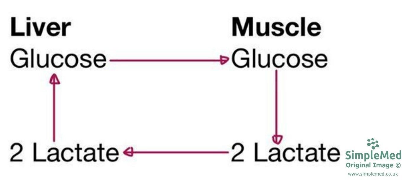
Diagram - The Cori Cycle, where the liver recycles lactate into glucose for the muscle to use
SimpleMed original by Dr. Maddie Swannack
Hormonal Changes During Extreme Exercise
During a marathon, there are a number of changes that occur in the hormonal profile of the body. This reflects the need of the body to increase the amount of glucose available in the blood.
- Insulin decreases - to prevent storage of glucose in the body.
- Glucagon increases - increased glycogenolysis, lipolysis, and gluconeogenesis.
- Increased adrenaline, growth hormone and cortisol - increased glycogenolysis, lipolysis, and gluconeogenesis.
Edited by: Dr. Thomas Burnell
Reviewed by: Dr. Marcus Judge
- 6649
