
Next Lesson - Muscles of the Thigh
Core
The muscles of the gluteal region are important in mobilising the lower limb at the hip joint. The 3 main movements that the gluteal muscles produce are abduction, extension and rotation of the hip. The muscles of the gluteal region can be divided into two compartments: superficial and deep.
Superficial Gluteal Compartment
There are 4 muscles within the superficial gluteal compartment. They are innervated (supplied by nerves) by either the superior gluteal nerve or the inferior gluteal nerve. Between them they mainly abduct and extend the hip: gluteus maximus is the main extensor, and the other three mainly abduct and medially rotate it.
This is the largest muscle in the body. Gluteus maximus originates (begins) from the ilium (part of the hip bone), coccyx and sacrum and attaches to the iliotibial tract (a band of connective tissue down the outer thigh) and to the gluteal tuberosity of the femur (a rough ridge on the back of the femur). It is innervated by the inferior gluteal nerve and acts to extend the hip joint (whilst also providing some aid in lateral rotation). The gluteus maximus is particularly important for powerful hip extension, for example when climbing or walking up stairs, and also contributes during normal gait (the way we walk).
This muscle is found between the Gluteus Maximus and the Gluteus Minimus. It originates from the ilium and attaches at the greater trochanter of the femur (the bony lump on the outer side of the upper thigh). It is innervated by the superior gluteal nerve. It acts to abduct and medially rotate the femur at the hip joint. It is also a key muscle for the stabilisation of the pelvis, with weakness or paralysis of this muscle (or of gluteus minimus, or damage to the superior gluteal nerve that supplies them) causing Trendelenburg's Sign (when standing on one leg, the pelvis drops on the opposite, unsupported side; see here for more details).
This muscle is the deepest and smallest of the superficial compartment and originates from the ilium. It attaches to the greater trochanter. Like the Gluteus Medius, it is innervated by the superior gluteal nerve and acts to abduct and medially rotate the femur at the hip.
This is a small superficial muscle that originates from the anterior superior iliac spine (ASIS, the bony point at the front of the pelvis that you can feel at the hip) and the front part of the iliac crest (the top edge of the hip bone). It inserts into the iliotibial tract (a band of connective tissue down the outer thigh), which attaches to the lateral condyle of the tibia (the outer knob at the top of the shin bone). The muscle therefore lies on the lateral (outer) side of the thigh. It is innervated by the superior gluteal nerve and acts to abduct and medially rotate the femur at the hip. It also helps to flex the hip.
There are five muscles in the deep gluteal group: piriformis, superior gemellus, obturator internus, inferior gemellus and quadratus femoris. They lie deep to the superficial gluteal muscles and mainly laterally rotate the femur. They are listed here from superior to inferior.
Piriformis is the most superior muscle in the deep gluteal group. It arises from the anterior surface of the sacrum, passes through the greater sciatic foramen (a large opening at the back of the pelvis) and inserts on the greater trochanter. It is supplied by the nerve to piriformis. It is mainly a lateral rotator of the femur (it turns the thigh outwards). It has a smaller role as an abductor (it moves the thigh away from the midline). What it does depends on the position of the hip, and with the hip flexed beyond 90 degrees it can turn the thigh inwards instead. Piriformis is an important landmark for structures leaving the pelvis. The superior gluteal nerve and vessels pass above it. The inferior gluteal nerve, sciatic nerve, posterior femoral cutaneous nerve (which supplies skin at the back of the thigh) and pudendal nerve (which supplies the perineum, the region between the genitals and anus) pass below it, and so do the nerves to obturator internus and quadratus femoris. Not every branch of the sacral plexus (the network of nerves in the pelvis) leaves the pelvis through the greater sciatic foramen: the nerve to levator ani (a pelvic floor muscle) goes straight to the pelvic floor within the pelvis.
This is one of a pair of muscles in the deep compartment of the gluteal region (the gemelli, from the Latin for twins). They sit either side of the tendon of obturator internus, so they are known as the 'thieving twins'. Each one 'steals' its innervation from the muscle below it. The superior gemellus lies just above the tendon of obturator internus, so it is innervated by the nerve to obturator internus. It originates from the ischial spine (a bony point at the back of the pelvis), and attaches to the greater trochanter. Both Gemelli act to laterally rotate and abduct the femur.
Originating from the pubis and ischium (parts of the hip bone) around the obturator foramen (a large opening in the hip bone), this muscle leaves the pelvis through the lesser sciatic foramen (a smaller opening at the back of the pelvis), attaches to the greater trochanter and is innervated by the nerve to obturator internus. It acts to laterally rotate and abduct the femur.
This is the other of the 'thieving twins', sitting inferior to the obturator internus tendon. Similar to the superior gemellus, the inferior gemellus 'steals' its innervation from the muscle below it (quadratus femoris), meaning it is innervated by the nerve to quadratus femoris. It originates from the ischial tuberosity (the 'sit bone'), and attaches to the greater trochanter. Both Gemelli act to laterally rotate and abduct the femur.
This muscle is the most inferior of the deep muscles and originates from the ischial tuberosity. It attaches to the intertrochanteric crest of the femur (a bony ridge running between the two bony lumps at the top of the femur). This muscle is innervated by the nerve to quadratus femoris and acts to laterally rotate the femur.
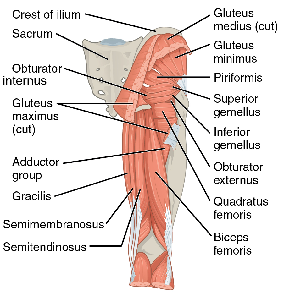
Diagram: the muscles of the gluteal region. Note that the gluteus medius and gluteus maximus have been cut away to show the deep muscles.
OpenStax College, Gluteal Muscles that Move the Femur, CC BY 3.0. Complete figure retained; the label “Obturator internis” has been corrected to “Obturator internus” for this version. Previous article adaptation by Sachin Sudhakaran.
Edited by: Dr. Maddie Swannack
Reviewed by: Dr. Ben Appleby and Dr. Marcus Judge
- 14092