

By Dr. Thomas Burnell and Dr. Bethany Turner
Next Lesson - Cardiac Conduction and Contraction
Core
The heart must change its output (cardiac output = CO) if there is a change in total peripheral resistance (TPR) in order to maintain blood pressure (BP).
Mean arterial BP ≈ TPR x CO
For example, when metabolism in the muscles increases during exercise, blood vessels dilate to supply muscles with more blood. This causes a fall in TPR which leads to a fall in arterial pressure (as per the above equation). The heart responds by increasing the cardiac output to raise the BP.
CO = stroke volume x heart rate
The above equation shows how CO is calculated to be able to calculate mean arterial BP.
- Stroke Volume (SV) - determined by:
- The Afterload - the blood/pressure that the ventricles pump against.
- The Preload - the amount that the ventricles fill in diastole.
- Heart Rate (HR) - determined by:
- The autonomic nervous system (ANS).
The Frank-Starling law describes how greater ventricular filling produces a stronger contraction and a larger stroke volume within the physiological range. This helps the healthy heart match its output to venous return.
The relationship between end-diastolic volume (EDV) and stroke volume (SV) depends on contractility and afterload. At a given contractile state and afterload, increasing EDV usually increases SV within the physiological range. The first figure below uses end-diastolic pressure (EDP) as an index of ventricular filling.
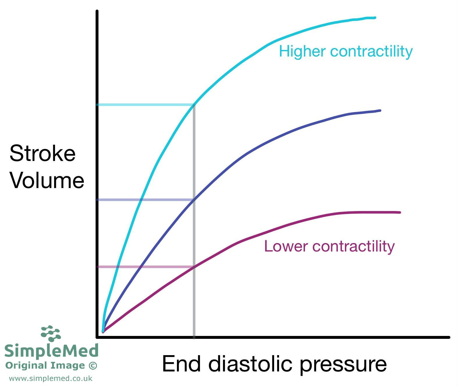
Diagram - How the stroke volume changes with the end diastolic pressure
SimpleMed original by Dr. Bethany Turner
Heart contractility can be altered by stimulation from the sympathetic nervous system. This causes the ventricles to pump harder, leading to an increased SV at a given EDV (as seen in the diagram above).
Filling stretches the sarcomeres before contraction. Within the physiological range, greater length increases force through length-dependent activation, including increased calcium sensitivity of the contractile proteins. A failing ventricle may gain little extra stroke volume from further filling. This impaired response should not be explained simply as actin and myosin losing contact.
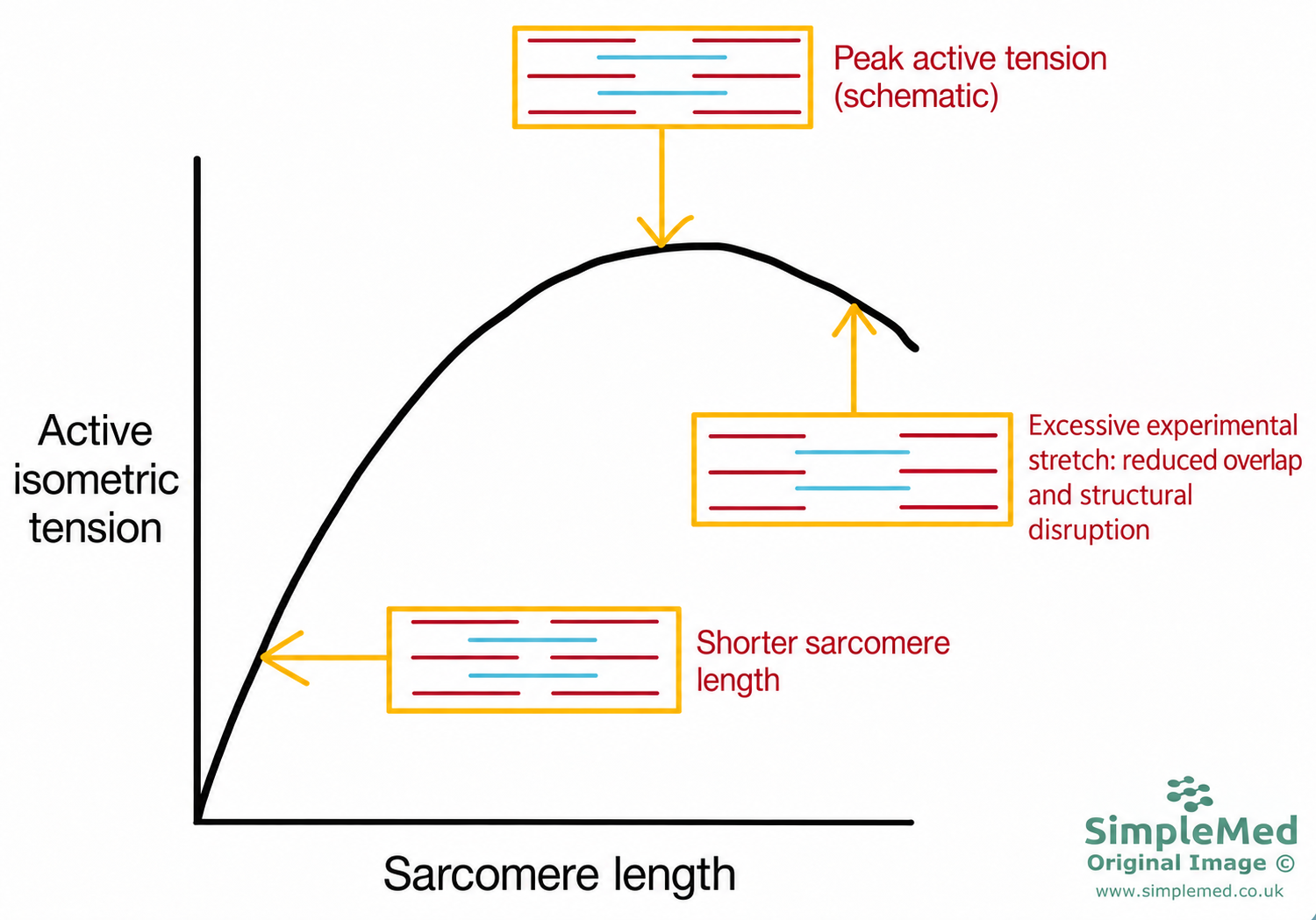
Diagram - Schematic muscle length-tension relationship. Active tension can fall with excessive experimental stretch through reduced filament overlap and structural changes. This is not a ventricular stroke-volume curve or an explanation of ordinary congestion. Physiological cardiac length-dependent activation includes increased calcium sensitivity.
SimpleMed original by Dr. Bethany Turner
Autonomic Control of the Heart
The autonomic nervous system is important for many physiological functions and for control of the body systems. In the CVS it controls the heart rate, force of contraction, and peripheral resistance of blood vessels.
The parasympathetic nervous system (PNS) control of the heart is via the Vagus nerve. The Vagus has pre-ganglionic fibres which synapse with the post-ganglionic fibres on the epicardial surface, or within heart walls at the SA and AV node. The post-ganglionics release Acetylcholine (ACh) which acts on M2-receptors causing a negative chronotropic effect (slowing of the heart rate).
- PNS causes a negative chronotropic effect by decreasing the slope of the pacemaker potential. It does this by increasing potassium ion conductance and decreasing cAMP.
The sympathetic nervous system (SNS) control of the heart is via post-ganglionic fibres from the sympathetic trunk. It innervates the SA node, AV node and myocardium. The post-ganglionics release noradrenaline (NA) which acts on β1-adrenoreceptors, causing a positive chronotropic (the heart beats faster) and positive inotropic effect (heart beats with more force).
Sympathetic stimulation increases cyclic AMP (cAMP) in SA node cells. cAMP binds directly to hyperpolarisation-activated, cyclic nucleotide-gated (HCN) channels, which carry the mixed Na+/K+ funny current (If), and shifts their activation towards more positive voltages. Greater inward pacemaker current helps the cells reach threshold sooner, increasing the rate at which they fire.
cAMP also activates protein kinase A (PKA). In ventricular myocytes, this enhances calcium entry and calcium release from the sarcoplasmic reticulum, increasing the force of contraction.
Sympathetic stimulation also speeds calcium reuptake into the sarcoplasmic reticulum and ventricular relaxation. Faster relaxation helps the ventricles fill when heart rate is higher. This is a separate effect: the faster pacemaker rhythm sets heart rate, rather than faster ventricular relaxation causing the SA node to fire sooner.

Diagram - The innervation to the heart and the effects it has on the heart
Creative commons source by OpenStax College [CC BY 3.0 (https://creativecommons.org/licenses/by/3.0)]
Autonomic Control of Vasculature
The ANS constantly releases NA at low levels to cause a small amount of vasoconstriction of blood vessels. This is the normal vasomotor tone of the blood vessels. This slight sympathetic activity allows the blood vessels to contract or dilate depending on the needs of the tissue by increasing or decreasing the level of NA. The NA acts on α1-adrenoreceptors present on most arteries and veins to cause some vasoconstriction to maintain the vasomotor tone.
Noradrenaline activates α1-adrenoceptors, stimulating calcium release mediated by inositol trisphosphate (IP3) from the sarcoplasmic reticulum. Calcium binds calmodulin, which activates myosin light chain kinase (MLCK). MLCK adds phosphate groups to myosin light chains, enabling smooth muscle contraction and vasoconstriction.
The same receptor pathway produces diacylglycerol (DAG), which activates protein kinase C (PKC). PKC can promote phosphorylation of the inhibitory protein CPI-17, reducing myosin light chain phosphatase (MLCP) activity through this downstream signal. MLCP normally removes phosphate groups from myosin light chains; it does not directly inhibit MLCK. Reducing MLCP activity helps sustain myosin phosphorylation and contraction at a given calcium concentration, a process called calcium sensitisation.
Adrenaline stimulates both α- and β-adrenoceptors. At low concentrations, β2-mediated relaxation can predominate in vascular beds with these receptors. As concentration rises, α-mediated vasoconstriction becomes more prominent and can dominate the overall vascular response. This changes the balance of effects; it does not switch off β-receptor activity.
In anaphylaxis, intramuscular adrenaline is the first-line treatment. Its α effects help reverse peripheral vasodilation and reduce tissue oedema, while its β effects support cardiac contraction and dilate the airways. These complementary actions explain its benefit, rather than a selective switch to α1 receptors.
Baroreceptors are located in the aortic arch and carotid sinus (located at the bifurcation of the common carotid arteries).
When blood pressure is increased, the baroreceptor nerve endings are stretched which increases their activity. This information is sent to the medullary control centre in the brainstem, which sends out a signal via the ANS to decrease heart rate and contractility, therefore decreasing BP.
With sustained hypertension, baroreceptors can adapt so that their response range shifts towards higher blood pressures. This is called resetting: it changes the relationship between blood pressure and the rate at which the receptors send nerve impulses, rather than switching the receptors off. They still fire and respond to short-term rises and falls in pressure, helping to limit sudden fluctuations. However, the reflex does not by itself restore persistently raised blood pressure to its previous level.
Reviewed by: Dr. Marcus Judge
- 19527

