

Next Lesson - Protein Function in Oxygen Transport
Core
Proteins are found throughout the human body and play an important role in many biochemical processes:
- Catalysts - enzymes
- Transporters - haemoglobin for transport of oxygen
- Structural support - collagen
- Immune system - immunoglobulins (antibodies)
- Ligands in cell signalling (molecules that bind to receptors) - protein and peptide hormones, such as insulin, and neuropeptides (neurotransmitters made of amino acids joined by peptide bonds)
- Ion channels and receptors - for hormones and neurotransmitters
Proteins are made of one or more polypeptides. A polypeptide is a chain of covalently linked amino acids (monomers). The amino acid sequence of a protein is encoded by a gene. Therefore, the nucleotide sequence of a gene determines the sequence of amino acids in the protein. The final polypeptide chain folds into a highly-specific and intricate three-dimensional structure, which is determined by the sequence of amino acids.
There are different names to describe the sizes of peptides & proteins:
- Peptides or oligopeptides = relatively small polymers with a low number of amino acid residues (amino acids once they are joined into a chain).
- Polypeptides or proteins = large polymers with large numbers of amino acid residues.
Amino Acids Chemical Structure
Amino acids are the building blocks of proteins as the monomers that the final protein structure is made out of. They all have a general structure of central carbon atom covalently bonded to an amino group (NH2), carboxyl group (COOH), a hydrogen atom and distinctive R group (the side chain).
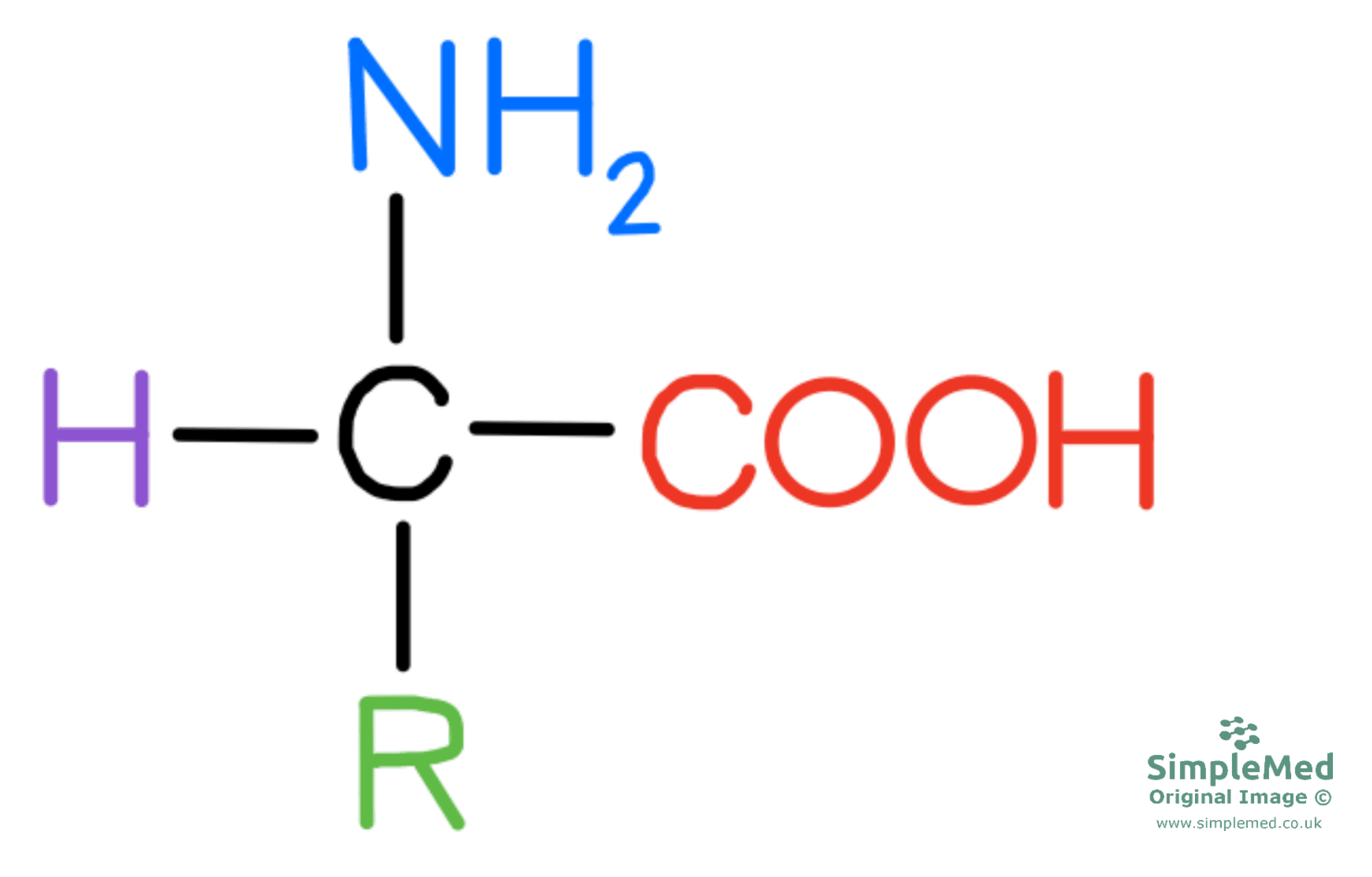
Diagram - The general structure of an amino acid
SimpleMed original by Dr. Peter Parkinson
Amino acids become ionised because the amino group can act as a base (proton acceptor) and the carboxyl group can act as an acid (proton donor). In water (an aqueous solution), whether each group carries an extra H+ (its protonation state) depends on the balance between its acid and base forms in the surrounding solution. The amino group can act as a proton acceptor because the nitrogen atom has a lone pair of electrons (a pair in its outer shell that is not involved in bonding), which can bond to the proton. This means that the amino group goes from NH2 to NH3+, and the carboxyl group transitions from COOH to COO-.
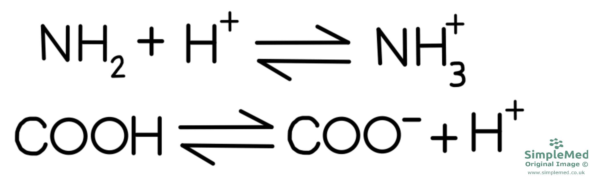
Diagram - How the amino and carboxyl groups exchange a H+ ion to become charged
SimpleMed original by Dr. Peter Parkinson
When the amino acid has one negative charge and one positive charge, the net charge of the amino acid is zero and this is known as a zwitterion. Depending on the pH of the solution, the amino acid can exist as a cation (overall positive charge) or as an anion (overall negative charge).
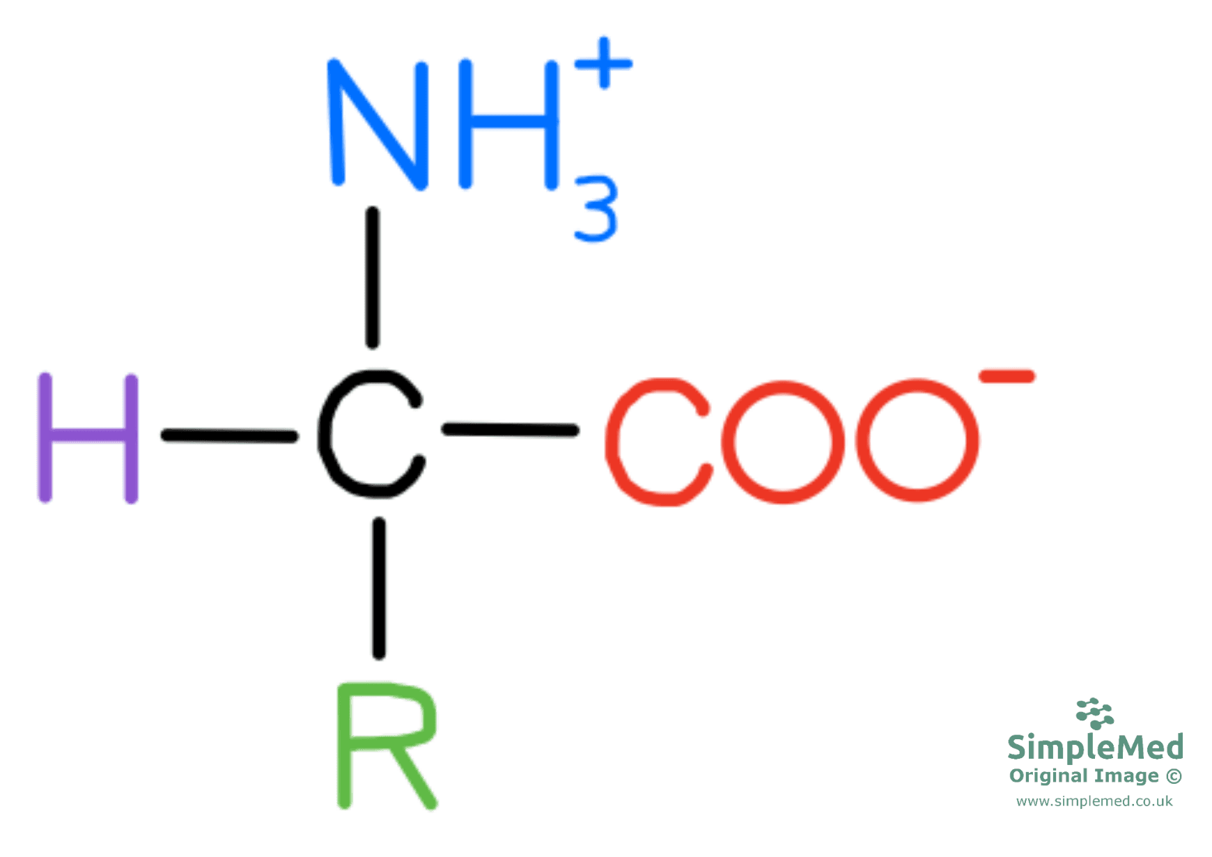
Diagram - The structure of a zwitterion
SimpleMed original by Dr. Peter Parkinson
A peptide bond forms between individual amino acids to connect them. The remaining part of each amino acid now left in the amino acid chain is called the amino acid residue. The peptide bond is formed in a condensation reaction as a single molecule of H2O is produced with each peptide bond formed.
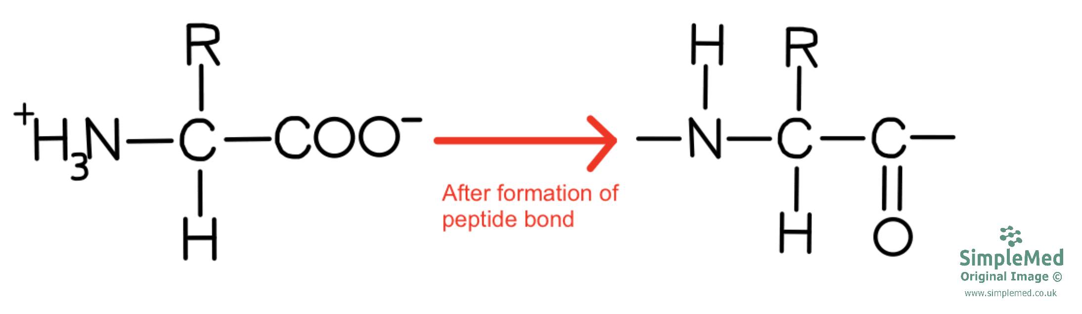
Diagram - The general appearance of an amino acid residue
SimpleMed original by Dr. Peter Parkinson
Amino acids are classified based on their R group which contributes to the overall function of the protein. The acid-base behaviour of the protein is largely determined by the R groups of its amino acids. The free amino group at one end of the chain and the free carboxyl group at the other end also carry charge.
Chemical properties of amino acid:
- Polar (hydrophilic) - able to form hydrogen bonds and can be further classified into positive, negative or neutral charge
- Non-polar (hydrophobic)
- Acidic
- Basic
- Neutral
Physical properties of amino acid:
- Aliphatic - R group just contains carbon and hydrogen atoms
- Aromatic - R group containing a flat, ring-shaped (cyclic, planar) structure that is very stable, e.g. the R group on phenylalanine
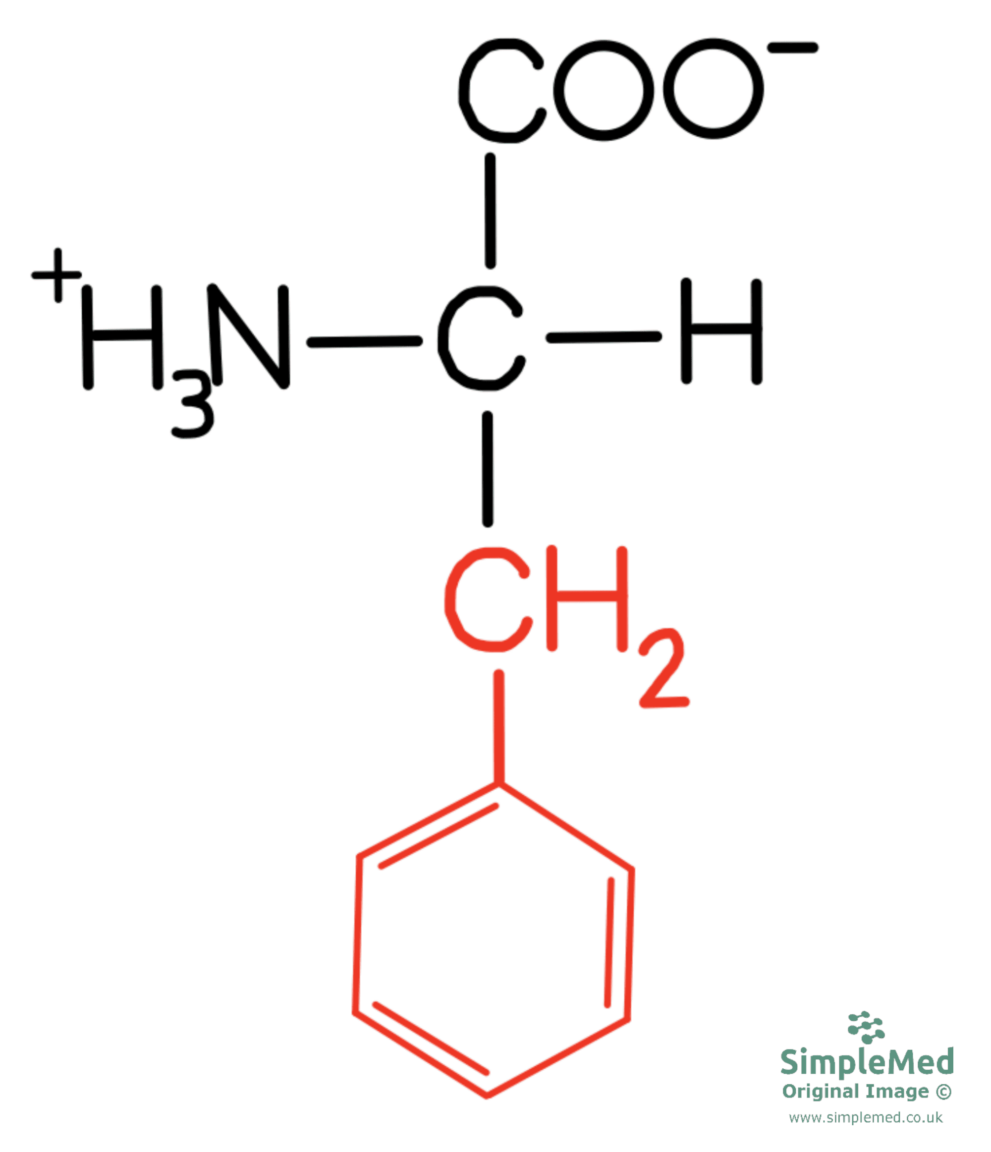
Diagram - The chemical structure of phenylalanine with its R group in red
SimpleMed original by Dr. Peter Parkinson
pKa and R Groups of Amino Acids
pKa is the pH at which an ionisable group is 50% protonated and 50% deprotonated, and is a measure used to indicate the strength of an acid. Whether an amino acid is positively or negatively charged, depends on the pKR (pK of the side group) value of the side group.
Positively charged R groups with pKR values:
- Lysine - 10.5
- Histidine - 6.0
- Arginine - 12.5
Negatively charged R groups with pKR values:
- Glutamate - 4.3
- Aspartate - 3.9
If the pH value of the solution < pKR value then the side group will be protonated (addition of hydrogen ions).
If the pH value of the solution > pKR value then the side group will be de-protonated (loss of hydrogen ions).
Examples:
- Lysine has a pKR of 10.5, therefore at physiological pH (7.4) the R group of lysine will be protonated.
- Aspartate has a pKR of 3.9, therefore at physiological pH (7.4), the R group of
aspartate will be de-protonated.
In summary: an ionisable group (one that can gain or lose H+) behaves differently on either side of its pKa. If the pH of the solution is lower than the pKa, there are many free protons. The group accepts them and becomes protonated. If the pH is higher than the pKa, there are fewer free protons. The group donates its protons and becomes de-protonated. Near its pKa, the group acts as a buffer (it resists changes in pH).
Definitions:
- Primary structure = linear amino acid sequence of polypeptide chain.
- Secondary structure = local folding of the polypeptide backbone (the repeating chain of atoms that runs along the polypeptide, without the R groups) due to interactions between atoms of the backbone. The two most common forms are the alpha helix and the beta-pleated sheet. Both are held in shape by hydrogen bonds formed within the structure.
- Tertiary structure = the overall three-dimensional configuration of a protein. It forms based on the interaction between the R groups of the amino acids within the protein.
- Quaternary structure = the interaction between multiple polypeptides and prosthetic groups (non-protein parts that stay attached to the protein) to form a multi-subunit protein. For example, haemoglobin is made up of four polypeptide chains, each with a haem prosthetic group that contains an iron ion.
The bond formed by linking two amino acids together is called a peptide bond and the reaction to form this bond is a condensation reaction - removal of a molecule of water.

Diagram - The formation of a peptide bond by a condensation reaction
SimpleMed original by Dr. Peter Parkinson
Characteristics of the peptide bond include:
- Planar - the carbon, hydrogen, oxygen and nitrogen atoms all lie in the same plane.
- Rigid - the carbon-nitrogen bond in the peptide bond has partial double-bond characteristics making the bond relatively rigid, with restricted rotation. This helps to keep the atoms in the same plane.
- Exhibits a trans conformation - the two alpha carbons (the carbons that carry the R groups) next to the peptide bond lie on opposite sides of it. The trans form is the usual one in proteins because it reduces steric hindrance (crowding between bulky side chains). The cis form, with both alpha carbons on the same side, is less common and is sometimes seen next to proline. Flexibility of the polypeptide arises from rotation around the bonds adjacent to the peptide bond.
The bonds either side of a peptide bond are able to rotate, allowing flexibility within the 3-D structure of the protein.
The way in which the polypeptide chain folds and the physical characteristic of the protein is determined by the sequence of the amino acids.
The overall structure of a protein determines the overall function of the protein.
Isoelectric point (pI) of a protein is the pH at which there is no overall net charge amongst the structure of the protein.
Basic proteins:
- pI > 7
- The protein contains many positively charged/basic amino acids - based on their R groups. These R groups will be protonated.
Acidic proteins:
- pI < 7
- The proteins contains many negatively charged/acidic amino acids.
Examples of proteins and their pI:
- Pepsin (a protease, an enzyme that breaks down proteins, found in the stomach) - pI of < 1.
- Haemoglobin (protein found in red blood cells that carries oxygen) - pI of 6.8.
- Cytochrome C (protein that carries electrons in the mitochondria in process of oxidative phosphorylation) - pI of 10.7.
Proteins can be joined to chemical components other than amino acids, either covalently (by strong bonds that share electrons) or non-covalently (by weaker attractions). Examples:
- Lipoproteins - lipid non-covalently associated with protein. E.g. LDL and HDL.
- Glycoproteins - carbohydrate covalently linked to protein E.g. Immunoglobulin.
- Phosphoproteins - phosphate group covalently linked to protein. E.g. casein in milk.
Secondary Structure of a Protein
The bonds involved in the secondary structure of a protein are hydrogen bonds.
- Polypeptide chain folded in a right-handed spiral conformation.
- The structure is stabilised by hydrogen bonds between N-H of one amino acid residue and C=O group of an amino acid residue four residues away.
- Not every polypeptide chain forms an alpha helix structure.
- Some residues favour a helix more than others. Alanine and leucine are strong helix formers, whereas glycine and proline are poor helix formers.
- Example: Ferritin (iron storage protein) - majority of the protein is alpha helix structure.
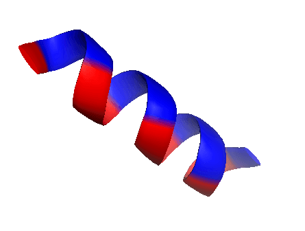
Diagram - The structure of an alpha helix
Creative commons source by Linnikh [CC BY-SA 4.0 (https://creativecommons.org/licenses/by-sa/4.0)]
- In beta-pleated sheets, the polypeptide chains are nearly fully stretched out which allows the R-groups to alternate between opposite sides of the chain.
- When there are two or more of these stretched out chains (beta strands) side by side, hydrogen bonds can form between the strands to give an almost 2-D sheet that is pleated.
- Neighbouring beta strands can run in the same direction (parallel) or in opposite directions (anti-parallel).
- Example: the receptor on a T cell (a type of lymphocyte, a white blood cell).
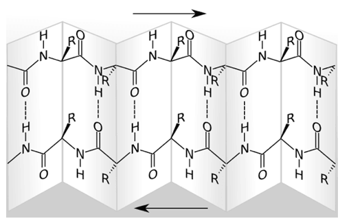
Diagram - An anti-parallel beta-pleated sheet
Creative commons source by Roland.chem [CC BY-SA 4.0 (https://creativecommons.org/licenses/by-sa/4.0)]
Tertiary Structure of a Protein
The tertiary structure of a protein is the three-dimensional structure of a single polypeptide chain. The chain folds so that the structure is as stable as possible. A number of forces hold the chain in this shape, and they form from interactions between the R groups of its amino acids.
The forces include:
- Hydrogen bonds.
- Ionic bonds - between R groups of opposite charge.
- Hydrophobic interactions - non-polar, hydrophobic R groups cluster together within the inside of the protein molecule.
- Disulphide bonds - covalent bonds formed between cysteine amino acid residues. These are the strongest bonds that contribute to the tertiary structure of a protein.
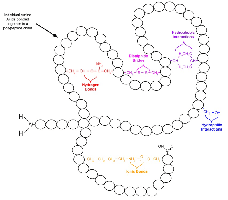
Diagram - The bonds formed in the tertiary structure of a protein
Creative commons source by WikiComTD, edited by Dr. Peter Parkinson [CC BY-SA 4.0 (https://creativecommons.org/licenses/by-sa/4.0)]
A protein’s overall tertiary structure indicates its function. Two broad classes are fibrous proteins and globular proteins. Membrane proteins, which sit in cell membranes, have their own section below.
- Have a role of support, protection and shape.
- Made up of single type of repeating secondary structure & long strands or sheets.
- Example: Collagen.
- Have a role of regulation and catalysis.
- Made up of several types of secondary structure.
- Form compact shapes.
- Example: Enzymes such as alcohol dehydrogenase.
When a polypeptide chain folds, the hydrophobic (non-polar) side chains are buried in the middle of the protein. The hydrophilic (polar) side chains are left on the surface. They interact with water molecules when the protein is placed in water (an aqueous solution).
Membrane proteins sit in or on the cell membrane. They make up about a quarter of human protein-coding genes. There are two main groups:
- Integral membrane proteins are firmly embedded in the lipid bilayer (the double layer of phospholipids that forms the membrane), and many span it from one side to the other. They have one or more hydrophobic regions that sit among the fatty acid tails of the phospholipids. The membrane-spanning part may be one or more alpha helices, or a beta sheet that forms a channel.
- Peripheral membrane proteins are attached to the surface of the membrane, either to integral proteins or to phospholipids.
This is the reverse of a water soluble protein. The hydrophobic regions face the fatty acid tails. The hydrophilic regions stick out of the membrane into the watery fluid on either side.
Membrane proteins act as receptors, channels and carriers (transporters). Receptors bind signals such as hormones and growth factors on the outside of the cell. Channels and carriers help substances to cross the membrane.
Quaternary Structure of a Protein
Some proteins are made up of two or more polypeptide chains that associate together; this is the quaternary structure of a protein.
One example of a protein with quaternary structure is haemoglobin. A single molecule of haemoglobin is made up of four subunits - 2 alpha chains and 2 beta chains that interact with each other. Each chain carries a haem group, which is a prosthetic group (a non-protein part attached to the protein). Haem is a carbon-based ring (a protoporphyrin ring) with a single iron ion at its centre. The iron ion is crucial for binding oxygen to be carried around the body.
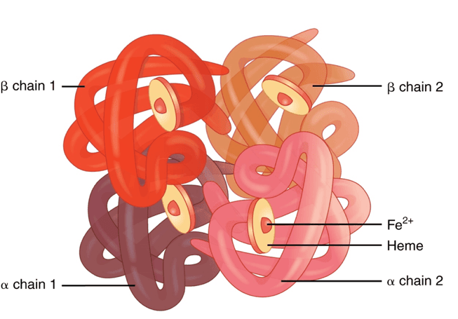
Diagram - The quaternary structure of haemoglobin
Creative commons source by OpenStax College [CC BY-SA 4.0 (https://creativecommons.org/licenses/by-sa/4.0)]
Another example is collagen. Collagen is a fibrous protein formed of three polypeptide chains wound around each other in a triple helix, with hydrogen bonds between the chains maintaining the structure. Glycine, whose side chain is a single hydrogen atom, is every third amino acid in each chain, so it fits into the middle of the helix. Proline and hydroxyproline (proline with an added hydroxyl group) make up many of the other positions. The many hydrogen bonds between the chains, some of them through water molecules, give the molecule great strength. The collagen molecules form covalent cross links with other collagen molecules to further increase the strength of the protein. The cross links are staggered to increase the stability of the molecule.
Collagen is further discussed in our Post-Translational Modification and Collagen Biosynthesis article.
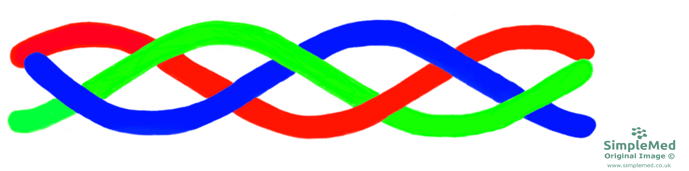
Diagram - The quaternary structure of collagen
SimpleMed original by Dr. Peter Parkinson
Any disruption to the protein’s structure is described as denaturation and involves breaking any of the forces that hold the structure of the protein together.
Heat
- Heat increases the kinetic energy within the molecule and leads to the molecules vibrating rapidly causing disruption to non-covalent interactions such as hydrogen and ionic bonds.
- Extreme heat is used in the process of sterilisation to denature proteins in microorganisms leading to their destruction.
pH
- A change in the pH of the solution the protein is placed in, will affect the ionisation states of the amino acids present within the polypeptide.
- This alteration will change the ionic and hydrogen bonds present within the protein.
Detergents and Organic Solvents
- Detergents, such as SDS (sodium dodecyl sulphate), have a hydrophobic (water-repelling) end and a hydrophilic (water-loving) end.
- The hydrophobic end binds the hydrophobic amino acids that normally sit inside the protein and help to keep its structure together. The detergent therefore acts to pull the protein apart and denature the protein’s shape. Strong detergents such as SDS do this efficiently. Milder detergents are used to lift membrane proteins out of the cell membrane, although they can still destabilise them.
- Organic solvents (carbon-based liquids) are a separate group of denaturants. They also disrupt hydrogen bonds and hydrophobic interactions.
Proteins within the human body can become misfolded for a number of reasons such as genetic defects and environmental factors. These misfolded proteins can then lead to diseased states.
In Alzheimer’s disease, one misfolded form of beta-amyloid protein is thought to be toxic when it builds up in the brain. Beta-amyloid is a fragment cut from a larger protein called the amyloid precursor protein. The fragments clump together to form amyloid plaques, which collect between neurones. There is also abnormal accumulation of tau protein. Tau normally binds to and stabilises microtubules (tiny tubes in the cytoskeleton that help guide nutrients and molecules along the neurone). Abnormal chemical changes make tau detach from the microtubules and stick together. It forms neurofibrillary tangles inside the neurones. These tangles disrupt the normal functioning of neurones, contributing to impaired transport mechanisms and synaptic communication. The amyloid plaques and tau tangles can affect neural function in the brain and eventually lead to neural cell death.
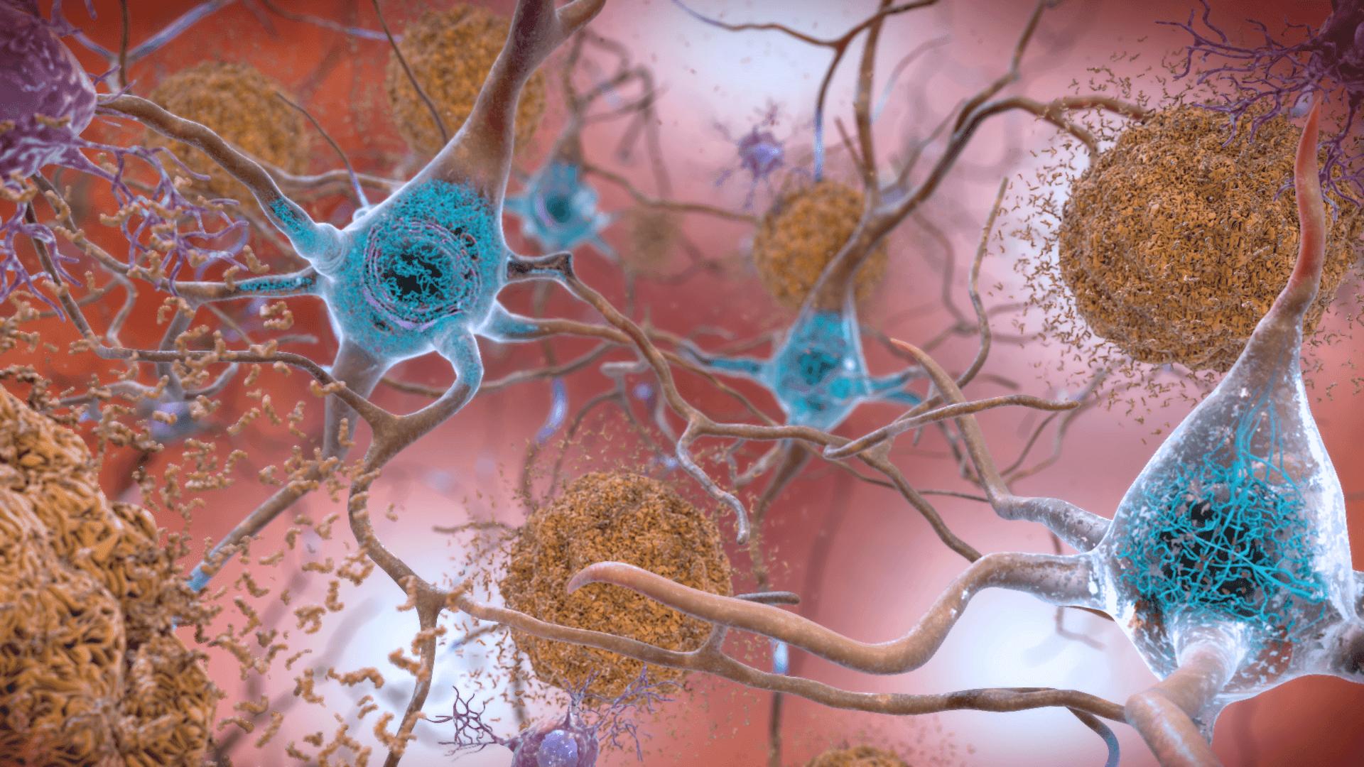
Image - Beta amyloid proteins (seen in brown) and tau proteins (seen in blue) in Alzheimer's Disease
Creative commons source by National Institute on Aging, NIH [CC BY-SA 4.0 (https://creativecommons.org/licenses/by-sa/4.0)]
Edited by: Dr. Ben Appleby
Reviewed by: Dr. Thomas Burnell and Dr. Marcus Judge
- 13994



