

By Dr. Thomas Burnell and Dr. Bethany Turner
Next lesson - Control of Cardiac Output
Core
Blood flow within a vessel can be described in two ways, either laminar or turbulent:
- Laminar flow is where all the blood is flowing in one direction within a blood vessel. The blood flowing in the centre of the blood vessel is flowing faster than the blood closer to the edge (a parabolic profile). This can be thought of as circular layers of blood in a vessel following at different speeds.
- Turbulent flow involves chaotic mixing rather than orderly layers. High flow velocity, low blood viscosity and disturbed vessel geometry favour turbulence. A narrowing can create a fast jet, while an irregular surface or branch point can disturb the flow. Increased resistance alone, including in hypertension, does not establish that flow is turbulent. Turbulence increases energy loss, so a greater pressure difference is needed to maintain a given flow.
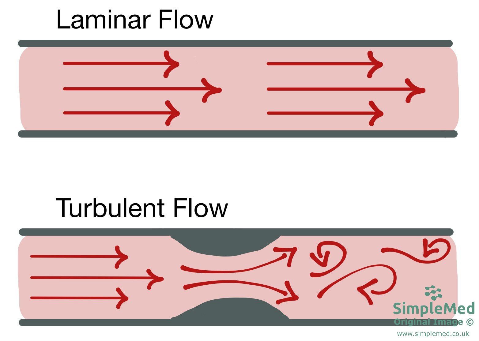
Diagram - Diagrammatic depiction of both laminar and turbulent blood flow - the arrows represent separate blood flow streams. In the second diagram, after the obstruction the flow becomes turbulent
SimpleMed original by Dr. Thomas Burnell and Dr. Bethany Turner
When feeling a pulse, we feel the pressure wave being transmitted before the blood arrives.
Type of pulse rhythm:
- Regular - lub dub lub dub...
- Regularly irregular - the beat is abnormal but the pattern is repeated at set intervals. E.g. 2nd degree heart block.
- Irregularly irregular - the beat is abnormal and does not follow a pattern. E.g. ventricular/atrial ectopics and atrial fibrillation.
Type of pulse rate:
- Bradycardia - where the heart rate is less than 60 beats per minute.
- Tachycardia - where the heart rate is more than 100 beats per minute.
Strength of the pulse:
- Pulse strength is influenced by pulse pressure, the difference between peak systolic arterial pressure and diastolic arterial pressure. E.g. a blood pressure of 120/80 mmHg has a pulse pressure of 40 mmHg (i.e. 120-80=40).
- In atrial fibrillation, disorganised atrial electrical activity replaces effective coordinated atrial contraction. Irregular impulse transmission through the atrioventricular (AV) node gives unequal intervals between ventricular beats. The varying filling time contributes to changes in stroke volume and pulse strength from beat to beat.
- In ventricular ectopics there is an early beat in the cycle caused by the ventricle spontaneously depolarising. This means that there is a longer pause before the next beat. This means the ventricle has more time to fill and so more blood is pumped out causing a stronger pulse.
Pulse Character/Quality:
- Thready pulse - a weak pulse that may accompany reduced stroke volume and a narrow pulse pressure.
- In LV failure the heart cannot beat as strongly so systolic pressure is lower.
- Bounding Pulse - felt in patients where the diastolic pressure is lower. It is felt as a strong pulse.
- Bounding pulse is associated with low peripheral resistance. A dilated vessel gives less resistance to blood flow so diastolic pressure is lower.
- In bradycardia the time between beats is longer allowing more time for diastole so the diastolic pressure drops further widening the pulse pressure.
Raising the arm above the level of the heart drains the venous blood in the arm quicker, lowering the peripheral resistance. This increases the difference between the diastolic and systolic pressures, resulting in an increased pulse pressure. This can be used to exaggerate a bounding pulse.
Locations of Common Pulses
Radial Pulse - felt on the radial side of the wrist.
Brachial Pulse - felt on the medial side of the biceps tendon at the inside of the elbow.
Carotid Pulse - under the jaw anterior to the sternocleidomastoid muscle. Palpate one side at a time only.
Femoral Pulse - felt just inferior to the mid-inguinal point (halfway between ASIS and the pubis).
Popliteal Pulse - felt deep in popliteal fossa (this is a hard pulse to find).
Pedal Pulses:
- Dorsalis Pedis Pulse - lateral to extensor hallucis longus tendon.
- Posterior Tibial Pulse - behind medial malleolus.
The mean arterial pressure can be calculated by either of the following equations:
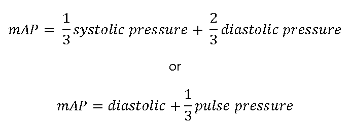
If this falls below around 70 mmHg then organ perfusion can be impaired, depending on the clinical context.
The jugular venous pressure (JVP) is used to measure the pressure in the venous system as the blood is returned to the right atrium. The right JVP is used as it has a straighter course to the right atrium and more reliably reflects right atrial pressure than the left.
This pulse can be seen in the neck when the patient lies at a 45° angle and looks to the left. It should be seen between the two heads of the sternocleidomastoid muscle, just above the clavicle. The JVP is measured as the height of the pulse to the sternal angle. It is measured in cmH2O. It may also be measured using a central line inserted into either the internal jugular vein or superior vena cava and allows a waveform to be measured.
Normally the JVP should not be more than about 3-4 cm above the sternal angle.
Conditions where the JVP can be raised include: right-sided heart failure, cardiac tamponade and volume overload.
- Right-sided heart failure and cardiac tamponade have a raised JVP as the heart is not working as an effective pump. This leads to blood building up in the venous system and raises the venous pressure.
- Volume overload causes an increased volume of blood in the venous system, which raises the venous pressure.
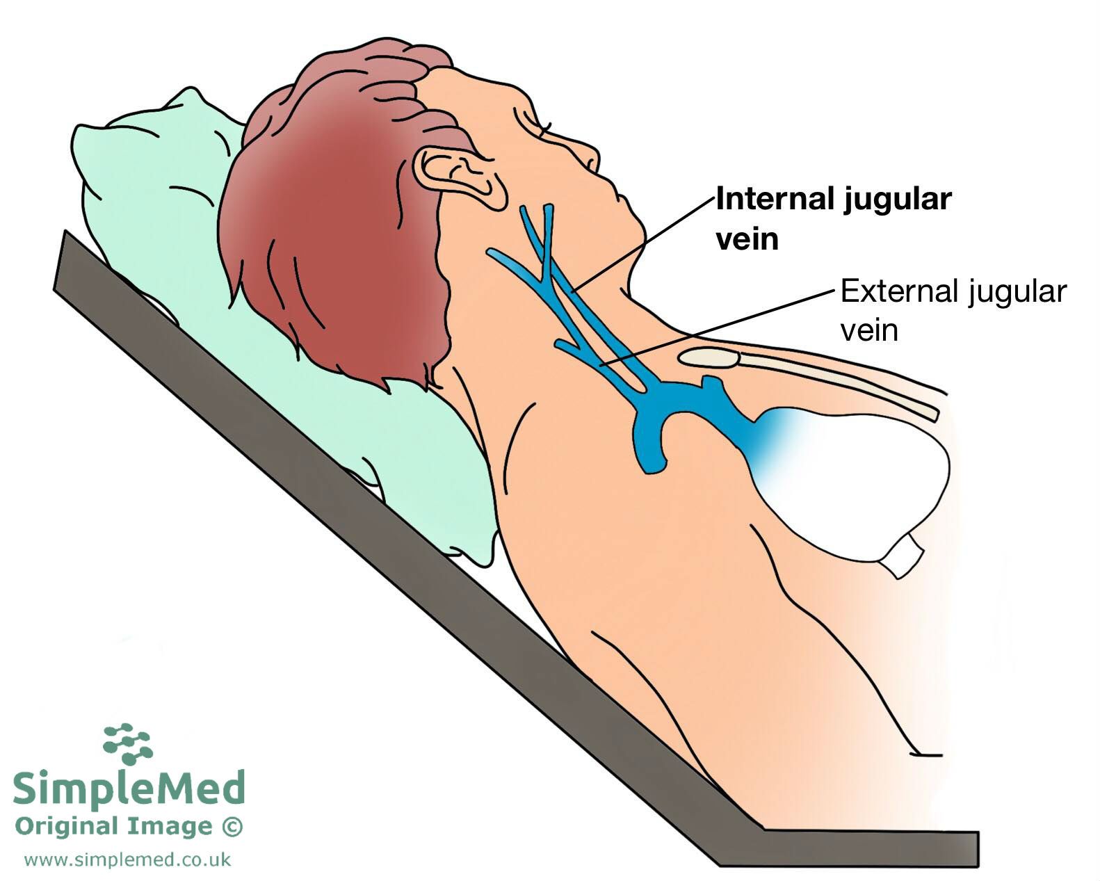
Diagram - The measurement of jugular venous pressure in a clinical setting. Note the 45 degree angle the patient is placed at
SimpleMed original by Dr. Bethany Turner
Reviewed by: Dr. Marcus Judge
- 24775
