

Next Lesson - Dermatomes and Myotomes
Core
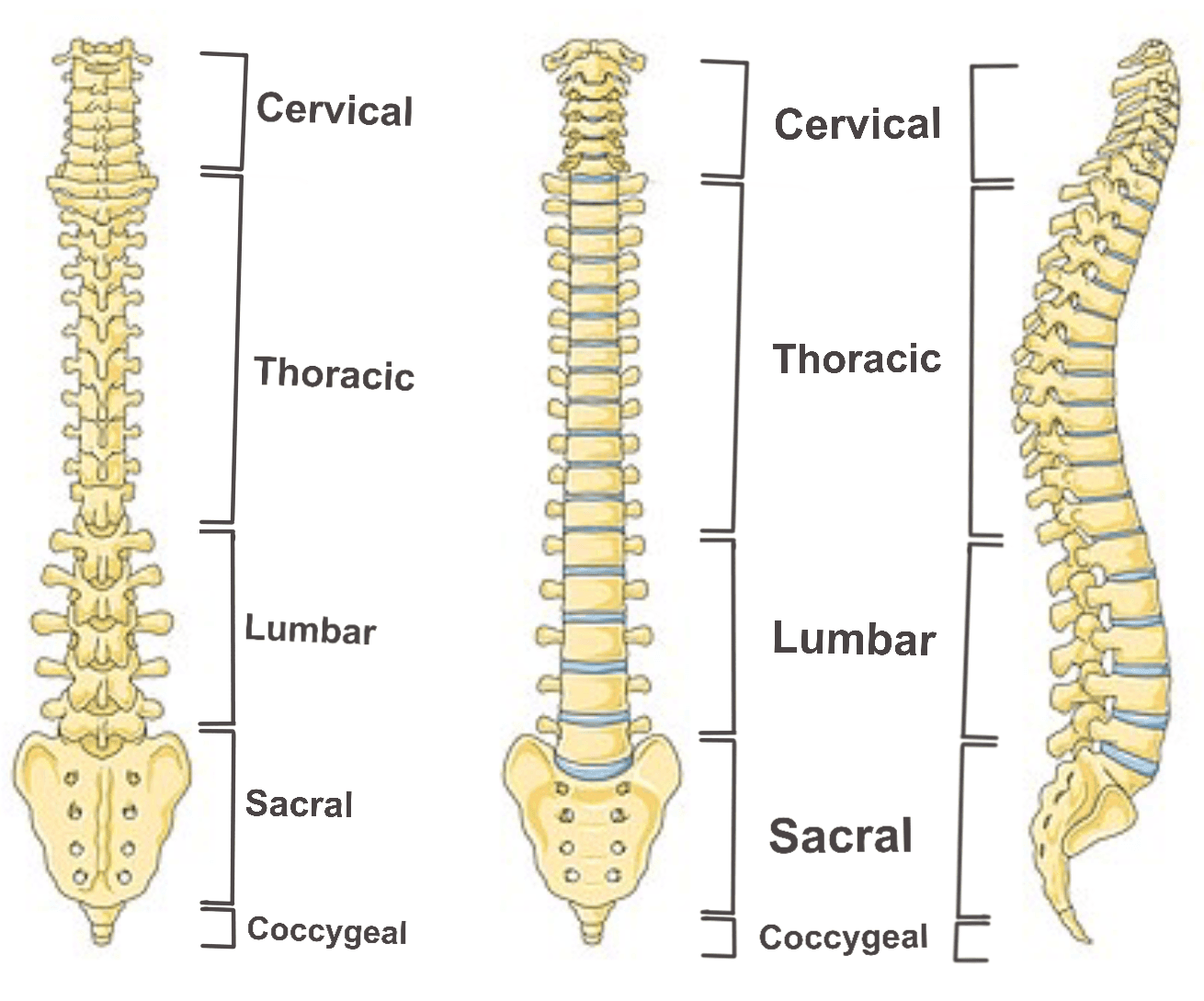 Diagram - Regions of the vertebral column
Creative commons source by Laboratoires Servier [CC BY-SA 4.0 (https://creativecommons.org/licenses/by-sa/4.0)]
The anterior part of the vertebrae is the vertebral body. Vertebral bodies increase in size as they lie more inferiorly in the spine, as the lower vertebrae support more weight. The vertebrae are separated by intervertebral discs.
The posterior part of a vertebra is formed by the vertebral arch. This arch forms the rear of the vertebral foramen. The foramina line up to form the vertebral canal which contains the spinal cord and cauda equina. There are also several bony landmarks to allow for attachment of ligaments and muscles.
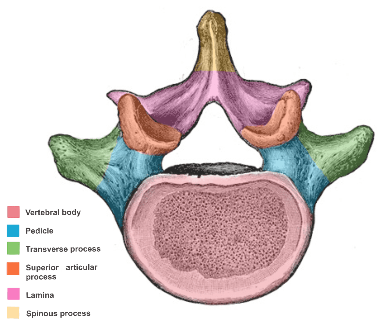
Diagram - Labelled structure of lumbar vertebrae
Public Domain Source Anatomy of the Human Body, by Henry Gray, edited by Dr. Emily Smith [Public domain]
Ligaments of the Vertebral Column
Ligaments around the vertebral body:
- The anterior longitudinal ligament runs along the anterior aspect of the vertebral bodies and prevents hyperextension of the vertebral columns.
- The posterior longitudinal ligament runs along the posterior aspect of vertebral bodies and prevents hyperflexion of the vertebral column.
Ligaments around the vertebral arch:
- Ligamentum flavum is located between the laminae of vertebrae.
- Interspinous ligaments are located between spinous processes.
- Supraspinous ligament runs along the tips of the spinous processes.
- Intertransverse ligaments are located between transverse processes.
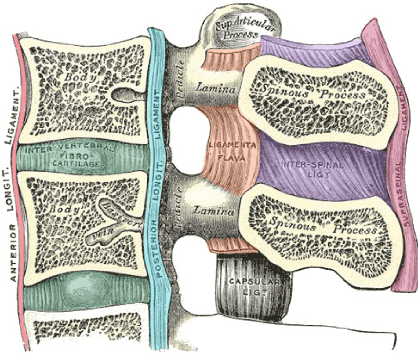
Diagram - The ligaments of the vertebral column
Public Domain Source Anatomy of the Human Body, by Henry Gray, edited by Dr. Emily Smith [Public domain]
Joints of the Vertebral Column
The joints between vertebral bodies are cartilaginous joints; they articulate with each other indirectly via intervertebral discs. These joints allow the weight bearing property of the vertebral column.
The intervertebral discs have two distinct regions:
- Annulus fibrosus is the outer region which is very strong and is a major shock absorber.
- Nucleus pulposus is the inner softer region.
Articular facets are joints between the vertebrae which allow movement. There is one on either side of the vertebrae.
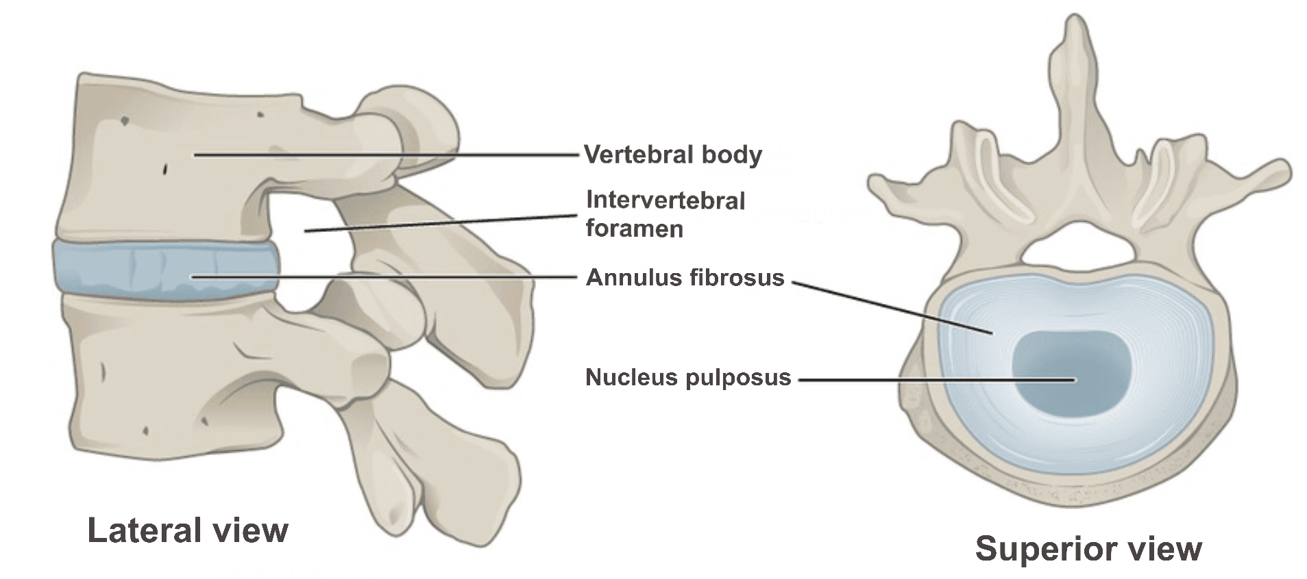
Diagram - The structure of intervertebral discs
Creative commons source by OpenStax College [CC BY-SA 4.0 (https://creativecommons.org/licenses/by-sa/4.0)]
The cervical spine is positioned in the neck, and is the most superior part of the spine. It is made up of 7 vertebrae (C1-C7). The c-spine is a very mobile region of the spine, with movements including flexion, extension, rotation and lateral flexion. The two most superior vertebrae, atlas and axis, have some unique features which distinguish them from the other vertebrae.
The nerve roots of the spinal cord at this level exit above their respective vertebra (for example the C1 nerve root is above the C1 vertebra), until the C7/T1 vertebrae. The C8 nerve root exits between C7/T1. Below this point the nerve roots exit below their respective vertebrae.
- Small vertebral bodies
- The vertebrae don’t support much weight, they are therefore smaller than in vertebrae lower down the spine.
- Bifid spinous processes
- In typical cervical vertebrae (C3-C6), the spinous process is commonly bifid, although this varies between individuals, especially at C6. C1 has no spinous process; C2 is atypical and commonly has a bifid process. C7 usually has a long, non-bifid spinous process.
- Transverse foramina
- Transmits the vertebral arteries (except for the C7 vertebrae).
- The facet joints articulate at an angle of approximately 45 degrees.
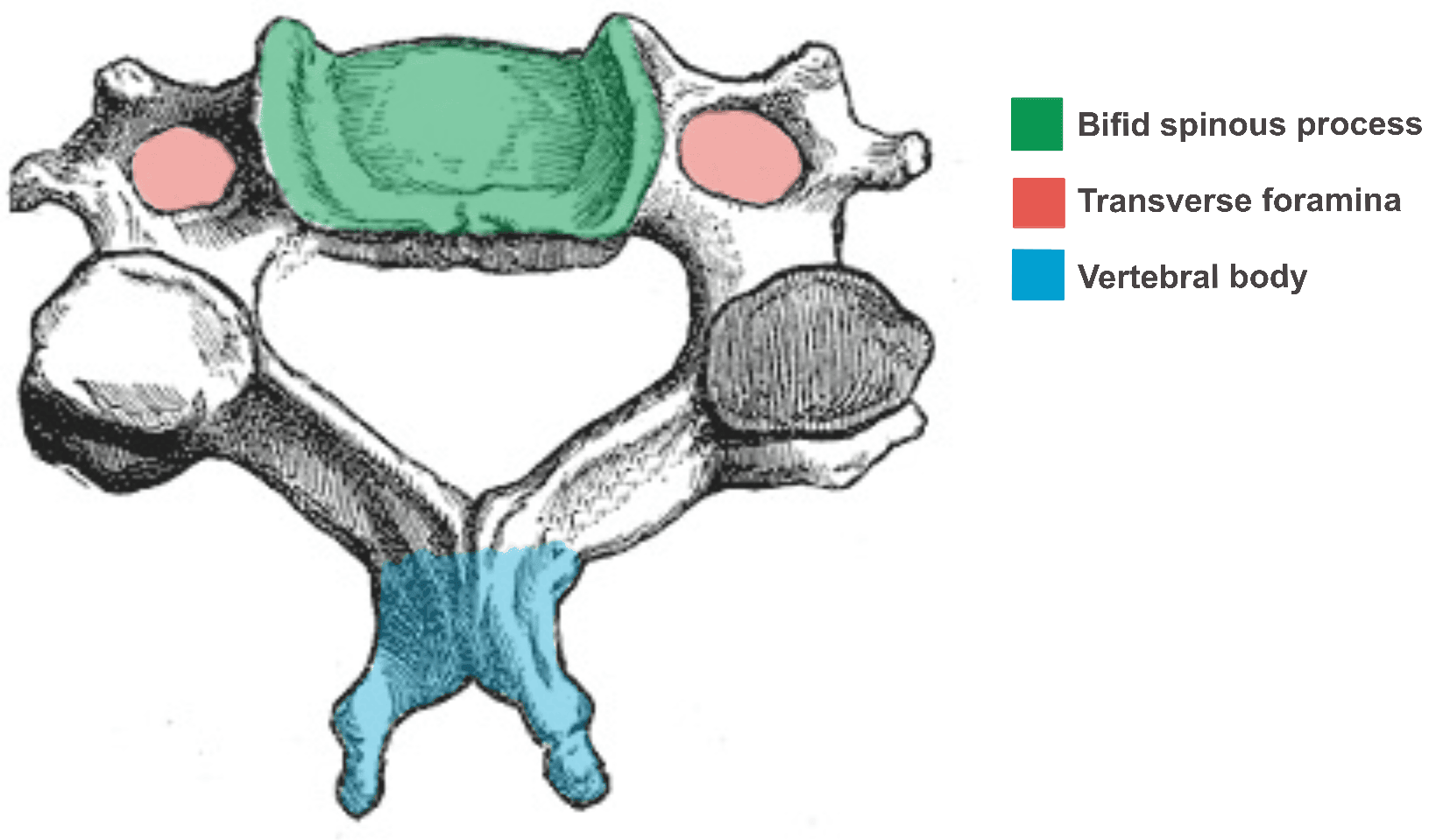
Diagram - Cervical Vertebra Structure
Public Domain Source Original from Gray's Anatomy, Derivative by Mikael Häggström, M.D, edited by Dr. Emily Smith [Public domain]
This is the first cervical vertebra. It articulates with the occiput of the skull superiorly (the atlanto-occipital joint - this joint permits the characteristic nodding flexion and extension of the head; lower cervical joints also contribute to flexion and extension of the neck), and axis inferiorly (the atlantoaxial joint - this joint is responsible for majority of rotation in the neck).
It has several features which make it different from the other vertebrae:
- No vertebral body.
- No spinous process.
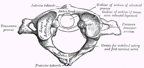
Diagram - The structure of the Atlas vertebra
Public Domain Source Anatomy of the Human Body, by Henry Gray, edited by Dr. Emily Smith [Public domain]
Axis is the second cervical vertebra, and is also atypical compared to the others. One of its features is the Odontoid process (or Dens) which allows it to articulate with C1. The dens is held steady by the transverse ligament where it articulates, preventing displacement of Atlas.
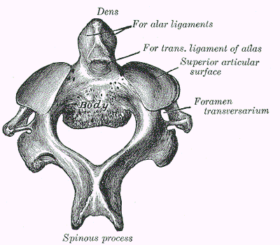
Diagram - The structure of the Axis vertebra
Public Domain Source Anatomy of the Human Body, by Henry Gray, edited by Dr. Emily Smith [Public domain]
The 7th cervical vertebra is also atypical. It has a single spinous process which is exceptionally long in the C spine and is palpable at the base of the neck. Although C7 has a pair of transverse foramina, the vertebral artery does not pass through them.
The thoracic spine is made up of 12 vertebrae (T1-T12), and, along with the sternum and ribs, provides protection to internal viscera such as the heart and lungs. It is a relatively immobile region of the spine, with the structure of the vertebrae allowing rotation and limiting flexion.
- T2-T8 have demi-facets and T9-T10 have whole facets.
- Articulation with the head of the ribs.
- T1-T10 have costal facets on the transverse processes.
- Articulation with the tubercle of the ribs.
- Spinous processes are long and slant inferiorly.
- The facet joints articulate at an angle of approximately 60 degrees.
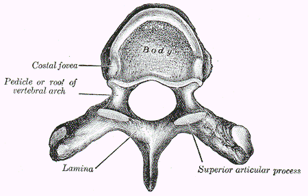
Diagram - Superior view of thoracic vertebrae, with key features labelled
Public Domain Source Anatomy of the Human Body, by Henry Gray, edited by Dr. Emily Smith [Public domain]
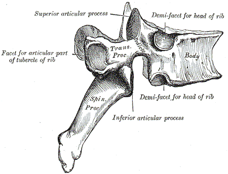
Diagram - Lateral view of thoracic vertebrae, with key features labelled
Public Domain Source Anatomy of the Human Body, by Henry Gray, edited by Dr. Emily Smith [Public domain]
The lumbar spine is located within the lower back, and is made up of 5 vertebrae (L1-L5). The spinal cord typically terminates at around the L1 to L2 level and the cauda equina continues from this point. The cauda equina is a bundle of lumbar, sacral and coccygeal nerve roots.
- Large, kidney shaped vertebral bodies.
- L5 is the largest vertebra as it needs to support the weight of the upper body.
- Triangular vertebral foramen.
- Mammillary processes on the superior articular processes for attachment of muscles within the back.
- Accessory processes on transverse processes for attachment of muscles within the back.
- The facet joints articulate at an angle of approximately 90 degrees.
Diagram - Labelled structure of lumbar vertebrae
Public Domain Source Anatomy of the Human Body, by Henry Gray, edited by Dr. Emily Smith [Public domain]
The sacrum and coccyx are located at the bottom of the vertebral column.
The sacrum is formed from five fused vertebrae (S1-S5) to create a triangular shape. The sacral nerve roots of the cauda equina pass through the sacral canal within the sacrum.
The coccyx (or tailbone) is formed from four vertebrae which fuse to form one bone of a triangular shape. It articulates with the sacrum at the sacrococcygeal symphysis. This joint allows limited, passive flexion and extension during labour and defaecation.

Diagram - Structure of the sacrum and coccyx, with key features labelled
Creative commons source by OpenStax College [CC BY-SA 4.0 (https://creativecommons.org/licenses/by-sa/4.0)]
Edited by: Dr. Ben Appleby
Reviewed by: Dr. Thomas Burnell
- 12573



