

Next Lesson - Control of Plasma Osmolarity
Core
The plasma is the clear fluid that supports the blood cells, and therefore the plasma volume is key in maintaining the supply of oxygen to the key organs of the body.
Sodium and Intravascular Fluid Volume
Sodium (Na+) is the main extracellular cation. Retaining sodium salts, when water is available, promotes water retention and expands extracellular fluid (ECF). The relevant quantity is total sodium content, not sodium concentration alone.
The ECF comprises interstitial fluid and plasma. Expansion of its plasma component can increase blood pressure, although cardiac function and vascular resistance also matter. Plasma sodium concentration describes sodium relative to water: hypernatraemia can occur with depleted, normal or expanded ECF volume, so it cannot by itself establish circulating volume.
The kidneys adjust sodium excretion to dietary intake. Thirst and antidiuretic hormone regulate water balance and interact with sodium balance to maintain volume and concentration. Sodium retention with water retention expands ECF; water loss alone can raise plasma sodium concentration without expanding the circulation. Sources: MSD fluid-volume physiology; water and sodium balance; hypernatraemia.
Water reabsorption along the nephron
Most filtered water is reabsorbed along the proximal tubule and thin descending limb of the loop of Henle. Water balance is then adjusted by regulated water reabsorption in the connecting tubule and collecting duct.
Antidiuretic hormone (vasopressin) regulates water reabsorption in the collecting system by increasing water-channel availability. The final amount of water excreted therefore varies with the body's needs. See renal water transport physiology.
SimpleMed Original by Dr. Maddie Swannack
A percentage of the original filtered water is different from a percentage of the fluid delivered to an individual segment. Segment values need a common denominator and physiological state before they can be added; total reabsorption cannot exceed the amount filtered.
Effectors which Change Sodium Excretion
There are a number of factors which influence renal sodium excretion.
Firstly, changes in osmotic and hydrostatic pressure alter the Na+ reabsorption in the proximal convoluted tubule (PCT), meaning if the filtrate is particularly concentrated, less Na+ will be reabsorbed. Na+ reabsorption in the PCT is also stimulated by the Renin-Angiotensin-Aldosterone System (RAAS), meaning increased RAAS activation results in more Na+ reabsorption. Finally, the principal cells of the late distal tubule and collecting ducts (CD) are targeted by aldosterone, which increases Na+ reabsorption.
Pressure Natriuresis and Diuresis
When BP in the renal artery increases, there is a reduction in the number sodium-hydrogen (Na-H) antiporters and reduced sodium-potassium (Na-K) ATPase activity. This means less Na+ is reabsorbed from the PCT - meaning less water is reabsorbed too. The result of this is the intravascular fluid volume decreases, as does BP.
This process of increased sodium excretion due to changes in blood pressure is called pressure natriuresis, and the increased water excretion is called pressure diuresis.
The process of sodium reabsorption is mainly active (i.e. needs ATP to occur), and is largely due to the action of Na-K-ATPase pumps on the basolateral membrane. Chloride (Cl-) reabsorption is closely linked to sodium handling, so in many parts of the nephron Na+ reabsorption is accompanied by Cl- movement.
Different segments of the nephron have different transporters for sodium in the apical (filtrate side) membrane:
- PCT:
- Na-H Antiporter
- Na-Glucose Symporter
- Na-AA (Amino Acid) Co-transporter
- Na-Pi (inorganic phosphate) co-transporter
- Loop of Henle
- Sodium-Potassium-2Chloride (NaKCC) symporter
- Early DCT
- NaCl symporter
- Late DCT and CD
- Epithelial Na+ channels (ENaC)
Please note, this is not a complete list, there are many other types of sodium transporters.
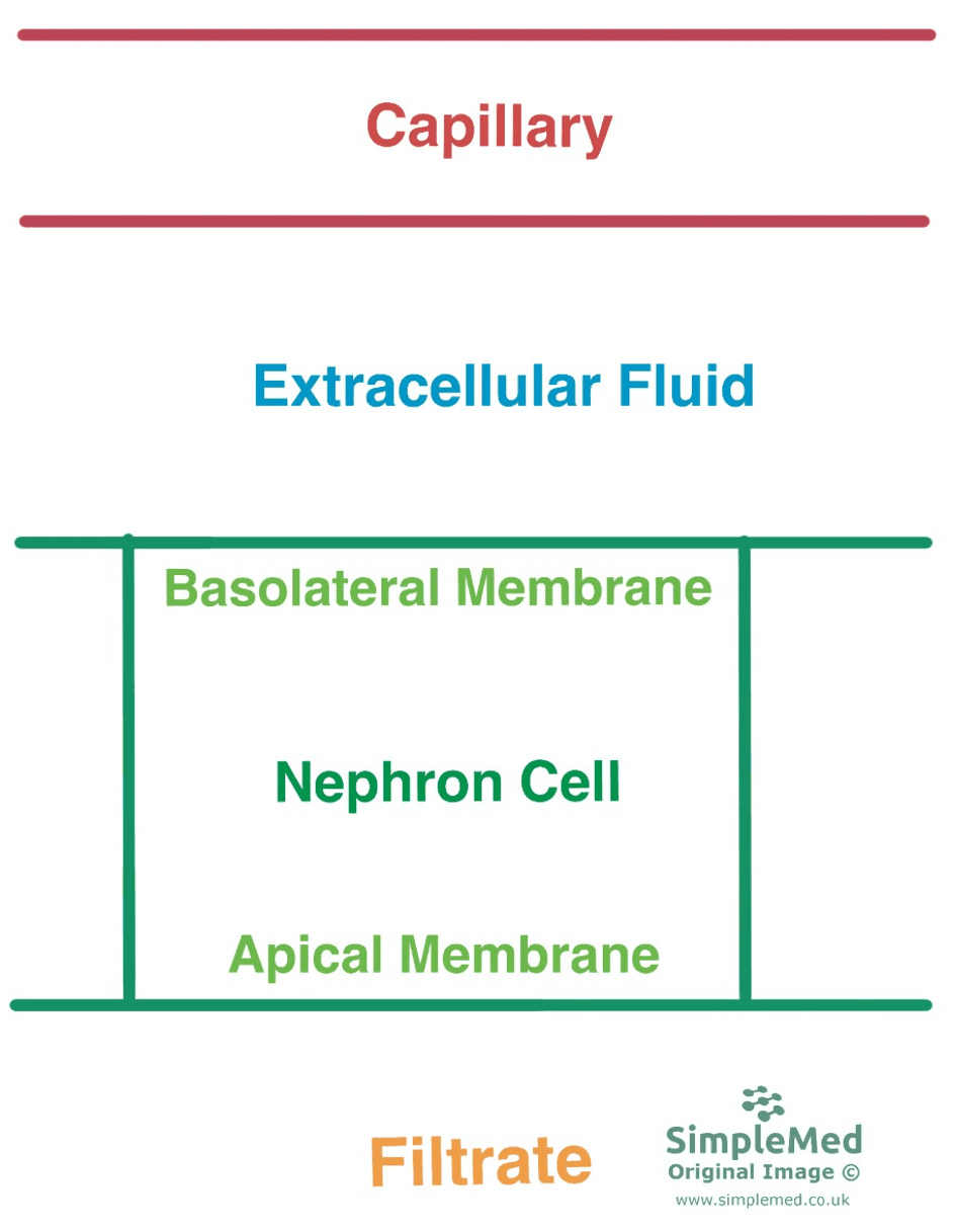
Diagram - The difference between the apical and basolateral membranes of the nephron cells. This is important to understand before trying to understand the transporters in the nephron
SimpleMed original by Dr. Maddie Swannack

Diagram - The different types of transport across cell membranes. Uniport moves one ion in one direction, symport moves two or more ions in the same direction, antiport moves two or more ions in more than one direction across the cell membrane
SimpleMed original by Dr. Maddie Swannack
Reabsorption in the Proximal Convoluted Tubule
The PCT is split into three segments, based on the transporters that are present in the membrane: S1, S2, and S3.
In S1, 1 Na+ ion is co-transported with glucose across the apical membrane through SGLT2. Sodium is then actively transported into the capillary across the basolateral membrane through Na-K-ATPase and glucose moves into the capillary in its own transporter.

Diagram - An overview of sodium transporters in S1 of the PCT. Note there are other transporters, and not all sodium and glucose move directly from the cells to the capillary (some move into the paracellular space first)
SimpleMed original by Dr. Joshua Sturgeon
The concentration of urea and Cl- in the filtrate in S1 increases to compensate for the loss of glucose. This creates a concentration gradient for the reabsorption of Cl- in S2 and S3.
In S2 and S3, there is again Na-K-ATPase on the basolateral membrane, however, the apical membrane differs significantly.
Na+ is reabsorbed through the Na-H exchanger and Cl- is reabsorbed in exchange for formate ions (HCOO-). The H+ and HCOO- ions are provided by methanoic acid (HCOOH), which readily diffuses across the apical membrane and breaks up into its constituent ions inside the cell, which can then be exchanged with Na+ and Cl-.
Cl- moves into the capillaries through co-transport with K+ which was brought into the cell via Na-K-ATPase on the basolateral membrane.
Aquaporin channels (AQP) are also present here, which allow water to move from the apical membrane through the cells and out of the basolateral membrane. Water is also able to move into the paracellular space (the space in between cells), and the water moved by these two methods can then move into the capillary.
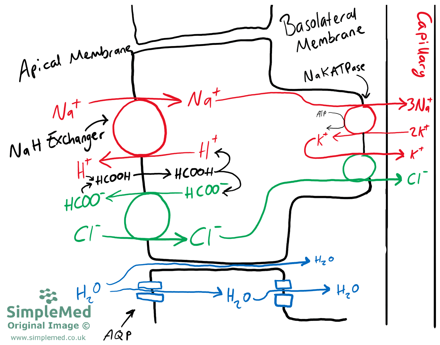
Diagram - An overview of sodium and chloride transporters in S2 and S3 of the PCT
SimpleMed original by Dr. Joshua Sturgeon
The PCT is responsible for 65% of water reabsorption. This is driven by a number of factors. Firstly, an osmotic gradient is generated by solute reabsorption so there is an increased osmolarity in the interstitial spaces. As a result, water moves out of the PCT into the interstitium down an osmotic gradient, and is then taken up into the capillaries due to their low hydrostatic pressure. Finally, oncotic forces in the capillary act to bring water into the capillary, as this capillary will still contain proteins and blood cells.
Reabsorption in the Loop of Henle
The descending limb of the Loop of Henle (LoH) is permeable to water, so water moves out of the filtrate and into the capillaries by osmosis. This is because there is a higher concentration of Na+ in the interstitial fluid and capillary due to PCT reabsorption. The descending limb is impermeable to Na+ and Cl-, meaning this loss of water creates an increased concentration of Na+ and Cl- in the lumen of the descending limb, ready for active transport in the ascending limb.
Initially, in the thin portion of the ascending LoH, the high concentration of Na+ allows for passive reabsorption of ions through the paracellular route.
In the thick ascending LoH this concentration gradient of Na+ has diminished as some has moved out already via passive transport, so Na+ is actively transported out of the lumen by NaKCC transporters. The Na+ ions can then move into the interstitium and capillaries by Na-K-ATPase.
Cl- moves into the interstitium in its own transporter. At this point, in the filtrate, there is a lack of K+ ions as they were transported with Na+ and Cl- in NaKCC. For this reason, it is vital that K+ ions diffuse back into the lumen of the LoH and this is done through the Renal Outer Medullary Potassium Channel (ROMK). This region of the nephron uses more energy (ATP) than any other, so it is particularly sensitive to hypoxia.
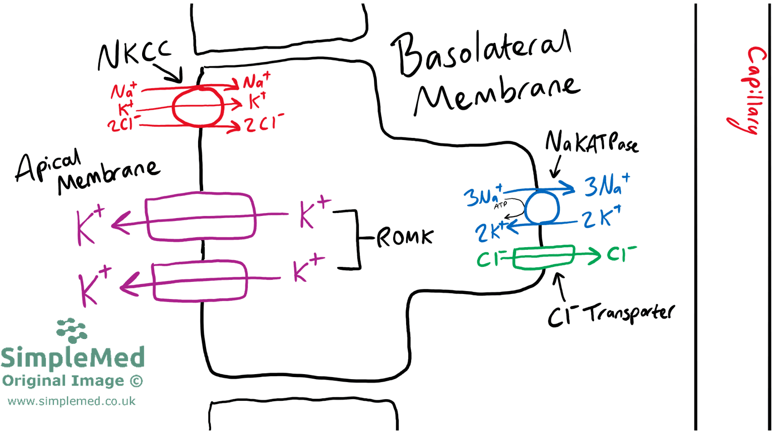
Diagram - An overview of the various transporters present in the thick ascending LoH. Remember solutes that end up in the interstitium will diffuse into the capillary
SimpleMed original by Dr. Joshua Sturgeon
Reabsorption in the Distal Convoluted Tubule
Due to the action of the thick ascending LoH, filtrate entering the DCT is hypo-osmotic (dilute) compared to plasma. The DCT is split into two regions: DCT 1 (the early part of the DCT) and DCT 2 (the late part of the DCT).
In DCT 1, NaCl enters across the apical membrane via a sodium-chloride co-transporter (NCC). This transporter is described as electroneutral as the 1+ charge from Na+ is cancelled-out by the 1- charge from the Cl-. Na+ is then transported into the capillary through Na-K-ATPase on the basolateral membrane, and the Cl- diffuses across a channel due to the high concentration in the cell. The NCC co-transporter is of clinical importance as it is the site of action of thiazide diuretics.
Calcium (Ca2+) ions are also reabsorbed in the DCT. Ca2+ is transported across the apical membrane, after which it is immediately bound by calbindin, a protein that needs vitamin D. This shuttles Ca2+ to the basolateral membrane where it is transported out into the interstitium through the sodium-calcium exchanger (NCX). This process is tightly regulated by hormones such as parathyroid hormone (PTH) and 1,25-dihydroxy-vitamin D (also known as activated Vitamin D and Calcitriol).
In the DCT, 2 Na+ enter across the apical membrane via ENaC and a small amount through NCC. Na+ is transported across the basolateral membrane by Na-K-ATPase. ENaC is another transporter of clinical importance as it is the site of action of amiloride (potassium-sparing) diuretics. The movement of Na+ through ENaC is not electroneutral as there is no Cl- that is being co-transported, so Cl- diffuses through the tight junctions between the cells (paracellular uptake) to maintain the electroneutrality.
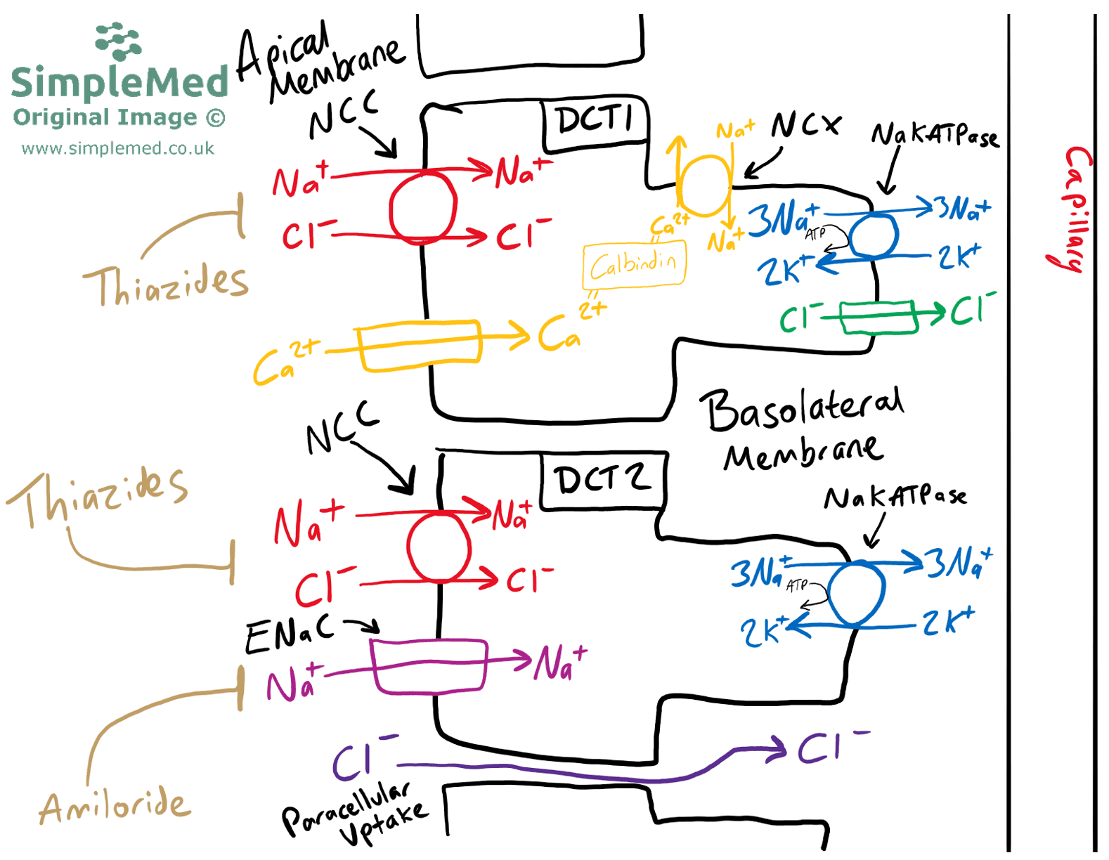
Diagram - An overview of the various transporters in the DCT with a DCT 1 typical cell above, and a DCT 2 typical cell below. Annotated in gold are the diuretics which have a site of action in the DCT. Remember that solutes which end up in the interstitium will eventually diffuse into the capillary
SimpleMed original by Dr. Joshua Sturgeon
Reabsorption in the Collecting Ducts
The DCT and the early part of the collecting duct (CD) share many similarities, and it is difficult to distinguish an exact transition from one cell type to another. The collecting duct can be divided into the cortical CD and the medullary CD - this portion focuses on the cortical CD.
There are two types of cells found in the cortical CD:
- Principal Cells - these make up 70% of the cells in the cortical CD and are responsible for the reabsorption of Na+ through ENaC (as was the case in DCT 2).
- Intercalated Cells - there are two types of intercalated cells. They are primarily involved in acid-base balance.
- Type A Intercalated Cells (acid-secreting cells) secrete H+ ions into the lumen via proton pumps, with bicarbonate returned to the blood across the basolateral membrane.
- Type B Intercalated Cells (base-secreting cells) secrete bicarbonate (HCO3-) into the lumen via a Cl-/HCO3- exchanger, resulting in chloride reabsorption.
Sodium moves out of principal cells through Na+/K+ ATPase.
K+ which moves into the cell through the basolateral membrane is transported into the lumen through ROMK.
Principal cells can take up variable amounts of water depending on the number of aquaporin channels present in the apical membrane - this is under the influence of antidiuretic hormone (ADH).
In both type A and B intercalated cells, Cl- moves across the basolateral membrane, into the capillary through various transporters.
Edited by: Dr. Maddie Swannack
Reviewed by: Dr. Thomas Burnell
- 5619
