
Next Lesson - Bones of the Hand
Core
There are four muscles in the upper arm. They can be compartmentalised into anterior and posterior compartments.
It is also very important when studying muscles that you are comfortable with anatomical terminology, as a lot is used in these articles. For more information, please check out our Anatomical Terminology Article.
There are three muscles in the anterior compartment of the arm. Biceps brachii and brachialis contribute to flexion at the elbow. Coracobrachialis does not cross the elbow; it flexes and weakly adducts the arm at the shoulder. The musculocutaneous nerve (a branch of the brachial plexus) is the main nerve supply to this compartment, with an additional radial nerve contribution to brachialis.
This muscle has two heads. The long head originates from the supraglenoid tubercle, found on the scapula, whilst the short head originates from the coracoid process. These heads converge to form a main belly which inserts into the radial tuberosity of the radius. The main function of the biceps is to supinate the forearm. It is also responsible for flexion at the elbow and shoulder joints as it crosses both of these joints.
Found deep to the biceps brachii, this muscle is the main elbow flexor. It originates from the humeral shaft and inserts into the ulna tuberosity.
This muscle originates from the coracoid process. It attaches to the medial humeral shaft. As this muscle does not cross the elbow joint, it doesn’t perform elbow flexion. It does, however, flex the arm at the shoulder joint.
The anterior muscles of the arm can be remembered by the acronym 'BBC'.
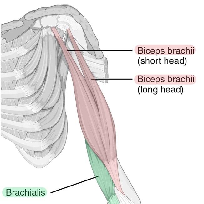
Diagram - Illustrates the location of biceps brachii and brachialis. The coracobrachialis cannot be seen in this illustration, however, it would be found deeper to these muscles. If trying to locate the coracobrachialis, try and work down from the coracoid process
Creative commons source by CFCF, edited by Dr. Sachin Sudhakaran [CC BY-SA 4.0 (https://creativecommons.org/licenses/by-sa/4.0)]
The posterior compartment is fairly straightforward; there is only one muscle to remember. The Triceps Brachii is a large muscle, situated posterior to the humerus. It has three heads:
- Long head - originates from the infraglenoid tubercle.
- Lateral head - originates from the humerus, just superior to the radial groove.
- Medial head - originates from the humerus, like the lateral head. However it originates just inferior to the radial groove. This head sits closer to the humerus than the others, meaning that in diagrams, it is not always visible.
As usual, these heads converge to form the belly of the triceps before attaching at the olecranon of the ulna. The triceps is responsible for extension of the elbow and is innervated by the radial nerve.

Diagram - This image illustrates the location of the triceps brachii. The medial head is hidden under the long and lateral heads.
Creative commons source by CFCF, edited by Dr. Sachin Sudhakaran [CC BY-SA 4.0 (https://creativecommons.org/licenses/by-sa/4.0)]
For Pathologies and Conditions of the Upper Arm, check out our article on Conditions of the Arm.
Reviewed by: Dr. Thomas Burnell
Edited by: Dr. Maddie Swannack
- 12964