

All done here! Check out our Genetic Testing and Counselling!
Core
Central Factors of DNA Damage Response
DNA damage response is a hugely complex area of genetics. Thankfully, there isn’t too much detail that is required for medical training.
In general, replication stress (where the replication fork slips, stalls or breaks; the fork is the point at which DNA is being copied) and mutagens (agents such as radiation and some chemicals that cause mutations) cause DNA damage. This activates a signalling cascade (a chain of molecular signals) which either results in the cell undergoing senescence (the cell permanently stops dividing) or apoptosis, or DNA being repaired at the next cell cycle checkpoint.
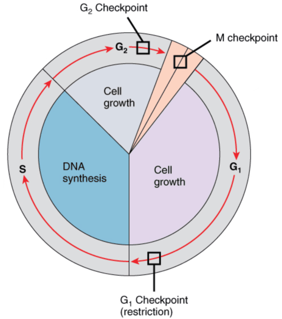
Diagram: The stages and checkpoints of the Cell Cycle
Creative commons source by OpenStax, edited by Dr. Joshua Sturgeon [CC BY 4.0 (https://creativecommons.org/licenses/by/4.0)]
The above image shows the cell cycle with each stage of interphase (the stages between one mitosis and the next) marked along with mitosis (in pink). This image also highlights where certain cell cycle checkpoints are located. Checkpoints are periods of temporary cell cycle arrest which provide time for DNA damage to be repaired.
The mitosis (M) checkpoint checks to make sure that all chromosomes are properly attached to the mitotic spindle (the protein fibres that pull chromosomes apart). If they are all attached correctly, the duplicated chromosomes can then be pulled apart.
The G1 (first gap phase) checkpoint checks to make sure the intracellular environment is favourable for DNA synthesis and that the DNA is undamaged. If so, then DNA synthesis can begin.
The G2 (second gap phase) checkpoint checks to make sure that all DNA is replicated and checks to see if all DNA damage has been repaired. If this is true, the cell can then undergo mitosis.
In healthy cells, these checkpoints work to ensure the cell remains healthy and can go through mitosis.
If DNA damage levels are too high and cannot be adequately repaired, signalling by p53 (a protein that stops damaged cells dividing) can cause the cell to arrest the cell cycle, allowing time for repair, or trigger apoptosis or senescence. Loss of p53 function allows damaged cells to progress through checkpoints inappropriately.
Ideally, all DNA damage will be repaired, and cell function will be maintained. However, this is not always possible.
There are multiple DNA repair pathways. The type of pathway used depends on the type of DNA damage and the cell cycle phase.
One way in which DNA can be damaged is through a double-strand break. As the name suggests, both strands of DNA have broken, so this is considered the most cytotoxic (cell-killing) type of DNA lesion (a damaged area of DNA). Although the fine details of repair pathways do not need to be memorised and only a general understanding is required, we have included this information for reference.
There are two main ways in which a double-strand break can be repaired:
Non-Homologous End Joining (NHEJ):
- DNA has a double-strand break.
- Proteins called Ku70/80 move to the exposed ends of DNA and act as caps to protect them.
- An enzyme called DNA-PKcs (the catalytic subunit of DNA-dependent protein kinase; a kinase is an enzyme that adds phosphate groups to other proteins) moves to the ends of DNA and forms a DNA-PK complex with Ku70/80. This complex activates Artemis, a nuclease (an enzyme that cuts DNA), which processes the ends of the DNA strands and removes damaged DNA if required.
- Other proteins, e.g. DNA ligase IV, are recruited to ligate (join) the two ends of DNA back together.
- NHEJ does not need a template and works throughout the cell cycle, including in G1. It often loses or adds a few DNA bases at the join, so it is error-prone.
- This is a more complicated repair mechanism and has multiple sub-pathways.
- Firstly, the exposed ends of DNA are resected (trimmed back to leave single-stranded ends).
- This mechanism utilises a homologous template (an undamaged copy of the DNA involved in the double-strand break), e.g. a sister chromatid (one of the two identical copies of a duplicated chromosome).
- In many models, strand invasion occurs: the broken end pairs with the matching sequence in the homologous template. This may involve formation of a Holliday junction (a four-way crossover where two DNA molecules are joined).
- The Holliday junction is then separated, either by being cut apart by enzymes called resolvases or by being dissolved, to complete the repair.
- Because it copies an undamaged template, homology-directed repair is mostly error-free. It works mainly in the S and G2 phases of the cell cycle, when a sister chromatid is available. The proteins BRCA1 and BRCA2 have key roles in this pathway.
The image below shows both mechanisms in much more detail than pre-clinical exams need, but it helps you to visualise the repair mechanisms.

Diagram: NHEJ and Homology-directed repair in detail
Creative commons source by Hannes Lans, Jurgen A Marteijn and Wim Vermeulen [CC BY 2.0 (https://creativecommons.org/licenses/by/2.0)]
DNA Repair: Relevance to Cancer
The two-hit hypothesis explains why both copies (alleles) of a tumour suppressor gene usually have to be damaged before a cell loses tumour suppression. A tumour suppressor gene is a gene whose protein normally stops cells dividing out of control. If a mutation damages one copy of a tumour suppressor gene such as p53, the cell is usually still protected because the second copy of the gene is unaffected. (Some mutant p53 proteins can also block the normal p53 protein made from the second copy. This is called a dominant-negative effect.) However, if a second ‘hit’ is given and there is no tumour suppression, the cell can go from being healthy to pre-malignant and then finally malignant. This process is called carcinogenesis.
Abnormal DNA damage responses can be inherited, which will predispose an individual to cancer in later life e.g. Lynch syndrome, previously called hereditary nonpolyposis colorectal cancer (HNPCC; nonpolyposis means without large numbers of polyps). Lynch syndrome is an autosomal dominant condition (one faulty copy of the gene is enough to cause it). It is caused by a germline mutation (a mutation present in every cell of the body, usually inherited from a parent) in a gene needed for DNA mismatch repair. Mismatch repair is the system that spots and corrects wrongly paired bases made when DNA is copied. When it fails, copying errors build up as mutations, which leads to a higher likelihood of cancer, mainly of the bowel (colorectal cancer) and the womb lining (endometrial cancer).
Once a cancer develops, cells can continue to mutate. This leads to intra-tumour heterogeneity (different cells within one tumour carry different mutations). Heterogeneity makes tumours harder to treat because it can lead to therapy resistance or metastasis.
Take for example, a highly heterogeneous tumour. Some cells in this tumour are highly sensitive to one form of chemotherapy (a medicine that kills cancer cells), whereas other cells are resistant to it. After chemotherapy, the cells which were sensitive will have died, but this leaves space for the resistant cells to grow. This means the chemotherapy is less effective overall as only the resistant cells are left in the tumour. This is called clonal expansion (the resistant cells multiply to form a larger group of identical cells, called a clone).
An alternative result to clonal expansion is chemotherapy-induced mutagenesis (the drugs themselves cause new mutations). This occurs when cells are not killed outright by the chemotherapy, but their DNA is damaged just enough to mutate. This is a problem as it further increases the heterogeneity of the tumour.
Radiotherapy (treatment with radiation) and many chemotherapy drugs treat cancer by causing more DNA damage. Cancer cells often already have defects in their DNA repair mechanisms, so extreme damage can push them into apoptosis. However, normal cells that divide quickly (for example hair follicle cells and the cells lining the gut) are also affected. This causes side effects, which depend on the treatment and the part of the body treated. Examples include:
- Radiation:
- Skin problems
- Fatigue
- Heart problems (mainly when the chest is treated)
- Chemotherapy:
- Hair loss
- Fertility problems
- Nausea and vomiting
- A weakened immune system, which makes infections more likely
- Anaemia, and bruising or bleeding more easily
- A sore mouth and throat (mucositis)
Cancer treatment can also promote tumour evolution. Radiotherapy and chemotherapy can also increase the risk of a new, separate cancer in the years after treatment (a second primary cancer, which is not the first cancer spreading). With radiotherapy the chance is small, and the benefits of treatment generally outweigh the risk.
A newer approach is to block a DNA repair pathway on which tumour cells depend, and this is now in routine NHS use. The best-known examples are PARP inhibitors, such as olaparib. PARP (poly-ADP ribose polymerase) is an enzyme that helps damaged cells to repair themselves. Cancer cells with faults in the BRCA1 or BRCA2 genes (the breast cancer genes, which are needed for homology-directed repair) already have a poor repair system. Blocking PARP then leaves them unable to repair themselves, and they die.
Normal cells still have their other repair pathways, so they are affected less. PARP inhibitors can still cause side effects, such as tiredness, feeling sick and a drop in blood cell counts (which raises the risk of infection and bleeding). The approach is also often limited to cancers with a matching repair defect. For example, NICE recommends olaparib to keep relapsed ovarian cancer under control after it has responded to platinum-based chemotherapy, but only if the person has a BRCA1 or BRCA2 mutation. This technique of targeting a second repair pathway in cells that have already lost the first is known as synthetic lethality: losing either pathway alone is survivable, but losing both kills the cell.
For more information about cancers, head to the Neoplasia articles in our Pathology series.
Edited by: Dr. Ben Appleby and Dr. Maddie Swannack
Reviewed by: Dr. Marcus Judge
- 8897


