

Next Lesson - Temperature Regulation
Core
Glucose is an important fuel for many cells in the body, although most cells can metabolise other substrates such as fatty acids or ketone bodies if glucose availability is low. Some cells rely heavily on glucose and on glycolysis for their energy. Red blood cells have an absolute requirement for glucose: they have no mitochondria and rely solely on glycolysis. Other cells that make heavy use of glycolysis include: neutrophils, kidney medulla cells (cells of the inner part of the kidney), and mature cells in the lens of the eye (the fibre cells that fill the lens; the single layer of cells on its surface has mitochondria and can also make ATP by oxidative phosphorylation). Stable glucose levels are also needed for normal brain function, but the brain can use ketone bodies as an energy source if it absolutely must.
Normal fasting blood glucose concentration = about 4.0-5.4 mmol/L (a fasting plasma glucose of 5.5 to 6.9 mmol/L is classed as high risk of type 2 diabetes).
Renal threshold = about 10 mmol/L. This is the blood glucose concentration above which the kidney can no longer reabsorb all the glucose filtered from the blood, so glucose appears in the urine. The renal threshold can be altered by physiological state (e.g. pregnancy) and other factors such as age.
Note: reference ranges can vary between textbooks and trusts. In practice and written exams always use the reference values provided.

Diagram - The chemical structures of glycogen as well as the α1-4 and the α1-6 glycosidic bonds (the bonds that join sugar molecules together)
SimpleMed original by Dr. Maddie Swannack
Glycogen is a molecule used to store glucose in cells. It is formed from chains of glucose molecules, linked into straight chains by α1-4 glycosidic bonds. Glycogen has a highly branched structure formed by α1-6 glycosidic bonds. These branched chains clump together to form granules inside certain cells, acting as a store of glucose.
These stores are mostly contained in cells of skeletal muscles and the liver. Around 80 to 100 g of glycogen is stored in the liver and can be used to raise the blood concentration of glucose. Approximately 300 to 400 g of glycogen is stored in skeletal muscle and can only be used by the muscle itself. The liver can release glucose into the blood because it possesses an important enzyme called glucose-6-phosphatase (an enzyme that removes the phosphate from glucose-6-phosphate, leaving free glucose).
Glycogen storage diseases are a rare group of diseases that involve a deficiency in an enzyme involved in glycogen storage.
Too much glycogen can build up if cells are unable to break it down, and this can damage tissues. One example is von Gierke’s disease (glycogen storage disease type I), where the enzyme glucose-6-phosphatase is missing, so glycogen cannot be broken down and accumulates in the liver and kidneys. Hypoglycaemia (low blood glucose) still occurs between meals because the glucose cannot be released.
Too little glycogen storage causes hypoglycaemia between meals, because there is no glycogen in storage that can be used to raise blood glucose levels. An example is glycogen storage disease type 0, where the liver cannot store glycogen. A muscle form also exists, in which too little glycogen in the muscle causes poor exercise tolerance.
Glycogenesis is the process of turning glucose into glycogen. It occurs through four reactions, two of which need energy: one uses ATP and one uses UTP (uridine triphosphate, an energy-carrying molecule similar to ATP). These reactions add glucose onto the end of one of the strands of a glycogen molecule. Glucose can be:
- Added to an existing branch through glycogen synthase forming α1-4 glycosidic bonds,
- Or begin a new branch through branching enzyme forming α1-6 glycosidic bonds.

Diagram - The process of glycogenesis
SimpleMed original by Dr. Maddie Swannack
Glycogenolysis is the process through which glycogen is broken down into glucose, so that the glucose can be used for respiration.
There are two different enzymes needed to break the different types of glycosidic bonds present in glycogen:
- Glycogen phosphorylase breaks the α1-4 glycosidic bonds.
- Debranching enzyme breaks the α1-6 glycosidic bonds.
Glycogen phosphorylase releases glucose-1-phosphate, which is converted into glucose-6-phosphate (glucose with a phosphate attached), while debranching enzyme releases free glucose at the branch points. The glucose-6-phosphate is then used in glycolysis in muscles, or if it is released by the liver, it can be used to raise blood glucose levels. This is because the liver possesses the enzyme Glucose-6-Phosphatase that is used to turn glucose-6-phosphate into glucose.

Diagram - The process of glycogenolysis
SimpleMed original by Dr. Maddie Swannack
Regulation of Glycogen Metabolism
Glycogen metabolism requires careful regulation to properly manage stores of glucose in the body. This is done through rate limiting enzymes (the slowest enzyme in a pathway, which sets the speed of the whole pathway), which are regulated by hormones.
Synthesis
Glycogen Synthase is the enzyme that is rate limiting in the synthesis of glycogen. Its action is inhibited by glucagon and adrenaline. This makes sense as glucagon is used to signal glycogen breakdown and adrenaline signals the fight or flight response meaning the body wants to increase the amount of glucose in the blood ready for use immediately rather than storing this glucose as glycogen.
Glycogen Synthase activity is activated by insulin.
Degradation
Glycogen Phosphorylase is the enzyme that is rate limiting in the degradation process of glycogen. Its action is activated when glucagon or adrenaline are released.
Glycogen Phosphorylase action is inhibited by insulin release.
Muscle Glycogen
Glycogen stored in muscles does not respond to glucagon, because glucose produced from the breakdown of muscle glycogen cannot be used to raise blood glucose. In the muscle adenosine monophosphate (AMP) is used instead of glucagon as the allosteric activator (a molecule that switches an enzyme on by binding somewhere other than its active site) of Glycogen Phosphorylase. This means that when there are many AMP molecules in the muscle, glucose is released to allow for increased ATP production within the muscle.
Gluconeogenesis is the production of new glucose from an originally non-glucose source; the three most common sources are amino acids, lactate and glycerol. This occurs throughout fasting, with its contribution increasing as hepatic glycogen stores become depleted and a new source of glucose is needed. It occurs mainly in the liver, but also in the kidney cortex.

Diagram - This diagram shows the Cori Cycle. This is the cycle between the muscle using up glucose in anaerobic respiration to produce lactate, which then travels to the liver and, through gluconeogenesis, is turned back into glucose
SimpleMed original by Dr. Maddie Swannack
Key enzymes that are involved are PEPCK (phosphoenolpyruvate carboxykinase) and fructose 1,6-bisphosphatase (which are both regulated), and glucose-6-phosphatase (which is also regulated, mainly through how much of the enzyme is made, and insulin reduces this). Pyruvate carboxylase, which converts pyruvate to oxaloacetate, is a further key enzyme.
The regulated enzymes have their actions increased by glucagon, adrenaline and cortisol (a stress hormone made by the adrenal glands) because the body needs to create glucose in times of starvation or stress, and decreased by insulin because the body does not need to create glucose when blood glucose is high.
Triacylglycerols are highly efficient storage molecules. For more information on triacylglycerol storage and fatty acid oxidation, please go to our Lipid Metabolism and Transport article.
For more information about Fatty Acid oxidation, check out our Lipid Metabolism and Transport article.
Fatty acid synthesis occurs mainly in the liver. The enzyme Acetyl-CoA Carboxylase produces malonyl CoA (a 3 Carbon [3C] molecule) from Acetyl CoA (2C). Next the Fatty Acid Synthase Complex builds fatty acids using malonyl CoA as a monomer (a single building block that is added repeatedly). This process requires ATP and NADPH (nicotinamide adenine dinucleotide phosphate, a molecule that carries electrons) to occur.
This process of fatty acid synthesis is increased by insulin. This is as when there is an increased amount of blood glucose available for metabolism (e.g. following a meal), there is also an increase in Acetyl CoA which can be stored as fatty acids. Citrate (the first molecule formed in the Krebs Cycle) also increases synthesis. Citrate made in the mitochondria is carried out into the cytoplasm, where the enzyme ATP-citrate lyase splits it to give Acetyl CoA, which can then be stored as fatty acid.
This process is decreased by glucagon and adrenaline. When these hormones are released, Acetyl CoA inside the cells needs to be available for the oxygen dependent pathway in respiration to produce more ATP. The process is also decreased by AMP (adenosine monophosphate), which rises when the cell is short of energy. High AMP activates AMP-activated protein kinase (AMPK, an enzyme that senses the cell's energy level). AMPK inactivates Acetyl-CoA Carboxylase, so fatty acid synthesis is switched off, while fatty acid oxidation and other pathways that generate ATP are switched on.
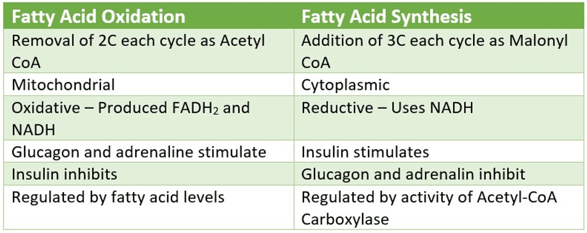
Table - The differences between fatty acid oxidation and fatty acid synthesis
SimpleMed original by Dr. Maddie Swannack
Blood glucose levels are normally tightly regulated, with fasting values typically around 4.0-5.4 mmol/L, through the balance of glycogenesis, glycogenolysis and gluconeogenesis. If blood glucose is not adequately maintained, this can lead to hypoglycaemia or hyperglycaemia, each with important clinical consequences. Low blood glucose can cause confusion, seizures and loss of consciousness, and persistently high blood glucose is how diabetes is diagnosed. In poorly controlled diabetes, severe hyperglycaemia (high blood glucose) can lead to diabetic ketoacidosis (a dangerous build-up of acid in the blood).
To test the ability of the body to regulate blood glucose, an oral glucose tolerance test can be performed. This involves asking the patient to fast for a defined period, after which blood glucose is measured. The patient is then given an oral glucose load, and blood glucose is measured again at set time points to see how well the body clears the glucose from the blood.
This table summarises the influences of insulin and glucagon/adrenaline on fatty acid synthesis and glycogen storage or breakdown.

Table - The influences of insulin and glucagon/adrenaline on energy storage
SimpleMed original by Dr. Maddie Swannack
Edited by: Dr. Ben Appleby
Reviewed by: Dr. Thomas Burnell and Dr. Marcus Judge
- 8909
