

Next Lesson - Connective Tissue
Core
This article explains the practical basics of how muscles work mechanically. By understanding the basics of muscle action the movement of any muscle can be predicted. For more information on the physiological make up of muscles, please see the article on The Structure of Muscles.
In this article, any use of the word ‘muscle’ refers to skeletal muscle. For more information on different types of muscle (smooth and cardiac), see the article on The Structure of Muscles.
Origin and insertion are two terms used to describe where a muscle attaches.
The ‘origin’ is conventionally the less mobile attachment, usually the more proximal one. For sartorius, the origin is the anterior superior iliac spine. During its usual open-chain action the pelvis is relatively fixed and the lower limb moves, but a different task can make this attachment move.
The ‘insertion’ is conventionally the more mobile attachment, usually the more distal one. Sartorius inserts on the medial side of the proximal tibia. In its usual open-chain action this attachment moves towards the origin. If the lower limb is fixed, the muscle can instead act on the pelvis without changing the anatomical names of its attachments.
Typically, all movements of muscles are described as beginning in the anatomical position. This is the position described as the ‘at rest’ position of the body: the whole body facing forward, looking forward, feet forward, palms forward (see our anatomical terms article for more detail).
Imagine that the hand is fixed in a pot of glue on a table. A forearm muscle can then pull its proximal attachment towards its fixed distal attachment: the pattern of movement reverses. This does not rename its anatomical origin and insertion. The conventional labels describe the attachments; the task and the external constraints determine which attachment moves.
The force generated by a contracting muscle is transmitted to its attachment points. Contraction produces tension, but the muscle need not shorten and the joint need not move. In an isometric contraction, muscle tension can hold a joint still against an opposing load. When movement is possible, the muscle’s line of pull relative to the joint determines the direction of its moment:
A muscle cannot ‘push’, only ‘pull’.
To predict a muscle’s action at a joint, identify the joint it crosses and the position of its line of pull relative to the joint axis. This predicts the direction of the moment it produces, rather than guaranteeing movement: an opposing load or another muscle may balance that moment. Skeletal muscles can also stabilise joints or act on soft tissues, so contraction does not always cause joint movement.
Muscle actions can be determined by answering these questions:
- Where are the origin and insertion of this muscle?
- How many joints does it cross?
- How does it relate to those joints?
- Is it anterior, to extend the knee?
- Is it posterior, to flex the knee?
- Which direction do the fibres run in?
- If the fibres run superior to inferior, then the muscle will shorten vertically, pulling the insertion superiorly.
- If the fibres run medial to lateral, then the muscle will shorten horizontally, pulling the insertion medially.
There are four main types of muscle found in the body, and they are categorised by the organisation of the myocytes.
Parallel muscles have myocytes that run all in the same direction, creating a thin, rectangular muscle. An example of this is the sartorius muscle.
Circular muscles have myocytes that run in a circular pattern. They make up the sphincters in the body, like the anal sphincter. On contraction, the opening in the centre of the circle will shrink.
Fusiform muscles have fibres running broadly along the long axis and a spindle-shaped belly that tapers at both ends. Biceps brachii is a familiar example. A bulging outline alone does not establish the physiological cross-sectional area or the force a muscle can generate. Force capacity depends on the total contractile fibre area, while fibre length influences the available shortening distance. Actual force and joint action also depend on activation and the line of pull.
Triangular muscles have a broad attachment with fibres converging towards a narrower tendon. Pectoralis major and temporalis are examples. Convergence describes their architecture, not a universal ranking of strength. Force depends on the amount of contractile fibre area recruited, fibre length and activation; the direction of pull and its moment arm determine the effect at a joint.
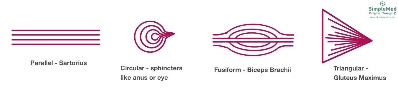
Diagram - The different arrangements of myocytes in different shapes of skeletal muscle
SimpleMed original by Dr. Maddie Swannack
There is another consideration when thinking about the direction of myocytes in a muscle, and this is whether the muscle is described as ‘-pennate’. This refers to the angle at which the myocytes attach to a tendon.
Unipennate muscles have fibres that attach obliquely to one side of a tendon. Merely running in one direction is not sufficient: parallel fibres can also share a direction without a pennate attachment. An example is flexor pollicis longus.
Bipennate muscles have fibres attaching obliquely to both sides of a central tendon, like the two sides of a feather. Rectus femoris is an example.
Multipennate muscles have several regions of fibres attaching obliquely to tendinous strands. The deltoid is a familiar example. This is a description of the fibre–tendon arrangement, rather than simply a muscle with more than two fibre directions.
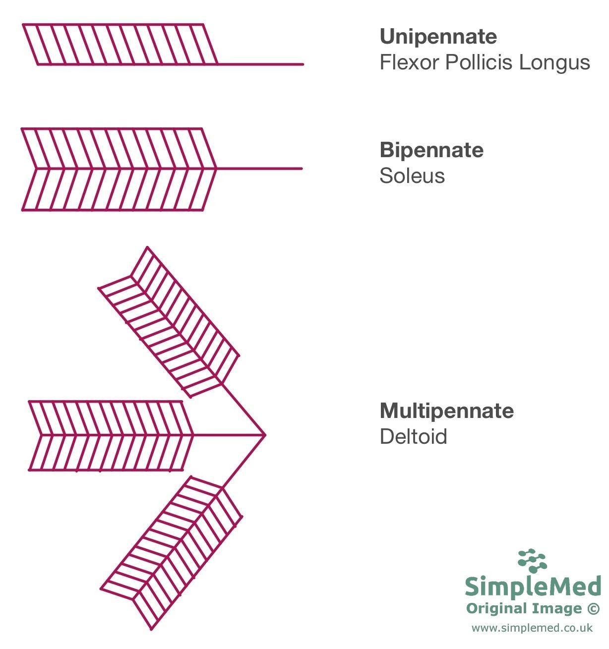
Diagram - The differences between unipennate, bipennate and multipennate muscles. The diagram has been simplified by not drawing in the full extent of the fibres in the multipennate muscle; the fibres of the strands would expand to meet each other, and fuse there to increase the strength of the muscle
SimpleMed original by Dr. Maddie Swannack
Edited by: Dr. Ben Appleby
Reviewed by: Dr. Thomas Burnell
- 7583
