

Next Lesson - Key Areas of the Upper Limb
Core
There are many muscles in the forearm, which mainly act at the elbow or wrist to bring about different movements. They can be categorised into anterior and posterior compartments, and can be challenging to learn.
It is also very important when studying muscles that you are comfortable with anatomical terminology, as a lot is used in these articles. For more information, please check out our Anatomical Terminology Article.
The anterior compartment can be split into three different layers; superficial, intermediate and deep. The muscles in the anterior compartment are all innervated by the median nerve, except for flexor carpi ulnaris and the ulnar (medial) half of flexor digitorum profundus which are innervated by the ulnar nerve.
There are four muscles in the superficial layer. They all originate from the medial epicondyle of the humerus:
Pronator Teres
This muscle attaches to the shaft of the radius and acts to pronate the forearm. Pronator teres is the most lateral of the four superficial anterior muscles.
Flexor Carpi Radialis
This muscle attaches to metacarpals II and III. It acts to flex at the wrist whilst also contributing to abduction. It lies immediately medial to pronator teres, with palmaris longus next medially and flexor carpi ulnaris most medial.
Palmaris Longus
This muscle attaches to the flexor retinaculum and acts to flex at the wrist. This is the only muscle in this layer that has a name that does not directly describe its action, but it attaches to the 'palm' through the flexor retinaculum, and this means it must flex the wrist.
Flexor Carpi Ulnaris
This muscle attaches to the pisiform bone in the hand. It contributes to both wrist flexion and adduction. It is the most medial of the muscles in this layer.
This layer of muscles can be visualised if the right hand is placed on the anterior side of the left arm, with the palm on the left medial epicondyle and the fingers pointing to the hand. The first finger illustrates the path of pronator teres, the middle finger the path of flexor carpi radialis, the ring finger the path of palmaris longus and the little finger the path of flexor carpi ulnaris.
Pronator teres, flexor carpi radialis and palmaris longus are all innervated by the median nerve. Flexor carpi ulnaris is innervated by the ulnar nerve (only one with ulnar in the name).
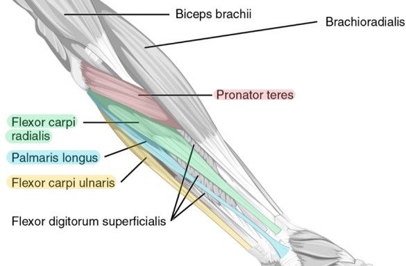
Diagram - This illustration highlights the muscles of the superficial layer. The flexor digitorum superficialis muscle can be seen underneath these muscles
Creative commons source by CFCF, edited by Dr. Sachin Sudhakaran [CC BY-SA 4.0 (https://creativecommons.org/licenses/by-sa/4.0)]
The intermediate layer is straightforward; there is only one muscle:
Flexor Digitorum Superficialis (FDS)
This muscle originates from the medial epicondyle of the humerus and also from the radius. It then splits into four tendons, which each attach to the middle phalanx of a finger. It acts to flex the fingers at the metacarpophalangeal joints (MCPJs) and the proximal interphalangeal joints (PIPJs). It also contributes to wrist flexion. It's also important to note that even though it has 'superficial' in the name, it is in the intermediate layer on the anterior forearm.
The deep layer has three muscles to know:
Flexor Digitorum Profundus (FDP)
This muscle originates from the ulna and, like FDS, splits into four tendons. These tendons, however, attach to the distal phalanx of each finger, allowing flexion of the distal IPJs as well as the MCPJs.
FDP has a slightly confusing innervation: the part of the muscle supplying the index and middle fingers is innervated by the median nerve (via the anterior interosseous branch), while the part supplying the ring and little fingers is innervated by the ulnar nerve. This is an important consideration when thinking about nerve lesions affecting finger flexion.
Flexor Pollicis Longus (FPL)
This muscle originates from the radius and attaches to the distal phalanx of the thumb. This muscle allows flexion of the IPJ and MCPJ of the thumb.
Pronator Quadratus
This muscle is found deep to the tendons of FDP and FPL, and lies in the distal forearm across the distal radioulnar joint. It originates from the ulna and attaches to the radius. This allows it to pronate the forearm.
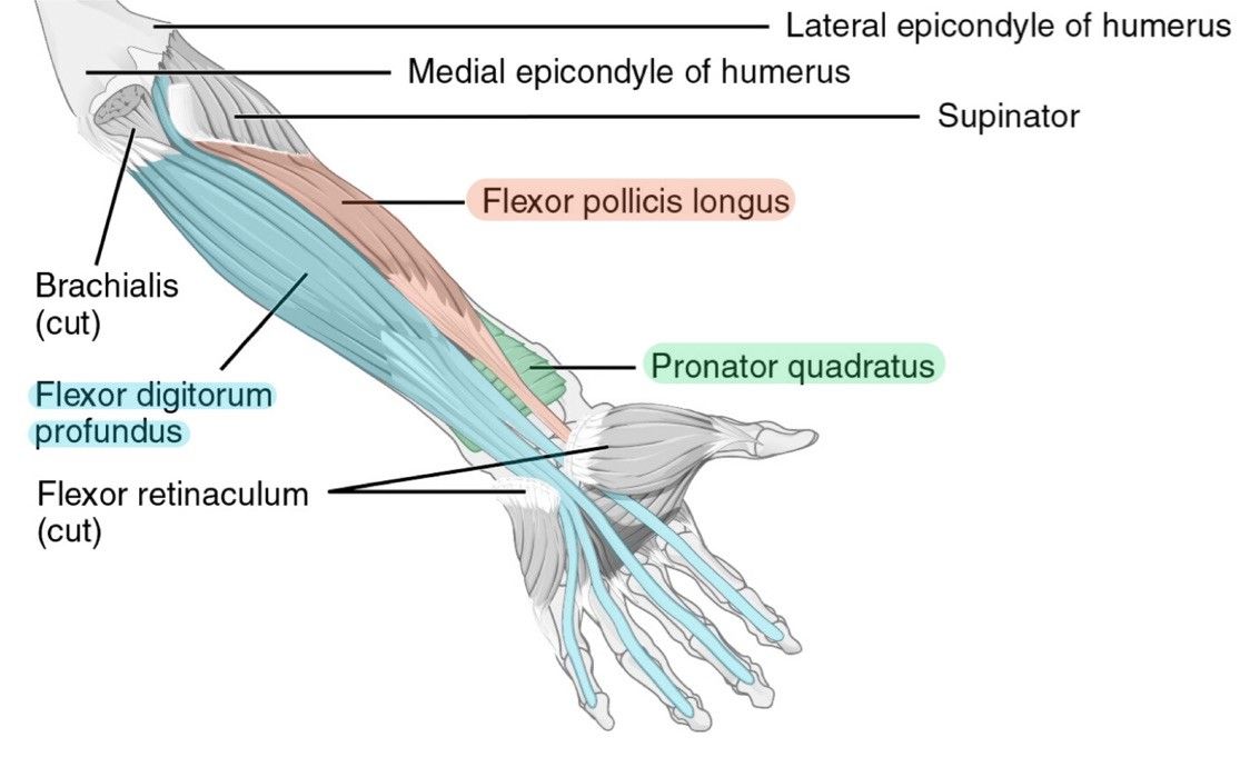
Diagram - This illustration highlights the muscles of the deep layer in the anterior compartment of the forearm
Creative commons source by CFCF, edited by Dr. Sachin Sudhakaran [CC BY-SA 4.0 (https://creativecommons.org/licenses/by-sa/4.0)]
The posterior compartment contains a few more muscles than the anterior compartment. However, it is easier to remember the innervation; all of these muscles are innervated by radial nerve. The posterior compartment can be split into superficial and deep layers.
The superficial layer has eight muscles to know (moving lateral to medial for the first seven), and they are all innervated by the radial nerve:
Brachioradialis
This muscle originates from the lateral humerus and attaches to the distal radius, just superior to the styloid process. It acts to flex at the elbow. Brachioradialis is sometimes known as the 'beer raising' muscle, because it is the muscle that helps to flex the elbow to lift a glass
Extensor Carpi Radialis Longus
This muscle originates from the lateral aspect of the humerus, just inferior to the brachioradialis and attaches to the metacarpal II. It acts to extend the wrist and also contributes to wrist abduction.
Extensor Carpi Radialis Brevis
This muscle originates from the lateral aspect of the humerus, just inferior to the brachioradialis and attaches to the metacarpal III. It acts to extend the wrist and also contributes to wrist abduction.
Extensor Digitorum
This muscle originates from the lateral epicondyle of the humerus, splits into four tendons and then attaches at the distal phalanx of each finger. It acts to extend the fingers at the MCPJs and IPJs.
Extensor Digiti Minimi
This muscle originates from the lateral epicondyle of the humerus and extends to the distal phalanx of the little finger. It acts to extend the little finger. This can be remembered by thinking of the classic pose of Dr Evil with his little finger at his mouth from the Austin Powers movies... 'I shall call him Mini-Me'...
Extensor Carpi Ulnaris
This muscle originates from the lateral epicondyle of the humerus and attaches to metacarpal V. It acts to extend at the wrist whilst contributing to wrist adduction.
Anconeus
This muscle is situated more superiorly than the other six superficial muscles. It originates from the lateral epicondyle of the humerus and attaches to the olecranon process. It acts to extend at the elbow joint.
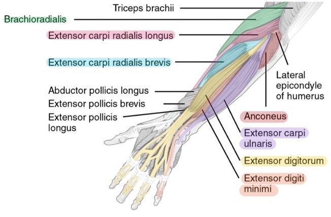
Diagram - This image highlights the muscles of the superficial layer of the posterior compartment of the forearm
Creative commons source by CFCF, edited by Dr. Sachin Sudhakaran [CC BY-SA 4.0 (https://creativecommons.org/licenses/by-sa/4.0)]
(Almost done guys, you're doing a great job!)
The deep compartment has five muscles, four of which act on the thumb and index finger.
Supinator
This muscle originates from the lateral epicondyle of the humerus and from the ulna. They insert into the posterior radius. As the name suggests, it supinates the forearm.
Extensor Pollicis Brevis
This muscle originates from the posterior radius and attaches to the proximal phalanx of the thumb, thereby acting to extend the thumb at the carpometacarpal and metacarpophalangeal joints.
Extensor Pollicis Longus
This muscle originates from the ulna and interosseous membrane. This muscle attaches to the distal phalanx of the thumb. It acts to extend the thumb.
Extensor Indicis
This muscle originates from the ulna and interosseous membrane, like the extensor pollicis longus. It attaches to the extensor expansion of the index finger and thereby acts to extend the index finger.
Abductor Pollicis Longus
This muscle originates from the interosseous membrane between the radius and ulna. It attaches to metacarpal I. As the name suggests, it abducts the thumb
The Supinator muscle is innervated by the deep radial nerve. As it passes the supinator, it changes name to the posterior interosseous nerve, which then goes on to supply the rest of the muscles in this compartment.
The muscles in the deep posterior compartment can be remembered with the acronym SEPIA: Supinator, Extensor Pollicis (longus and brevis), (extensor) Indices, Abductor (pollicis longus).
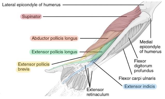
Diagram - This image highlights the muscles of the deep layer of the posterior forearm
Creative commons source by CFCF, edited by Dr. Sachin Sudhakaran [CC BY-SA 4.0 (https://creativecommons.org/licenses/by-sa/4.0)]
A way to remember the posterior forearm muscles: The Rule of Threes
3 to the wrist: extensor carpi radialis brevis, extensor carpi radialis longus, extensor carpi ulnaris.
3 to the digits: extensor digitorum, extensor digiti minimi, extensor indices.
3 to the thumb: extensor pollicis longus, extensor pollicis brevis, abductor pollicis longus.
3 others: brachioradialis, supinator, anconeus.
For Pathologies and Conditions of the Forearm, check out our article on Conditions of the Forearm and Wrist.
Reviewed by: Dr. Thomas Burnell
Edited by: Dr. Maddie Swannack
- 37831



