

Next Lesson - Muscles of the Shoulder
Core
This article will discuss five key bones that provide structure to the upper limb. These bones are often formed to allow for maximum manoeuvrability, allowing for complex movements to be performed. The following will be discussed:
- Scapula
- Clavicle
- Humerus
- Ulna
- Radius
This is a large, flat, triangular bone found posteriorly in the thorax. It articulates with the humerus and clavicle to form the shoulder girdle and allow for the movement of the shoulder joint. It is the site of attachment for many muscles and is therefore vital for normal function of the shoulder. The scapula articulates with the head of the humerus via the glenohumeral joint and articulates with the clavicle via the acromioclavicular joint.
There are three main surfaces of the scapula:
- Costal surface - region facing the ribcage
- Lateral angle - region closest to the humerus that bears the glenoid fossa
- Posterior surface - region that faces outwards
Some key bony landmarks of the scapula include:
Coracoid Process - found on the costal surface, this is a projection that lies just inferior to the clavicle. It is the site of origin for many muscles, such as the coracobrachialis and biceps brachii.
Glenoid Fossa - found on the lateral surface, it articulates with the head of the humerus to form the shoulder joint.
Supraglenoid and Infraglenoid Tubercle - regions found immediately superior and inferior the glenoid fossa respectively. These are sites of attachment for heads of muscles in the upper limb.
Spine of the Scapula - found posteriorly, it divides the posterior surface of the scapula into two regions.
Supraspinous and Infraspinous Fossae - regions found just superior and inferior to the spine of scapula respectively. They give origin to supraspinatus and infraspinatus respectively, two of the rotator cuff muscles.
Acromion - a projection/elongation of the spine of the scapula which articulates with the clavicle to form the acromioclavicular joint.

Diagram - Costal aspect of the left scapula
Creative commons source by Atlas of Human Anatomy by Professor Dr. Carl Ernest Bock, edited by Dr. Sachin Sudhakaran [CC BY-SA 4.0 (https://creativecommons.org/licenses/by-sa/4.0)]
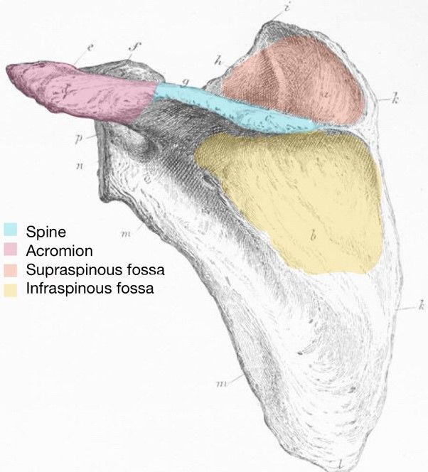
Diagram - Posterior aspect of the left scapula
Creative commons source by Atlas of Human Anatomy by Professor Dr. Carl Ernest Bock, edited by Dr. Sachin Sudhakaran [CC BY-SA 4.0 (https://creativecommons.org/licenses/by-sa/4.0)]
Also known as the collarbone, this is a long bone which articulates with both the sternum and the acromion of the scapula. It allows for movement by forming the shoulder girdle with the scapula. It provides protection by directly shielding the neurovascular structures that sit behind it, and by transmitting shock received by the hands (ie in a fall) through the chest.
There are a few landmarks to know:
- Medial end (sternal end) - a faceted region that articulates with the manubrium of the sternum.
- Shaft - allows for attachment of different muscles and is also the region most prone to fracture.
- Lateral end - location of a small facet which articulates with the acromion.

Diagram - Anterior aspect of the left clavicle. The highlighted region in the middle illustrates the most common fracture site. The articulating surfaces are highlighted on either side, with the acromial end on the left and the sternal end on the right
Creative commons source by Atlas of Human Anatomy by Professor Dr. Carl Ernest Bock, edited by Dr. Sachin Sudhakaran [CC BY-SA 4.0 (https://creativecommons.org/licenses/by-sa/4.0)]
This is a long bone within the upper limb found between the shoulder and the elbow. It articulates with the scapula to form the shoulder joint and articulates with the ulna to form the elbow joint.
Some proximal bony landmarks include:
Head of the Humerus - region that articulates with the glenoid fossa of the scapula.
Greater Tubercle - a bony ridge that is found laterally on the humerus. It is the site of attachment for three of the rotator cuff muscles and can be palpated externally.
Lesser Tubercle - a smaller ridge, that is found more medial to the greater tubercle.
Intertubercular Sulcus - a groove that runs between the two tubercles that allows for the tendon of the long head of biceps brachii to descend.
Anatomical and Surgical Neck - these necks separate the head from the shaft. The anatomical neck is found proximal to the tubercles, whilst the surgical neck is found distal to the tubercles. The surgical neck is a common site for fractures of the humerus.

Diagram - Anterior view of proximal left humerus
Creative commons source by Atlas of Human Anatomy by Professor Dr. Carl Ernest Bock, edited by Dr. Sachin Sudhakaran [CC BY-SA 4.0 (https://creativecommons.org/licenses/by-sa/4.0)]
The shaft of the humerus is the site of attachment for many muscles, and fractures of the shaft of the humerus can lead to damage to the radial nerve due to its incredibly close relation to the shaft. In fact, the radial nerve runs through a spiral groove found on the posterior humerus.
The distal end of the humerus is responsible for articulating with the ulna and the radius to form the elbow joint. Therefore, there are a few bony landmarks to know:
Capitulum - an anterior articulating region which articulates with the head of the radius. It is found lateral to the trochlea.
Trochlea - a large articulating region that is found medially and extends posteriorly. It articulates with the trochlear notch of the ulna to form the elbow joint.
Lateral and Medial Epicondyles - found proximal to the capitulum and trochlea, these are projections of the humerus that allow for sites of attachment of different muscles. The medial epicondyle is larger than the lateral epicondyle and is easily palpable. Running posterior to the medial epicondyle is the ulnar nerve (this is important because damage to the medial epicondyle can damage the ulnar nerve, which is why hitting the elbow can cause tingling in the hand).
Olecranon Fossa - found on the posterior humerus just proximal to the trochlea.
Coronoid Fossa - found on the anterior humerus and just proximal to the trochlea.
Radial Fossa - found on the anterior humerus and just proximal to the capitulum.
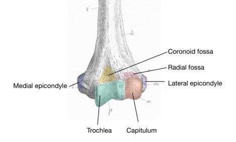
Diagram - Anterior view of distal left humerus
Creative commons source by Atlas of Human Anatomy by Professor Dr. Carl Ernest Bock, edited by Dr. Sachin Sudhakaran [CC BY-SA 4.0 (https://creativecommons.org/licenses/by-sa/4.0)]
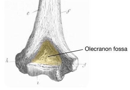
Diagram - Posterior view of distal left humerus
Creative commons source by Atlas of Human Anatomy by Professor Dr. Carl Ernest Bock, edited by Dr. Sachin Sudhakaran [CC BY-SA 4.0 (https://creativecommons.org/licenses/by-sa/4.0)]
One of two bones in the forearm, this long bone lies medially to the radius. It acts to provide leverage for elbow movement.
Proximally, there are a few key landmarks to allow for effective articulation with the humerus to form the elbow joint:
Trochlear Notch - a large depression that articulates with the trochlea.
Olecranon - a large projection that forms part of the trochlear notch. It can be easily palpated (known as the tip of the elbow) and is the site of attachment for the triceps brachii muscle.
Coronoid Process - an anterior projection that also forms part of the trochlear notch.
Radial Notch - located laterally, this articulates with the radius to form the proximal radioulnar joint.
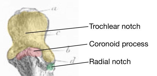
Diagram - Anterior surface of proximal left ulna
Creative commons source by Atlas of Human Anatomy by Professor Dr. Carl Ernest Bock, edited by Dr. Sachin Sudhakaran [CC BY-SA 4.0 (https://creativecommons.org/licenses/by-sa/4.0)]
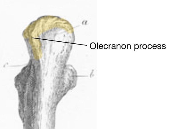
Diagram - Posterior surface of proximal left ulna
Creative commons source by Atlas of Human Anatomy by Professor Dr. Carl Ernest Bock, edited by Dr. Sachin Sudhakaran [CC BY-SA 4.0 (https://creativecommons.org/licenses/by-sa/4.0)]
The ulna tapers off to its distal end, and whilst the shaft is the site of attachment for a few muscles, it also has an interosseous border for attachment of the interosseous membrane.
The distal end of the ulna is significantly smaller than the proximal end. The head of the ulna can be found here and it articulates with the ulnar notch of the radius, forming the distal radio-ulna joint. There is also a large bony projection found medially, called the ulnar styloid process.
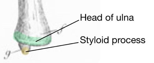
Diagram - Anterior surface of distal left ulna
Creative commons source by Atlas of Human Anatomy by Professor Dr. Carl Ernest Bock, edited by Dr. Sachin Sudhakaran [CC BY-SA 4.0 (https://creativecommons.org/licenses/by-sa/4.0)]
Like the ulna, this is another long bone in the forearm and is found laterally. It rotates around the ulna, allowing supination and pronation of the forearm at the proximal and distal radioulnar joints.
Proximally is the head of the radius - a disk-like structure, which articulates with the capitulum of the humerus. It also articulates with the radial notch of the ulna. The radial head rotates within the annular ligament at the proximal radioulnar joint. The radial tuberosity is the insertion of biceps brachii, which contributes to active forearm supination.
The shaft provides attachment for muscles and the interosseous membrane, which connects the radius and ulna.
Distally, the radius articulates with the scaphoid and lunate bones in the hand, forming the wrist joint. On the lateral surface, there is a radial styloid process. The medial surface is concave to allow for articulation with the head of the ulna via the ulnar notch.
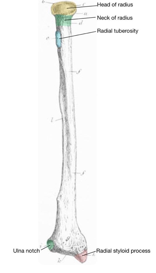
Diagram - Anterior surface of left radius
Creative commons source by Atlas of Human Anatomy by Professor Dr. Carl Ernest Bock, edited by Dr. Sachin Sudhakaran [CC BY-SA 4.0 (https://creativecommons.org/licenses/by-sa/4.0)]
For Pathologies and Conditions of the Upper Limb, check out our articles on Conditions of the Shoulder and Clavicle, Arm, Elbow and Forearm and Wrist.
Reviewed by: Dr. Thomas Burnell
Edited by: Dr. Maddie Swannack
- 8872
