

Next Lesson - Urinary Tract Infections
Core
Causes of Urinary Tract Obstruction
A urinary tract obstruction is any obstruction that occurs to the outflow of urine. This can happen at any level in the urinary tract, from the kidneys to the urethral opening.
The causes of urinary tract obstruction can be categorised into supra-vesical (above the bladder, i.e. ureteric) or infra-vesical (below the bladder i.e. urinary retention) pathologies. The causes are outlined below and will be discussed in more detail later.
Supra-vesical obstruction can be caused by either an extraluminal (external compression of the ureter) or intraluminal (pathology within the ureter or ureteric walls) abnormality.
- Retroperitoneal malignancy - malignancy originating in the retroperitoneal region or lymph node metastases commonly from breast, gynaecological, and prostate cancer.
- Direct obstruction by a tumour - bladder cancer can obstruct the ureters at the vesicoureteric junction (VUJ). Locally advanced prostate cancer can also obstruct the urethra.
- Retroperitoneal fibrosis
The most common intraluminal cause of ureteric obstruction is renal colic. This is usually caused by a stone within the urinary tract, but can also be caused by blood clots (clot colic) or sloughed papilla (necrosis of the renal papillae in which dead cells are discarded into the lumen of the ureter).
This is a rare condition in which excess connective tissue forms in the retroperitoneal space of the abdomen.
There are many causes:
- Idiopathic
- Malignancy - breast, lung, stomach, lymphoma, and others
- Autoimmune
- Drugs
- Abdominal Aortic Aneurysm (AAA)
This fibrosis can cause compression of the ureters as they (along with the kidneys) are retroperitoneal structures. This is therefore a cause of extra-luminal, supra-vesical obstruction.
Treatment for this includes surgical decompression of the ureters, exclusion of a possible underlying malignancy (which may require biopsies) and steroids/immunosuppression.
Renal colic is a very common cause of acute ureteric obstruction. Classically, it will present with colicky pain (pain which comes on in waves) with a loin to groin radiation, and haematuria, which may be non-visible (apparent only on urinalysis).
By far the most common cause of renal colic is kidney stones - though it may be caused by blood clots, and sloughed papillae. This usually occurs unilaterally, although it can be bilateral. Pyonephrosis can develop if there is an infection of the collecting system proximal to the obstruction.
Risk Factors for Kidney Stones
The following are risk factors for the development of kidney stones:
- Male Gender - men have a greater incidence of kidney stones compared to women
- Age - there are two peak ages for developing kidney stones: in the 20’s and 50’s
- Metabolic - hypercalcaemia can predispose a patient to develop kidney stones
- Family History
- Dehydration
- Hypertension
- Increased BMI
- Anatomical Abnormalities - for example: polycystic kidney disease, horseshoe kidney, and pelvoureteric junction (PUJ) stenosis
Kidney stones can be differentiated based on their chemical make-up:
- Calcium Oxalate - the most common type of stone
- Calcium Phosphate - includes apatite and brushite minerals
- Struvite Stones - magnesium ammonium phosphate stones associated with urease-producing bacteria and alkaline urine
- Uric Acid Stones - favoured by persistently acidic urine; ammonium urate stones form under different biochemical conditions
- Hydroxyapatite - a calcium phosphate mineral, so this overlaps with the calcium phosphate category above
- Cystine Stones - occur in cystinuria, an inherited transport disorder causing excessive urinary cystine; cystine is less soluble in acidic urine
One imaging technique is KUB Radiography (x-ray of the kidneys, ureters, and bladder). This will allow for the identification of calcium containing stones as these are radiopaque (so will appear white on x-ray). On plain KUB radiography, uric acid stones are radiolucent, whereas cystine stones have poor radiopacity and may be difficult to see. A negative KUB does not exclude a stone. Non-contrast CT KUB is more sensitive for stone detection; the choice of imaging depends on the clinical situation.
Kidney stones are likely to obstruct the ureter in three places, where the renal tract is the narrowest:
- Pelviureteric Junction (PUJ) - the junction between the renal pelvis and the ureter. The ureter is significantly smaller in diameter than the renal pelvis so stones can become stuck here.
- Pelvic Brim - this is where the ureters cross over the top of the common iliac arteries. This is an area of narrowing so again stones can become lodged here.
- Vesicoureteric Junction (VUJ) - the junction between the ureters and the bladder. The acute angle of entry of the ureters into the bladder causes a ‘u-bend’ which is a common site for stones to become lodged.
Some kidney stones will pass through the urinary tract without significant problems due to their small size, and the only management needed may be adequate analgesia and good hydration.
Surgical management may be needed in some cases:
- Stone is larger than 5mm
- Patient is in uncontrolled pain
- Bilateral kidney stones causing obstruction
- The patient only has one kidney which is obstructed by a stone
Stones can be treated surgically via laser lithotripsy (using a laser inside the ureters to break up the stone) or extracorporeal shockwave lithotripsy (ESWL [a non-invasive procedure aiming shockwaves at the location of the kidney stone]). ESWL has a number of risks, and is contraindicated in acute urinary tract infection or urosepsis, uncorrected bleeding disorders or coagulopathies, pregnancy, and abdominal aortic aneurysm.
Pyonephrosis is the accumulation of pus within an obstructed renal collecting system, and this is a urological emergency. Failure to decompress the obstruction may lead to death from sepsis or a permanent loss of renal function. Pyonephrosis should be suspected in patients with an obstructed kidney which develop signs of sepsis.
Pyonephrosis requires urgent drainage of the obstructed collecting system by a ureteric stent or percutaneous nephrostomy, alongside immediate antibiotics and sepsis management. Obtain urgent senior urological input to arrange source control; antibiotics alone do not relieve the infected obstruction.
Pelviureteric Junction (PUJ) Obstruction
PUJ obstruction is impaired drainage from the renal pelvis into the ureter at their junction. It may result from anatomical narrowing or external compression, or functional impairment of peristalsis in the junctional segment. It is usually present from birth, but may appear or be recognised later. This can present with antenatal hydronephrosis which is picked up on an ultrasound scan, but it can also present at any stage of life. It can be asymptomatic, so only found incidentally on imaging, but the classical presentation is loin pain that is worse after heavy fluid intake or alcohol consumption.
When surgical correction is needed, pyeloplasty can be performed using a laparoscopic or open approach. This is the surgical reconstruction of the renal pelvis in an attempt to relieve the PUJ obstruction.
Infra-vesical Obstruction - Urinary Retention
Urinary retention can be classified as acute or chronic, with chronic urinary retention further split into high-pressure and low-pressure retention.
Some causes of urinary retention are specific to male and female anatomy, and some causes are common to both.
Causes of urinary retention in men:
- Benign prostatic hyperplasia (BPH)
- Prostate cancer
- Urethral strictures
- Urinary tract infection
- Constipation
- Neurological dysfunction
- Recent surgery
- Drugs
Causes of urinary retention in women:
- Urethral stenosis
- Pelvic masses
- Urinary tract infection
- Constipation
- Neurological dysfunction
- Recent surgery
- Drugs
Acute urinary retention presents with a painful inability to void and a residual volume (volume left in bladder after attempting to void) of 300-1500ml. This is incredibly painful for patients, so catheterisation is essential both to relieve pain and to prevent complications resulting from the backing up of urine.
Causes of acute urinary retention include:
- BPH - most common cause in men
- Urethral strictures
- Prostate cancer
- Urinary tract infection
- Constipation
- Severe pain
- Medication - antimuscarinic medications and spinal anaesthesia can affect the innervation of the bladder which can cause retention
- Neurological - iatrogenic damage during pelvic surgery, peripheral neuropathy, upper motor neurone diseases (e.g. Multiple Sclerosis and Parkinson’s disease), and Bladder-Sphincter Dyssynergia (a disturbance in the co-ordination of the contraction of the detrusor muscle and the relaxation of the urethral sphincter) can all cause acute urinary retention
Treatment of acute urinary retention involves:
- Catheterising and recording the volume of urine drained on catheterisation.
- A full examination including abdominal exam, external genitalia exam, and a digital rectal examination (DRE).
- Urine Dip.
- Urea and Electrolytes (U&Es).
- Treat any obvious underlying causes - conditions like constipation can cause acute urinary retention. Not treating the underlying cause can result in another episode of retention.
- Alpha-blockers - these can be useful in men for the relaxation of the prostate.
Patients with chronic urinary retention have a long-standing reduced ability to void their bladders. This stretches the detrusor muscle over time and desensitises it, which means that this type of retention is painless. Patients may still be able to void small amounts of urine but are likely to suffer from weak stream and hesitancy. Larger volumes of urine can be retained, from 300-4000ml.
In men, it can be commonly caused by BPH, urethral strictures, or prostate cancer. In women, common causes include pelvic prolapse or pelvic masses. Neurological causes can affect both men and women and include peripheral neuropathy as well as upper motor neurone diseases, like MS and Parkinson's disease.
Chronic urinary retention can be categorised into high-pressure or low-pressure retention.
In high-pressure retention, the pressure of the retained fluid is so high that it can cause damage to the kidneys, resulting in hydronephrosis and abnormal urea and electrolytes (U&Es). Low-pressure retention will not impact renal function and does not cause hydronephrosis.
The management of chronic urinary retention involves long-term catheterisation (especially in high-pressure retention). Intermittent self-catheterisation (where patients are taught to place and change catheters themselves) can be used for patients who want to avoid a long-term catheter.
After treatment, patients with urinary retention should be monitored for complications resulting from post-obstructive diuresis. This starts as a physiological response to retention as accumulated salt and water are removed from the body. In a few cases, this can become excessive and severe, leading to dehydration and electrolyte imbalance.
Careful monitoring of fluid input and output and oral fluid replacement are usually enough to manage post-obstructive diuresis, however, some patients may need intravenous fluids.
Patients with chronic urinary retention can develop acute retention. This can be caused by an acute deterioration in the condition causing the chronic retention, or a new condition on top of the previous one. These patients will have very large residual volumes but still have minimal discomfort due to the desensitisation of the bladder. They should be treated as per the acute retention guidelines, but these patients are more at risk of post-obstructive diuresis so will need careful monitoring.
Obstructive uropathy is a structural or functional hindrance to normal urine flow. Kidney dysfunction resulting from obstruction is termed obstructive nephropathy; renal impairment is not required to define obstructive uropathy. Situations in which obstruction can impair overall kidney function include:
- Bilateral ureteric obstruction
- Unilateral ureteric obstruction in a patient with one kidney
- High-pressure chronic retention
Obstruction complicated by infection, anuria or acute kidney injury requires urgent assessment and relief of the obstruction as indicated. Obstructive nephropathy can cause post-renal AKI and hyperkalaemia.
Treatment involves correcting the underlying cause of the obstruction, and monitoring for the complications of AKI and hyperkalaemia. Upper tract obstructions can be drained using a JJ-stent (a tube going from the renal pelvis to the bladder) or a nephrostomy (a tube from the renal pelvis that comes out through a port in the skin).
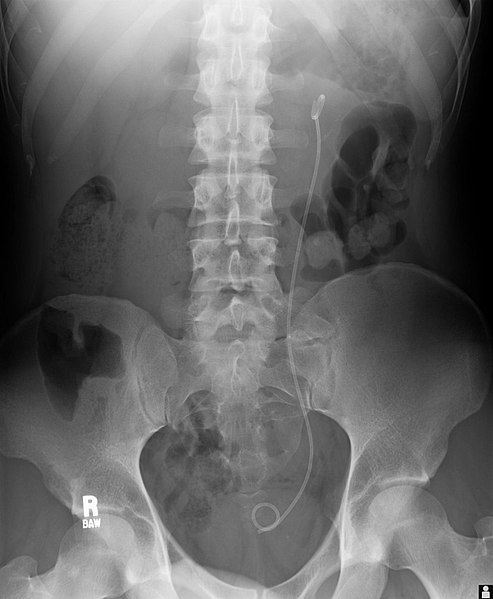
Image - An abdominal x-ray with a left JJ stent
Public Domain Source by Lucid Smog [Public domain]
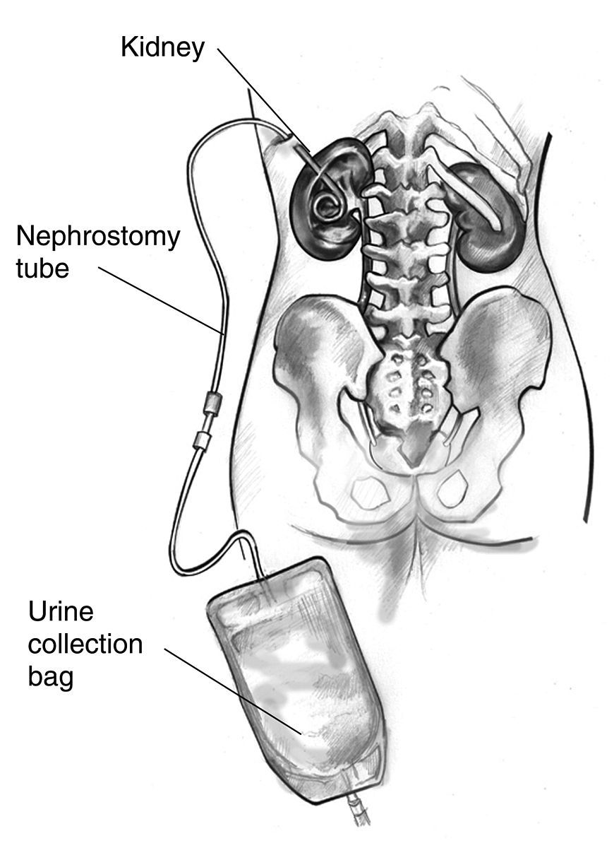
Diagram - A nephrostomy tube in the left kidney, connected to a urine collection bag
Public Domain Source by United States Department of Health and Human Services [Public domain]
Edited by: Dr. Maddie Swannack
Reviewed by: Dr. Thomas Burnell
- 5450


