

Next Lesson - Bones of the Lower Limb
Core
There are four key areas within the lower limb. Moving inferiorly, they are connected with each other (this may prove to be helpful when it comes to remembering the contents of each area). From most superior to most inferior, we have:
- The femoral triangle
- The femoral canal
- The adductor canal
- The popliteal fossa
This is a large region located on the anterior aspect of the thigh. It is important to understand the location of this site as doing so allows for identification of the femoral pulse and access to the femoral artery, which is critical for procedures such as coronary angiography.
Borders of the Femoral Triangle
There are three main borders to know:
- Superior border - Inguinal ligament
- Lateral border - Medial aspect of Sartorius
- Medial border - Medial aspect of Adductor Longus
These can be remembered by the acronym SAIL: Sartorius, Adductor (longus), Inguinal Ligament
The femoral triangle also has a roof and a floor:
- Roof - Fascia lata
- Floor - Pectineus, Iliopsoas and Adductor Longus (the muscles indicate just how superficial the femoral triangle is relative to other key structures within the thigh)
Contents of the Femoral Triangle
There are four main structures found within the femoral triangle. Moving lateral to medial:
- Femoral Nerve
- Femoral Artery
- Femoral Vein
- Femoral Canal (containing lymphatic vessels and lacunar lymph nodes)
The femoral artery, vein and canal are all enclosed within a fascial layer - the femoral sheath.
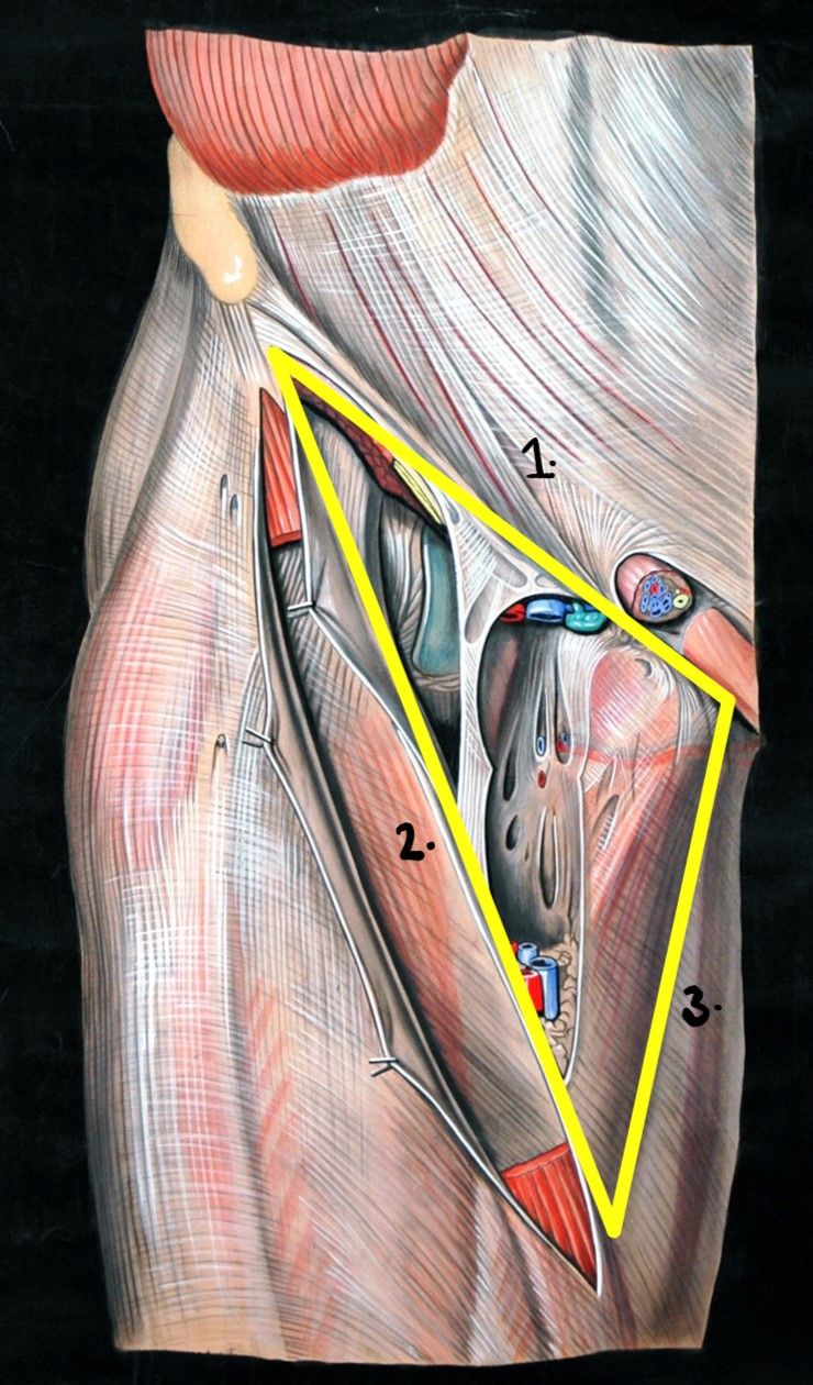
Diagram - The yellow triangle highlights the location of the Femoral triangle. The numbers indicate the relative borders (1. Inguinal Ligament, 2. Medial aspect of Sartorius, 3. Medial aspect of Adductor Longus)
Creative commons source by Elisa Schorn, circa 1900, edited by Dr. Sachin Sudhakaran [CC BY-SA 4.0 (https://creativecommons.org/licenses/by-sa/4.0)]
An easy way to remember the contents of the femoral triangle is with the acronym NAVEL (Nerve, Artery, Vein, Empty space, Lymphatics (the latter 2 of which both make up the femoral canal)).
Another helpful acronym is NAVY - Nerve, Artery, Vein, Y-fronts (this helps you to remember that the nerve is most lateral, furthest from the genitalia).
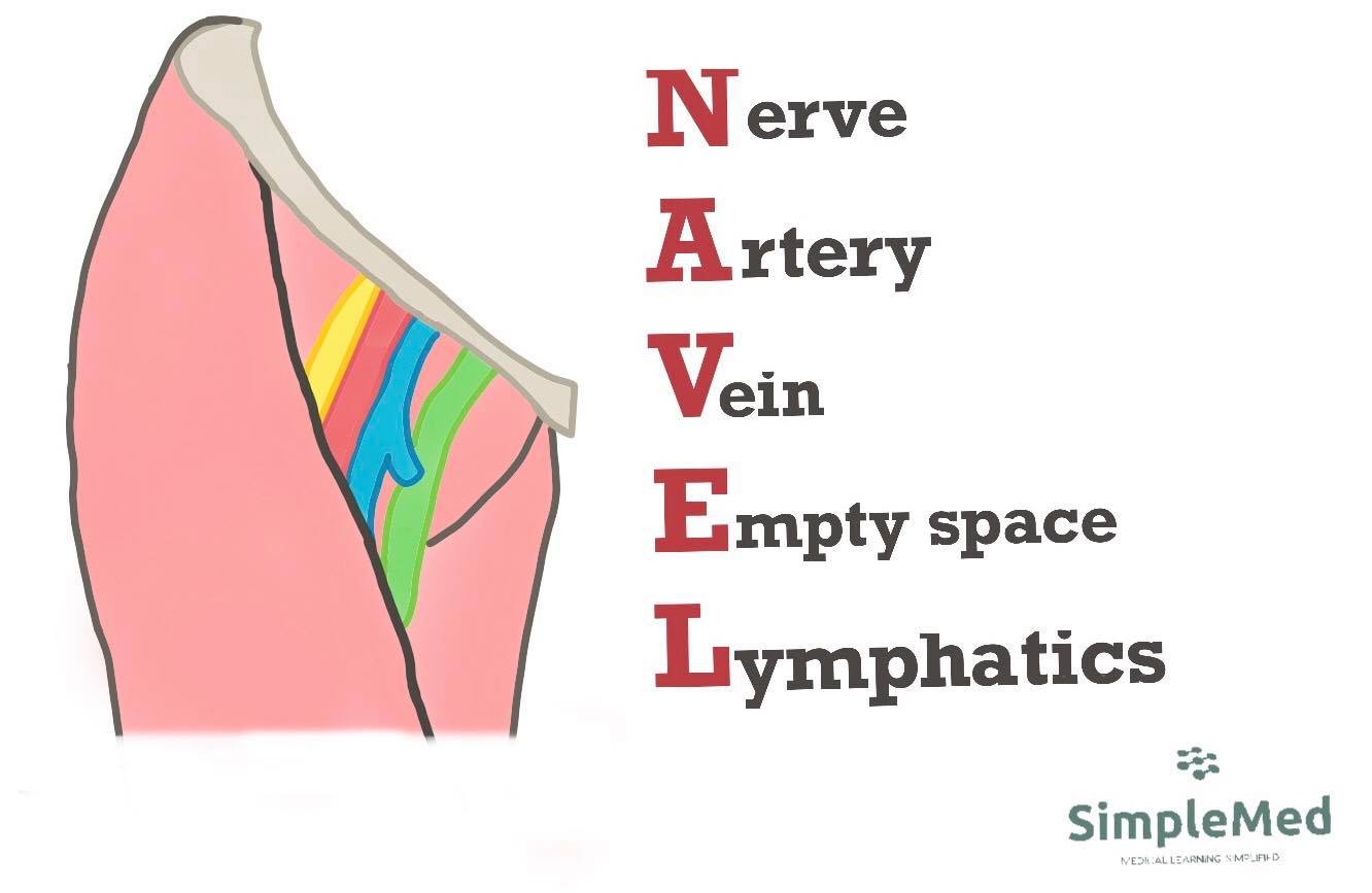
Image - Visual representation of the NAVEL mnemonic, from left to right (lateral to medial)
SimpleMed original by Dr. Emily Smith
This is located medially within the femoral triangle and is a common site of femoral hernias, whereby the abdominal contents move down into the femoral canal. This is because the canal also contains a region of potential space into which structures can enter. Due to the rigidity of the borders forming the femoral canal, herniated abdominal contents are at high risk of strangulation, where compromised blood supply can occur. This is a surgical emergency.
Borders of the Femoral Canal
The femoral ring is the proximal entrance to the canal. Its four boundaries are:
- Anterior border - Inguinal ligament
- Posterior border - pectineal ligament over the superior pubic ramus, with pectineus behind
- Medial border - Lacunar ligament
- Lateral border - Femoral vein
Contents of the Femoral Canal
As mentioned before, the femoral canal contains multiple lymphatic vessels and deep lymph nodes (including the lacunar node). The empty space within the femoral canal allows for slight distention without complete obstruction to the surrounding vessels - this is useful because it allows the lymph nodes to swell in infection without obstructing blood flow.
This region is found inferiorly to the femoral triangle - it is a continuation of the bottom point of the femoral triangle, and so sits in the anterior thigh.
Borders of the Adductor Canal
There are three borders:
- Anteromedial border - sartorius
- Lateral border - vastus medialis
- Posterior wall - adductor longus proximally and adductor magnus distally
Contents of the Adductor Canal
As this canal is a continuation of the Femoral triangle, there are similarities in terms of content:
- Femoral Artery
- Femoral Vein
- Saphenous Nerve (cutaneous branch of the femoral nerve)
- Nerve to Vastus Medialis
Once the Femoral artery and vein leave the adductor canal, they become the popliteal artery and vein.
It is important to understand the location of the adductor canal, because hypertrophy of the muscles forming the borders of this canal can lead to Adductor Canal Compression Syndrome. This can result in symptoms associated with obstructed arteries (claudication) or nerves (e.g. paraesthesia).
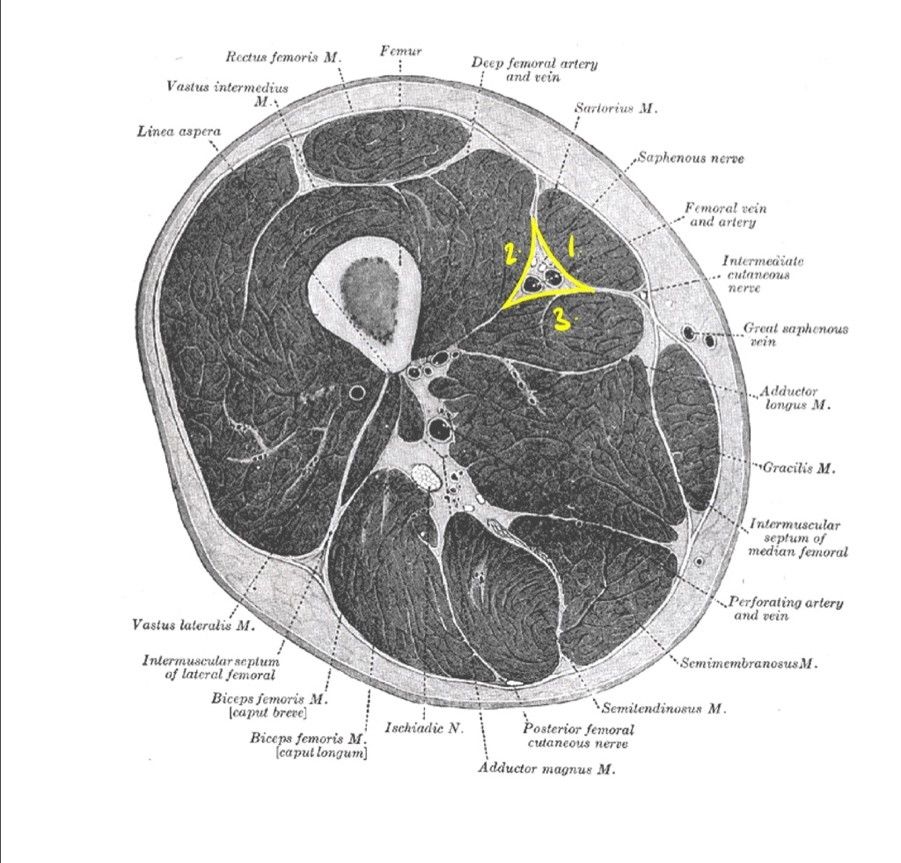
Diagram - This image shows a cross section of the left thigh. The highlighted triangle indicates the location of the adductor canal. The numbers indicate the muscles forming the borders of this canal (1. Sartorius, 2. Vastus Medialis, 3. Adductor Longus)
Public Domain Image by Henry Vandyke Carter, edited by Dr. Sachin Sudhakaran [Public domain]
This is a diamond-shaped region located posterior to the knee. This site allows access to the popliteal artery, meaning it is the best site to palpate the popliteal pulse. Complications concerning the popliteal fossa include Baker’s cysts and popliteal aneurysms.
Borders of the Popliteal Fossa
There are two superior borders and two inferior borders:
- Superomedial border - Semimembranosus (semimembranosus is medial)
- Superolateral border - Biceps femoris
- Inferomedial - Medial head of Gastrocnemius
- Inferolateral - Lateral head of Gastrocnemius
The floor of the popliteal fossa is the posterior aspect of the knee joint, whilst the roof is composed of popliteal fascia and skin.
Contents of the Popliteal Fossa:
The main central bundle is ordered superficial to deep:
- Tibial nerve (branch of the sciatic nerve)
- Popliteal vein
- Popliteal artery
Their side-to-side relationships vary along the course. The common fibular nerve (also known as the common peroneal nerve) follows the lateral margin with biceps femoris.
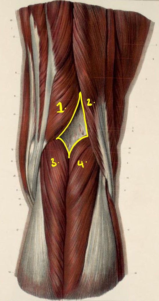
Diagram - This image illustrates the posterior aspect of the right lower limb. The yellow diamond highlights the region of the popliteal fossa. The numbers indicate the muscles that form the borders (1. Semimembranosus, 2. Biceps Femoris, 3. Medial head of Gastrocnemius, 4. Lateral head of Gastrocnemius)
Public Domain Source, edited by Dr. Sachin Sudhakaran [Public domain]
Edited by: Dr. Marcus Judge and Dr. Maddie Swannack
- 9789
