

Next Lesson - The Nephron
Core
The development of the urinary system is closely related to the development of the reproductive system, as they both begin their development as a portion of intermediate mesoderm called the urogenital ridge.
The development of the kidneys in the embryo involves the formation of three separate systems, with the degeneration of the last marking the onset of development of the next. They are all derived from the urogenital ridge, the portion of intermediate mesoderm that also gives rise to the reproductive system. This portion of mesoderm is specifically known as the nephrogenic cord.
The pronephros begins development around the start of the fourth week of gestation in the cervical region of the embryo.
Divisions of intermediate mesoderm form between 6 and 10 pairs of tubules named nephrotomes. These nephrotomes join the pronephric duct, which is a duct that extends from the cervical region to the cloaca (the membrane at the distal end of the embryo).
The pronephros is never functional, and regresses completely by the end of the fourth week of gestation.
The mesonephros is the second embryonic kidney system to develop, which starts development caudally (inferiorly) to the pronephros. The pronephric duct induces the development of the mesonephros from the nearby mesoderm in the thoracolumbar region.
These tubules receive a blood supply from the aorta allowing for filtration of blood. They drain into the mesonephric duct, a continuation of the pronephric duct. This mesonephric duct goes on to form the internal genitalia of the male under the influence of testosterone.
The mesonephros acts as the excretory system for the embryo for weeks four to eight. Around 2 months of gestation, the ureteric bud develops on the caudal end of the mesonephros, prompting the development of the metanephros (the definitive kidney).
The metanephros is the definitive kidney, meaning this is the same kidney that exists into adulthood.
It appears in around the 5th week of development and becomes functional at around 12 weeks of gestation. Its formation is prompted when the ureteric bud comes into contact with an area of intermediate mesoderm.
The metanephros has two components:
Collecting System
The ureteric bud begins as a structure branching off the mesonephric duct. It begins forming when the mesonephric duct comes into contact with the urogenital sinus (a part of the cloaca - see later).
The collecting system is derived from the ureteric bud, which dilates to form the ureter, renal pelvis, calyces and the collecting tubules.
Excretory System
The excretory system is formed from the intermediate mesoderm.
It develops to form the nephron - the Bowman’s Capsule, proximal convoluted tubule, loop of Henle, and distal convoluted tubule.
Anatomical Migration
The metanephros develops at the caudal end of the embryo. This means that to get to the anatomical position in an adult (the lumbar region) the kidney needs to migrate superiorly. As it does this, it forms multiple new branches of the aorta to provide its blood supply. This is different to the testes, which retain their original arterial supply during descent, leaving a long testicular artery compared with the renal arteries.
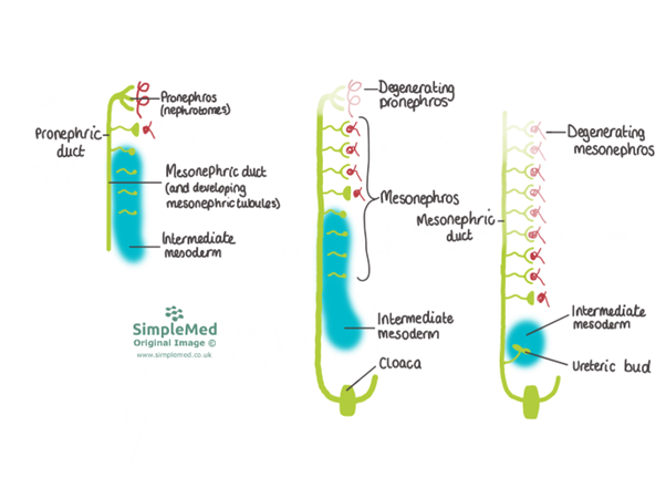
Diagram - The Three Stages of Embryological Kidney Development
SimpleMed original by Dr. Maddie Swannack
Development of the Bladder and Urethra
The bladder and urethra are primarily formed from the cloaca - the structure that is the common outflow for the urinary, reproductive and gastrointestinal tracts.
Between the fourth and seventh weeks of development, the cloaca divides into two portions separated by the uro-rectal septum. This septum ruptures through the cloacal membrane further into development, allowing the hindgut to open.
The posterior portion of the cloaca becomes the anal canal.
The anterior portion becomes the urogenital sinus. This further divides into three:
- The superior portion forms the bladder.
- The pelvic portion forms the urethra in females, and the prostatic and membranous portions of the urethra in males. It also contributes to the female reproductive system.
- The caudal portion forms the spongy (penile) urethra in males, and contributes to the female reproductive system.
Initially, the bladder is drained by the allantois, a structure inside the connecting stalk that links the bladder to the yolk sac. This structure later becomes the urachus and eventually fibroses to form the median umbilical ligament, which can be found in adults.
As the bladder grows, it begins to grow into the mesonephric ducts. This gives rise to the bladder trigone, a triangle of tissue on the inside of the bladder made up of the two ureteric orifices superiorly and the internal urethral orifice inferiorly.
What happens to the rest of the mesonephric duct from this point varies between sexes: in males, due to hormones produced by the testicles, the mesonephric ducts persist and go on to form the internal genitalia of the male. In women, without the hormones produced by the testicles, they degenerate.
Abnormalities in Development of the Urinary System
As with the development of any system, there are a number of things that can go wrong. This is by no means an exhaustive list.
Renal Agenesis - if the ureteric bud fails to properly interact with the mesoderm, the kidney tissue will not develop. If this happens on both sides, it is incompatible with life as the blood cannot be filtered.
Migration Errors - as the metanephros forms in the pelvic region, there may be difficulties in its migration to the lumbar region. This can lead to a perfectly functional kidney, just in the pelvis.
Horseshoe Kidney - this is a congenital abnormality, where the inferior pole of both kidneys are fused together, forming one big kidney.
Duplication Defects - if the ureteric bud splits early in development, a kidney may have more than one ureter attaching to it.
Accessory Renal Arteries - when the kidneys ascend into the lumbar region, they normally form new arteries and degenerate their old ones. In this condition, the old ones do not degenerate, meaning the kidney has multiple arteries supplying it.
Persistent Cloaca - a defect in the uro-rectal septum formation or in the splitting of the urogenital sinus. A persistent cloaca describes a common outflow tract for two or more of the urinary system, the reproductive system or the gastrointestinal tract.
Edited by: Dr. Marcus Judge
Reviewed by: Adrian Judge
In this article
The embryo develops three different kidney systems, each degenerating before the formation of the next.
- 742
