

Next Lesson - Cells and Components of the Nervous System
Core
Neuroanatomy is a very complicated thing, as the brain is the most complicated organ in the human body. It is also closely related to the anatomy of the head and neck, and articles on this can be found in our Head and Neck unit here.
Basic Components of the Nervous System
The largest division in the nervous system is between the central and peripheral nervous systems.
The central nervous system includes the cerebral hemispheres (brain), brainstem, cerebellum, and the spinal cord.
The peripheral nervous system includes the dorsal and ventral roots of the spinal nerves and any peripheral nerves - basically anything that is not part of the brain or the spinal cord!
Grey matter and white matter are terms used to describe different portions of the central nervous system depending on which cell types are located within that portion. For more information on the different cell types of the nervous system, please see here.
Grey matter is made up of cell bodies and dendrites within the central nervous system and is highly vascular (meaning it has a good blood supply). It also contains axons for communication with the white matter, but these are low in number.
White matter is made up of axons and their supporting cells only. The name ‘white matter’ occurs due to the fatty myelin sheath of the axons, which appears white on the cross section.
Peripheral Nervous System ‘Matter’
The terms grey matter and white matter are specific to the central nervous system, so cannot be used when referring to the peripheral nervous system.
The peripheral nervous system equivalent to grey matter is a ‘ganglion’, because this is a collection of cell bodies in the peripheral nervous system.
The equivalent term to white matter in the peripheral nervous system is a peripheral nerve, as this is the name for an axon or collection of axons.
The spinal cord is composed of 31 spinal segments, each supplying a specific region of skin with sensory innervation (dermatome) and a specific muscle group with motor innervation (myotome) on each side of the body. Each segment connects with a spinal nerve through two nerve roots that originate in the spinal cord: the dorsal (sensory) root and the ventral (motor) root.
The spinal cord has a central core of grey matter with an outer shell of white matter.
There are a number of divisions of white matter in the spinal cord based on where the axons in the white matter are travelling to or from.
A tract is an anatomically and functionally defined portion of white matter that connects two distinct regions of grey matter. Impulses in a tract travel down that collection of axons in one direction, and it can only be either motor or sensory.
A fasciculus (plural fasciculi) is a subdivision of a tract supplying one distinct portion of the body.
A funiculus (plural funiculi) is a segment of white matter that contains multiple distinct tracts. Because there are multiple tracts present, impulses can travel in different directions.
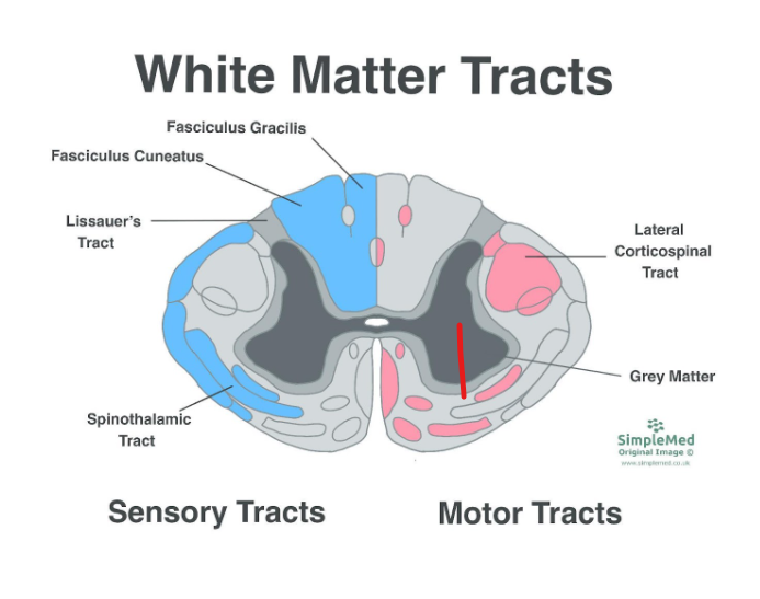
Diagram - Cross Section of the Spinal Cord
SimpleMed original by Dr. Maddie Swannack
In the above diagram, the sensory tracts are shown on the left (in the blue) and the motor tracts are shown on the right (in the pink). It is important to remember that all tracts occur on both sides of the spinal cord, as marked with the outlines.
Similarly to the white matter in the spinal cord, the grey matter is organised into regions which are the equivalent of tracts. These columns are numbered, called Rexed’s Laminae.
The most important region of grey matter in the spinal cord is the ventral horn, and it is easiest to imagine this as a column of ventral horns that link the ventral horns of all spinal levels. Different levels of the ventral horn ‘column’ supply different specific muscles, for example the level L1-L2 supplies the iliopsoas muscle.
A nucleus is a grey matter structure comprised of a collection of functionally related cell bodies.
The cortex is a folded sheet of cell bodies found on the surface of the brain. It is between 1 and 5 mm thick and is a grey matter structure (because it is comprised of cell bodies).
A fibre is a term used to describe an axon and its supporting cells. In the CNS, the supporting cells are oligodendrocytes.
An association fibre connects cortical regions within the same hemisphere.
A commissural fibre connects the left and right hemispheres of the brain, or the left and right halves of the cord.
A projection fibre connects the cerebral hemispheres with the cord or brainstem, and vice versa.
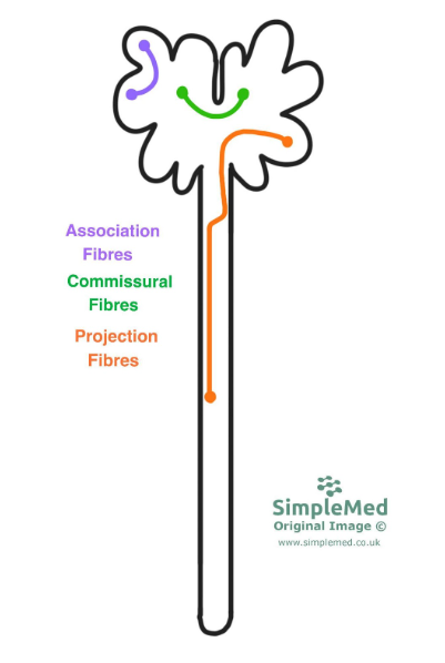
Diagram - The Three Main Types of Fibre
SimpleMed original by Dr. Maddie Swannack
The brainstem is split into three portions:
The midbrain is the most superior portion of the brainstem. It is responsible for eye movements and reflex responses to sound and vision.
The pons is the central portion of the brainstem. It is involved in the regulation of sleep and arousal.
The medulla is the most inferior portion of the brainstem. It is responsible for the co-ordination of a major motor pathway (relying on the medullary pyramids), and contains the respiratory and cardiovascular centres.
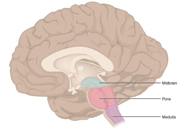
Diagram - The Midbrain Structures
Licensed under Creative Commons, Source.
The gyri are the raised areas of the cerebral cortex, with the sulci between them. Together, they make up the bumpy surface of the brain that is familiar.
There are a number of important landmarks:
Central Sulcus - sits in the coronal plane and separates the frontal and parietal lobes.
Precentral Gyrus - contains the primary motor cortex.
Postcentral Gyrus - contains the primary sensory cortex.
Lateral or Sylvian Fissure - separates the temporal lobes from the parietal and frontal lobes.
Calcarine Sulcus - the primary visual cortex surrounds this sulcus.
Parahippocampal Gyrus - visible on the inferior aspect of the brain, the parahippocampal gyrus is a key cortical region for memory coding.
Cingulate Gyrus - visible on the midline, the cingulate gyrus is responsible for emotion and memory.
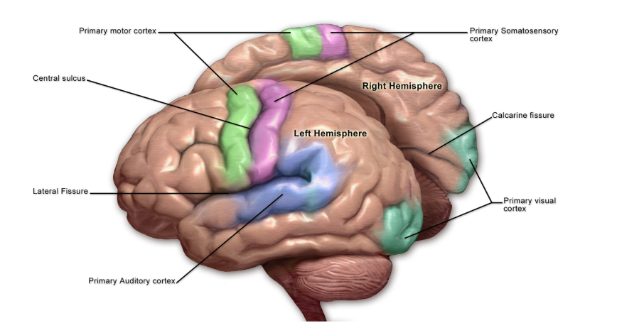
Diagram - The Cortices of the Brain
Licensed under Creative Commons, Source.
The optic chiasm is the site in which the fibres of the visual system cross.
The uncus is a region of the cerebral cortex of the temporal lobe that can herniate through the tentorium cerebelli in conditions of raised intracranial pressure and compress the midbrain structures.
The medullary pyramids are paired structures within the medulla oblongata, visible on the inferior aspect of the brain, and contain the descending corticospinal motor tracts.
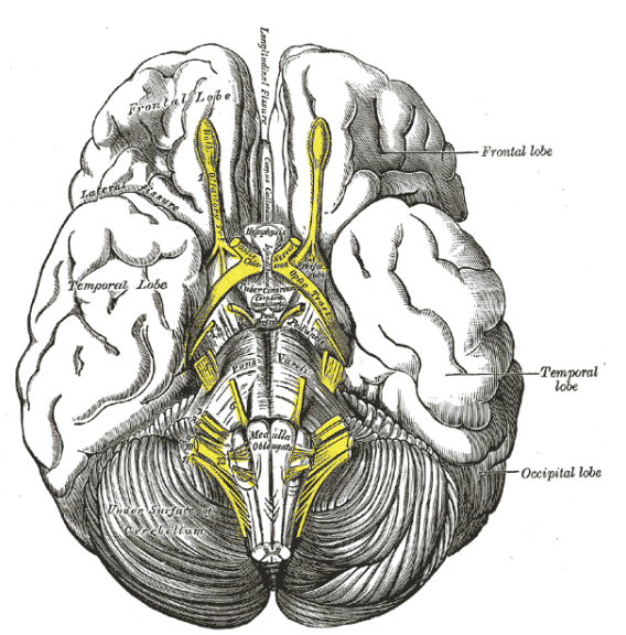
Diagram - The Inferior Surface of the Brain
Licensed under Creative Commons, Source.
The corpus callosum is made up of commissural fibres that connect the two hemispheres of the brain.
The thalamus is a sensory relay station that connects to the sensory cortex.
The hypothalamus is essential for homeostatic control in the body.
The fornix is a portion of the brain that acts as the major outflow pathway from the hippocampus, which is important for memory.
The cerebellar tonsils are a region of the cerebellum that can herniate through the foramen magnum at the base of the skull, similarly to the uncus of the temporal lobe, and compress the medulla.
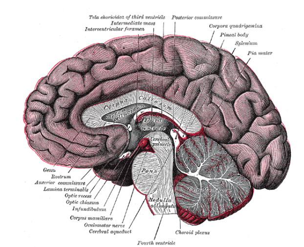
Diagram - The Midline Structures of the Brain
Licensed under Creative Commons, Source.
Edited by: Dr. Marcus Judge
Reviewed by: Adrian Judge
In this article
The CNS includes the brain, brainstem, cerebellum, and spinal cord. The PNS includes dorsal and ventral roots of spinal nerves and peripheral nerves.
- 777
