

Next Lesson - Anatomy of the Ear
Core
The autonomic nervous system involves the sympathetic and parasympathetic nervous systems, which maintain the internal environment. They regulate body functions that are not under conscious control.
Both systems usually use a two-neurone chain: a preganglionic neurone begins in the central nervous system and synapses in a peripheral ganglion, where a postganglionic neurone carries the signal to the target. Sympathetic preganglionic cell bodies lie in the thoracolumbar spinal cord. Parasympathetic preganglionic cell bodies lie in the brainstem or sacral spinal cord; cranial parasympathetic outflow does not originate in the spinal cord.
The sympathetic response is known as a ‘fight or flight’ response and is the typical response to stress.
As a result of sympathetic stimulation, the following happens within the head and neck:
- Pupillary dilation
- Lid retraction using the tarsal muscle
- Vasoconstriction of blood vessels
- Increased sweating

Diagram - Illustration of sympathetic innervation pathways in the body. Notice the cell bodies of the preganglionic neurones originating from T1-L2 segments of the spinal cord, and that the innervation to the head and neck synapse in the cervical chain.
Creative commons source by Blausen.com staff (2014). "Medical gallery of Blausen Medical 2014". WikiJournal of Medicine 1 (2). DOI:10.15347/wjm/2014.010. ISSN 2002-4436. [CC BY 3.0]
Pathways of Sympathetic Innervation
Cell bodies of the pre-ganglion neurones reside within specific segments of the spinal cord (T1-L2). This can be described as thoracolumbar outflow.
The sympathetic ganglia are located within sympathetic chains, two structures that lie lateral and parallel to the spinal column.
When considering the sympathetic pathway for head and neck structures, the superior cervical ganglia (found at the top of each sympathetic chain) contain the cell bodies of the post-ganglionic neurones innervating effector tissues.
The axons of the post-ganglionic neurones are closely related with blood vessels to reach their relative effector tissues:
- Pupillary Dilation - dilator pupillae. The post-ganglionic neurone is closely related to the internal carotid artery.
- Assisted Lid Retraction - superior tarsal muscle. The post-ganglionic neurone is, once again, closely related to the internal carotid artery.
- Smooth muscle of blood vessels and sweat glands supplied by post-ganglionic neurones. The post-ganglionic neurones are associated with the external carotid artery.
A disruption to this sympathetic nervous supply can lead to partial ptosis (drooping of the upper eyelid), miosis (constricted pupil) and anhidrosis (loss of sweating). Causes of this could include a Pancoast tumour (commonly a non-small cell lung cancer located at the apex of the lung) or carotid artery pathology.
The parasympathetic response is otherwise known as ‘rest and digest’.
In the head and neck, the following happen due to parasympathetic innervation:
- Constriction of the pupil
- Increased secretion from lacrimal and salivary glands

Diagram - Parasympathetic pathways have a craniosacral origin: preganglionic neurones arise in the brainstem or sacral spinal cord. The cranial component travels in nerves III, VII, IX and X; the sacral component supplies pelvic targets. Sympathetic outflow is thoracolumbar.
Creative commons source by Blausen.com staff (2014). "Medical gallery of Blausen Medical 2014". WikiJournal of Medicine 1 (2). DOI:10.15347/wjm/2014.010. ISSN 2002-4436. [CC BY 3.0]
Pathways of Parasympathetic Innervation
The parasympathetic nerves originate from the ‘craniosacral’ outflow, meaning that it is the brainstem and the sacral spinal cord that provide the innervation.
The cell bodies of pre-ganglionic neurones are found within nuclei that are located within the brainstem. These pre-ganglionic neurones track along one of four cranial nerves (oculomotor, facial, glossopharyngeal or vagus) before synapsing with a post-ganglionic neurone in a ganglion.
From here, the post-ganglionic neurone can progress, along a branch of the trigeminal nerve, to target tissues.
There are five key parasympathetic pathways, shown in the table below. Autonomic targets include glands, smooth muscle and cardiac muscle in the heart. Cardiac muscle is a distinct tissue type; it is not smooth muscle. Vagal parasympathetic signalling influences the heart, airways and gastrointestinal tract.
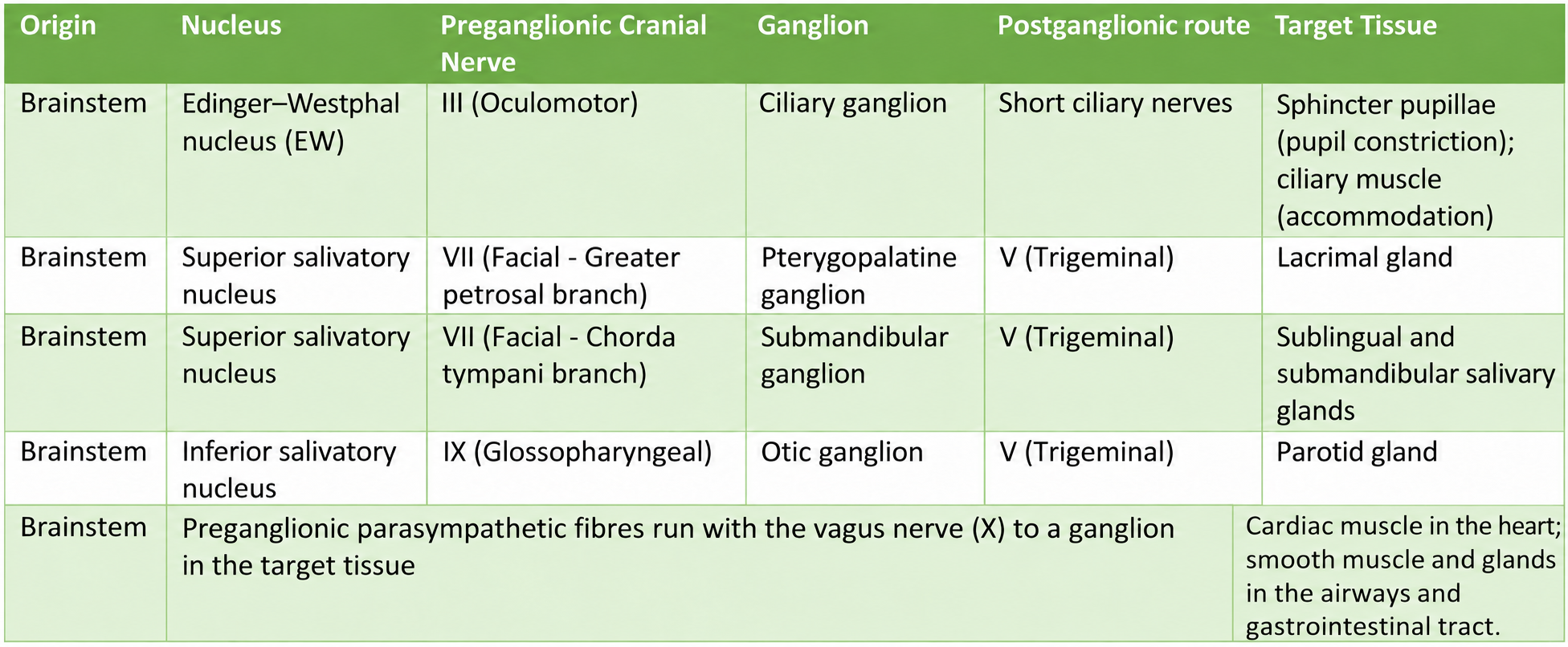
Table - Five cranial parasympathetic pathways and their targets. Sphincter pupillae constricts the pupil; the ciliary muscle changes lens shape for accommodation. Vagal targets include cardiac muscle in the heart and smooth muscle and glands in the airways and gastrointestinal tract.
Consensual Pupillary Light Reflexes
The consensual light reflex is an example of parasympathetic innervation in action.
The consensual light reflex is a phenomenon that occurs when a light is shone only in one eye, and the pupils of both eyes constrict in response, one due to the direct response to the light, and one due to parasympathetic innervation.
When a light is shone in the left pupil, the sensory afferents from the left optic nerve (cranial nerve II) transmit the signal from the left retina. Some branches of the left optic nerve enter the midbrain, some connect with the left Edinger Westphal nucleus (EDW) and some connect with the right EDW. The parasympathetic nerves in the EDW send impulses that track towards the eye with the third cranial nerve (oculomotor) towards the ciliary ganglion, where they synapse and innervate the sphincter pupillae muscle, causing it to constrict.
Because both EDW nuclei have received the impulse about the light shone in the left eye, they both send impulses that cause the pupils to constrict. This causes the direct light reflex in the left eye (responding directly to the light) and the consensual light reflex in the right eye (responding only to the parasympathetic innervation).
When light is shone into the left pupil, the optic nerve detects the light and some branches of this nerve enter the midbrain. This stimulates the EDW nuclei (both left and right) which has parasympathetic fibres extended along CN III. These pass through the ciliary ganglion and innervate the sphincter pupillae. The result is a constricted pupil in the left but because the right EDW nucleus was also stimulated, there is a ‘consensual’ light reflex in the right eye.
Edited by: Dr. Maddie Swannack
Reviewed by: Dr. Thomas Burnell
- 5423


