

Next Lesson - Diabetes Mellitus
Core
The pancreas is a large gland that develops embryologically as an outgrowth of the foregut, and can be found posterior to the stomach. The body of the pancreas is situated in the left upper quadrant of the abdomen.
99% of pancreatic tissue is exocrine tissue, which acts to produce digestive enzymes that are then secreted directly into the duodenum. The exocrine function of the pancreas will be covered in greater detail in the gastrointestinal unit; this series focuses on the endocrine function of the pancreas.
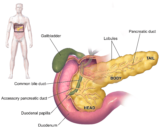
Diagram: Shows the location and basic structure of the pancreas
Creative commons source by Blausen.com staff (2014). "Medical gallery of Blausen Medical 2014" [CC BY-SA 4.0 (https://creativecommons.org/licenses/by-sa/4.0)]
The endocrine function of the pancreas is to produce hormones from Islets of Langerhans. Islets of Langerhans are predominantly made up of α and β cells, which secrete glucagon and insulin respectively. The functions of insulin and glucagon are to regulate the metabolism of carbohydrates, proteins and fats. Insulin works to lower blood glucose levels, whilst glucagon works to raise them.
The brain uses glucose at the fastest rate in the body, and relies on the blood for its supply. The plasma glucose reference range when fasting is normally around 4.0 to 5.9 mmol/L, and values may rise to below 7.8 mmol/L after a meal. The renal threshold for glucose is approximately 10 mmol/L, but this varies between individuals and is lower during pregnancy and higher in the elderly. Glycosuria is the point at which glucose begins to appear in the urine. Insulin and glucagon have opposing effects on blood glucose and together help regulate its concentration.

Diagram: Table showing the differences between insulin and glucagon
SimpleMed original by Dr. Jenny Hubball
Insulin is the hormone of energy storage. It is anti-gluconeogenic, anti-lipolytic and anti-ketogenic, meaning insulin acts to promote the storage of glucose. It is a peptide hormone with an α-helix structure, consisting of two unbranched peptide chains connected by two disulphide bridges. Insulin is synthesised in pancreatic β-cells via the following pathway:
- The single polypeptide preproinsulin is synthesised in the nucleus of β-cells.
- A signal peptide on the preproinsulin directs the polypeptide chain to the rough endoplasmic reticulum.
- Preproinsulin is cleaved to proinsulin.
- The proinsulin is folded and the disulphide bridges are formed.
- Proinsulin is transported to the Golgi apparatus where it is cleaved to produce insulin and C-peptide, which are both packaged into a secretory granule that then waits for a signal to be released.
Insulin is then released from the pancreatic β-cells via the following pathway:
- Food intake results in an increase in blood glucose concentration.
- Glucose diffuses into the pancreatic β cell via GLUT2 transporters.
- The increase in intracellular glucose causes an increase in glycolysis and ATP production.
- The increase in ATP production increases the intracellular ATP:ADP ratio.
- The increased ATP:ADP ratio causes the ATP-dependent K+ channels on the cell membrane to close.
- The closure of the K+ channels causes the membrane to depolarise.
- Voltage-gated Ca2+ channels on the cell membrane open, and there is a calcium influx into the cell.
- The increase in intracellular calcium causes secretory granules to be released via exocytosis.

Diagram: Outlines the mechanism of release of insulin from pancreatic β-cells
SimpleMed original by Dr. Jenny Hubball
The release of insulin then increases the uptake of glucose into cells of target tissues (adipose tissue and skeletal muscle) via the insertion of GLUT4 transporters and an increase in glycogen synthesis. Insertion of GLUT4 channels allows glucose to be transported across the target cell membrane. Insulin receptors are heterotetramers composed of two α subunits and two β subunits linked by disulphide bonds. The α chain is on the exterior of the cell membrane, and the β chain spans the membrane in a single segment.
Glucagon is the hormone that opposes insulin. It acts to raise glucose levels, meaning that it is glycogenolytic, gluconeogenic, lipolytic and ketogenic.
Glucagon is a 29 amino acid structure comprised of only a single polypeptide, meaning its synthesis is more simple than insulin’s. It is synthesised in pancreatic α-cells, where it is packaged into granules and secreted in response to low glucose levels.
Glucagon is most active in the liver, in which it functions to increase the rate of glycogenolysis, stimulate gluconeogenesis and stimulate lipolysis to increase plasma fatty acid concentration. If a diabetic patient is experiencing hypoglycaemia and cannot take sugar orally, glucagon can be given (for example, by intramuscular injection), or intravenous glucose may be required in a clinical setting.
Reviewed by: Dr. Thomas Burnell
- 7734

