

Next Lesson - Somatosensory System
Core
The cerebrospinal fluid (CSF) is a fluid that surrounds the brain and spinal cord. It is colourless and clear, and there is approximately 150 ml of CSF within the central nervous system at any one time. The CSF has a number of functions, the most important of which are mechanical support and metabolic processes. The CSF acts as a cushion to the brain, helping to protect it from damage against the hard skull, and helping to support it inside the cranial cavity to prevent it from sagging into the spinal canal. The CSF also has metabolic functions, acting to supply the central nervous system with glucose.
The brain develops as a tube, meaning that in a fully developed brain, there is a hollow space in the centre. These cavities within the brain are called ventricles, and four of them together form the ventricular system: the left lateral ventricle, the right lateral ventricle, the third ventricle and the fourth ventricle. For more information about the development of the brain, please see here.
The CSF is produced from structures contained within the ventricular system called the choroid plexus. Portions of choroid plexus are found in all of the ventricles, allowing for even production and flow of CSF through the ventricular system, but the lateral ventricles contribute more than any other ventricle.
The lateral ventricles are the largest ventricles in the brain, and these have portions that enter the frontal, temporal, and occipital lobes.
The two lateral ventricles are connected at the midline by the interventricular foramen, which drains into the third ventricle which sits on the midline, between the two thalami.
The third ventricle connects to a long tube called the cerebral aqueduct, which drains into the diamond-shaped fourth ventricle.
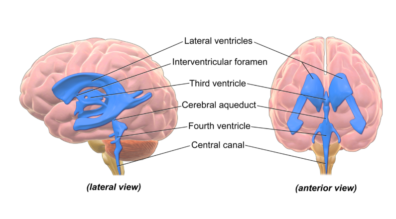
Diagram - The Ventricular System
Licensed under Creative Commons, Source.
The CSF drains from the fourth ventricle in a number of ways.
The first mechanism is through the central canal of the spinal cord. This is a very narrow space, so not very much CSF is removed this way.
The majority of the CSF drains from three apertures in the fourth ventricle, which are shown on the image above, and allow CSF to drain from the ventricles of the brain to the subarachnoid space. There are two paired, lateral apertures which are the two arm-like structures seen lateral to the fourth ventricle on the anterior view above, and a third, median aperture on the dorsal side of the fourth ventricle, seen on the lateral view above.
These apertures have eponymous names; the lateral apertures are known as the foramen of Luschka, and the median aperture is known as the foramen of Magendie. This is helpful because Lateral and Luschka both start with ‘L’ and Median and Magendie both start with ‘M’.
From the subarachnoid space, the CSF moves through arachnoid granulations into the superior sagittal sinus, and into the venous system. The image below shows the circulation of CSF through the ventricular system and around the brain in more detail.
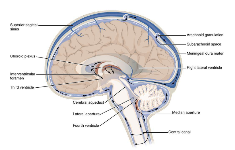
Diagram - The Circulation of the Cerebrospinal Fluid through the Ventricular System and Subarachnoid Space
Licensed under Creative Commons, Source.
Edited by: Dr. Marcus Judge
Reviewed by: Adrian Judge
In this article
The cerebrospinal fluid (CSF) is a fluid that surrounds the brain and spinal cord.
- 614
