

Next Lesson - Glomerular Disease
Core
Urinary incontinence (UI) is defined as the complaint of any involuntary leakage of urine. This can have a huge impact on a person’s quality of life through social exclusion and a sense of shame. Despite this, many patients “just put up” with urinary incontinence without seeking medical help, as it is seen as “a normal part of ageing”.
There are four main types of urinary incontinence:
- Stress Urinary Incontinence (SUI) - the complaint of involuntary leakage on increased abdominal pressure, such as exertion, sneezing or coughing.
- Urge Urinary Incontinence (UUI) - the complaint of involuntary leakage accompanied by or preceded by the urge to initiate voiding (passing urine).
- Mixed Urinary Incontinence (MUI) - the complaint of involuntary leakage associated with urgency, and also with exertion, effort, sneezing, or coughing.
- Overflow Incontinence - the complaint of involuntary leakage associated with the bladder being overly full, either due to detrusor weakness (weakness of the bladder wall muscle) or blockage, with no urge to micturate (pass urine).
The prevalence of UI increases with age, and SUI is a common subtype, particularly in females. A history is the most important investigation when determining the type of urinary incontinence present in a patient.
The main risk factor for developing UI is older age. Pregnancy and childbirth, pelvic organ prolapse (when the womb, bladder or bowel bulges into the vagina) and menopause are other possible risk factors. UI is more common in females.
Some other risk factors for UI include:
- Race
- Family predisposition
- Anatomical abnormalities
- Neurological abnormalities
- Obesity
- Increasing age (the main risk factor, as above)
- Increased intra-abdominal pressure
- Cognitive impairment (for example dementia)
- UTIs
- Drugs (for example diuretics and some antidepressants)
- Pelvic surgery
Lower Urinary Tract Symptoms (LUTS)
LUTS may be associated with urge urinary incontinence.
LUTS can be classified into three types based on which part of the micturition process they are associated with storage, voiding, and post-micturition. The table below shows the symptoms associated with each type of LUTS.
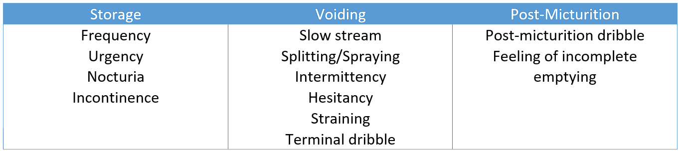
Table - Three types of lower urinary tract symptoms
The types of LUTS experienced can give clues about the underlying diagnosis, and can be seen in other conditions, not just UI. For example, hesitancy (a delay before urine starts to flow) can be a symptom of Benign Prostatic Hyperplasia (BPH, a non-cancerous enlargement of the prostate). Because the prostate is enlarged, a greater intra-vesical pressure (pressure inside the bladder) is needed to void (pass urine). This pressure takes time to build up, so it shows as hesitancy.
A thorough history is key when a patient presents with UI as this can be helpful in categorising the type of UI the patient is experiencing. For example, a sudden urge preceding the incontinence is typical of urge incontinence, whereas leakage on coughing is typical of stress incontinence.
An examination of a patient should include measurement of their BMI, an abdominal examination with particular attention paid to trying to palpate (feel for) a full bladder, a Digital Rectal Examination (DRE) (to assess anal tone as a marker of sacral nerve function, and to check for prostate enlargement in males; the sacral nerves come from the base of the spine and supply some of the muscles used for passing urine), and for females an examination of the external genitalia and a vaginal examination should also be performed.
All patients should receive a urine dipstick (a test strip dipped in a urine sample) in order to rule out a urinary tract infection, and to also check for haematuria (blood in the urine), proteinuria (protein in the urine), and glycosuria (glucose in the urine). Any of these may point to an underlying problem that needs further assessment.
Basic first tests can be done in primary care. The patient is asked to keep a bladder diary (a record of drinks, visits to the toilet and leaks, kept for at least 3 days). A frequency-volume chart (a record of the times and volumes of urine passed) is a similar record. The patient’s post-micturition residual volume (the amount of urine left in the bladder after passing urine, measured by a bladder scan or catheter) is checked if voiding dysfunction (difficulty emptying the bladder) or recurrent UTIs are suspected. These can all be performed between general practice appointments, as no specific secondary care need exists without these.
Further optional investigations could include invasive urodynamics (like pressure-flow studies in which a computer measures the pressure in the patient’s abdomen and in the bladder (the vesical pressure) to calculate the detrusor pressure, which is the pressure made by the bladder wall muscle), pad tests (where a patient wears a pre-weighed pad for a set period, sometimes after taking an agent to colour the urine, to confirm that leaked fluid is urine), or cystoscopy (direct visualisation of the patient’s bladder).
The management of UI depends on the symptoms the patient is experiencing, the degree of bother the symptoms are causing the patient, the effect the treatment will have on the patient’s symptoms, and any previous or current treatments the patient may have had. In any case, the management plan should be individualised and based on a systematic approach.
A conservative approach (one that does not use medicines or surgery) to the treatment of UI starts with lifestyle modifications, together with pelvic floor muscle training (exercises that strengthen the muscles supporting the bladder and urethra) for stress incontinence and bladder training for urge incontinence (both described below). Patients should be encouraged to modify their fluid intake, lose weight (if their BMI is over 30), and stop smoking (if applicable). Modification to fluid intake should be explained carefully to the patient. Patients who drink too much should drink less, and patients who drink too little should drink more, because not drinking enough creates more concentrated urine, which can irritate the bladder.
Avoiding constipation may also help, because constipation can contribute to UI. Patients can also undergo bladder training, which means learning to increase the length of time between feeling the need to void (pass urine) and doing so. The patient voids at planned times, and the gaps are gradually lengthened.
Decreasing caffeine intake is recommended as a trial for overactive bladder (a bladder whose detrusor muscle contracts too often), and drinking too much alcohol or caffeine is a possible cause of urge incontinence.
Contained incontinence is a form of therapy which does not aim to treat the cause of the UI, but to collect the leaked urine and allow for disposal at a time that is convenient. This is generally used for patients who are unsuitable for surgery or have failed conservative or medical management.
This can be done in the form of an indwelling catheter (a catheter which is put in place and can be left in long-term). NICE asks for careful consideration before a catheter is used long-term. Reasons include chronic urinary retention (being unable to empty the bladder fully) when the patient cannot manage to drain the bladder with a catheter themselves, skin wounds or pressure ulcers contaminated by urine, distress from bed and clothing changes, or the patient’s own preference. A catheter may not stop the leaking in urge incontinence. A catheter can be inserted through the urethra or above the pubic bone (suprapubic catheter), which may cause fewer urinary tract infections and urethral complications. Suprapubic catheter insertion requires a full bladder at the time of insertion and carries a small risk of bowel injury, which can be serious.
Other forms of contained incontinence devices are sheath devices (for men), essentially an adhesive condom attached to catheter tubing and a bag, or incontinence pads.
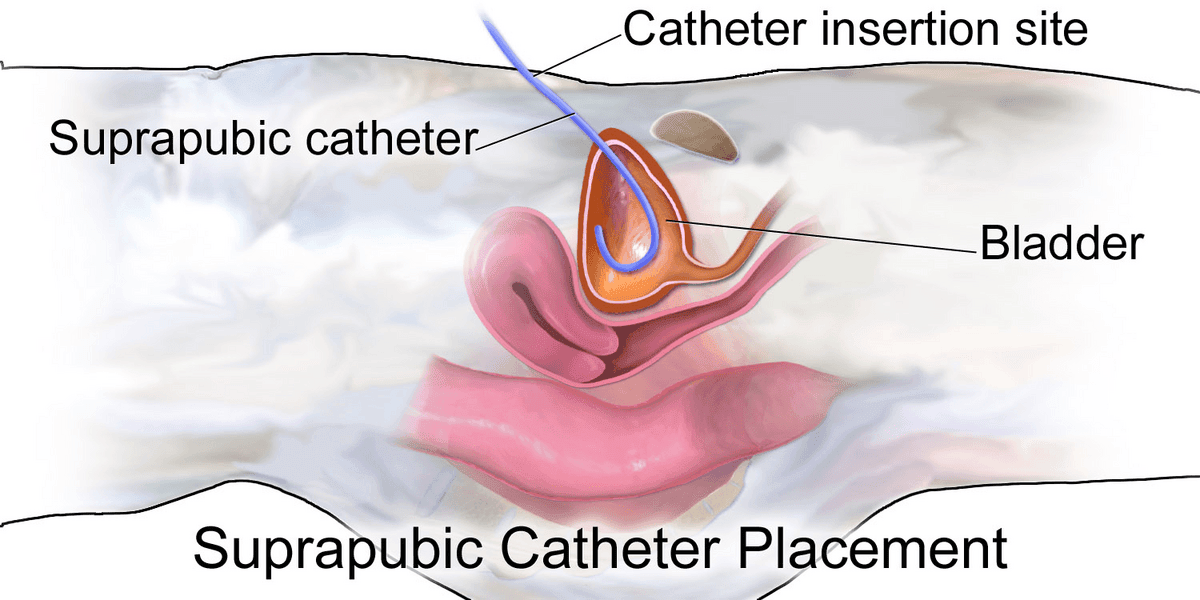
Diagram - Placement of a suprapubic catheter in a female
Creative commons source by BruceBlaus [CC BY-SA 4.0 (https://creativecommons.org/licenses/by-sa/4.0)]
Initially, patients presenting with stress incontinence should be offered a trial of supervised pelvic floor muscle training (exercises that strengthen the muscles around the bladder and urethra, which help to keep the urethra closed). These exercises consist of at least eight contractions of the pelvic floor, three times a day. This should be done for at least three months before reassessing the patient, and a referral to specialist physiotherapists can be made to supervise the training.
Duloxetine is an option for the pharmacological (drug) management of SUI in women. It is a combined noradrenaline and serotonin uptake inhibitor (it stops nerve endings taking these chemicals back up, so more stays available). This increases activity in the striated (skeletal) muscle portion of the urinary sphincter during the filling phase of the bladder. NICE does not recommend duloxetine as a first-line treatment (the one tried first) or as a routine second-line treatment (the one tried next if the first fails). It may be offered as second-line therapy if the woman prefers drug treatment to surgery or is not suitable for surgery. She should be counselled about its adverse effects, such as nausea.
Surgical options differ for males and females. For females, the choice of operation depends on several things, including whether the patient plans to have future pregnancies. Pregnancy and childbirth can sometimes cause surgical treatments to fail, so some patients choose to wait until after they have had children.
Low-tension vaginal tapes (a strip of synthetic mesh placed under the urethra to support it) were for many years the most common surgical procedure for the permanent treatment of SUI. In England, tape insertion almost stopped from 2019, after the “mesh pause” and concerns about complications, and bulking agents (described below) became the mainstream treatment. The tape is made of a polypropylene mesh (NICE advises a type 1 macroporous mesh, which has large pores), which helps to support the mid-urethra. The tape can be placed using minimally invasive techniques. In one 13-year follow-up study of 70 women, 81.4% were objectively cured (assessed with a stress test). Mesh is controversial in the UK. A few patients have had serious complications, such as long-lasting pain, permanent nerve damage and mesh exposure through the vaginal tissues, and the true long-term risk is unknown. The mesh is a permanent implant and complete removal might not be possible. In England, a period of “high vigilance restriction” was announced in 2018 for a group of mesh procedures for stress incontinence and prolapse, and the NHS says that vaginal mesh surgery for incontinence may not currently be possible on the NHS.
Retropubic suspension procedures (also called colposuspension, in which the neck of the bladder is lifted and stitched in the lifted position) aim to correct the anatomical position of the proximal urethra and improve urethral support. A fascial sling is a different operation. A sling made from the patient’s own tissue (usually the rectus fascia, the layer of tissue covering the abdominal muscles) is placed around the neck of the bladder to support the urethra and increase bladder outflow resistance (how hard it is for urine to leave the bladder). NICE offers colposuspension or this sling, and also includes the mesh tape in that choice.
Intramural bulking agents (substances injected into the wall of the urethra) are often chosen for patients who may want to have a future pregnancy. In one international survey of doctors, they were the commonest choice of operation for women who had not completed their families, although most advised postponing surgery until after childbearing. They are a temporary measure to treat SUI: the effect wears off over time, repeat injections may be needed, and they are less effective than the other operations, so NICE advises considering them if those are not suitable or acceptable to the patient. Injections of substances such as silicone (polydimethylsiloxane) or polyacrylamide hydrogel (collagen was used in the past but has been discontinued because of immune-related adverse reactions) are placed into the wall of the urethra, at the bladder neck (where the bladder joins the urethra) or further along the urethra, under general or local anaesthetic. This improves the ability of the urethra to resist abdominal pressure.
For males, the standard surgical treatment is an artificial urinary sphincter. These help correct sphincter deficiencies caused by neurological damage or surgery. A cuff is placed around the urethra and applies pressure to keep it closed. The patient can temporarily deflate the cuff using a hydraulic system, which allows voiding. NICE advises considering an artificial sphincter only for men whose symptoms have not responded to conservative management and drug treatments. Another option for males is a male sling procedure (a sling placed under the urethra to support it), which has shown good results in men with mild to moderate stress incontinence.
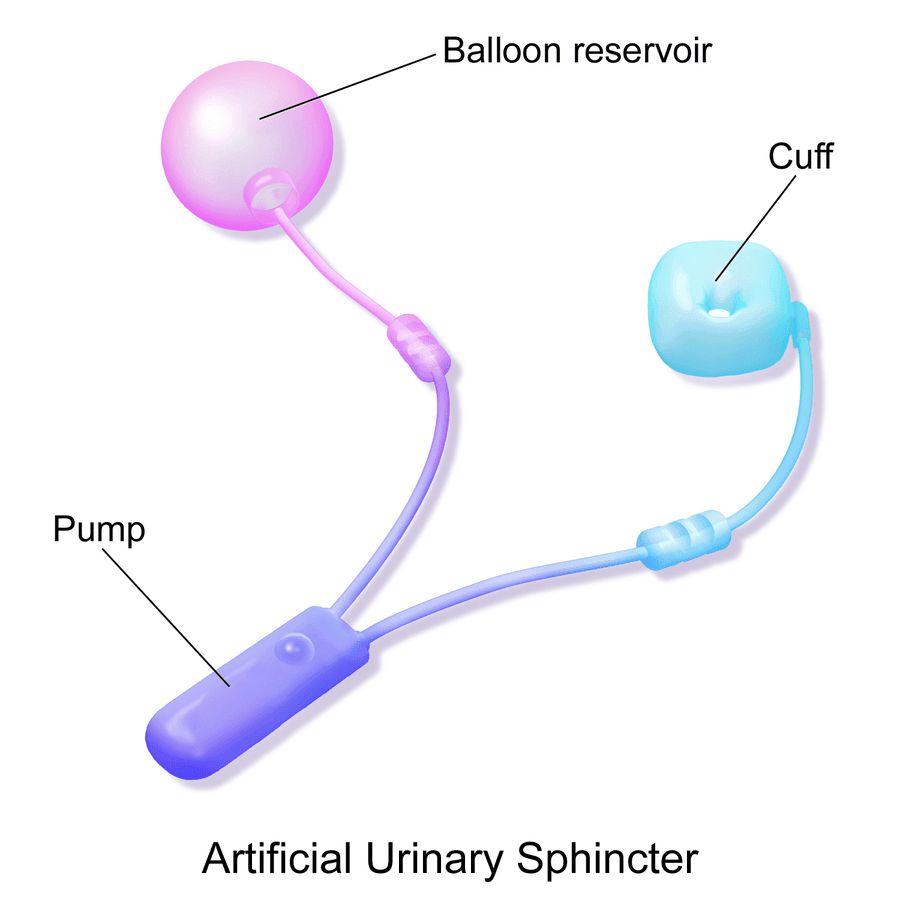
Diagram - An artificial urinary sphincter
Creative commons source by BruceBlaus [CC BY-SA 4.0 (https://creativecommons.org/licenses/by-sa/4.0)]
The first-line treatment for UUI is bladder training. This consists of a voiding schedule, initially every hour of the day, to train the bladder out of producing the urge. The patient should try not to void in between, using distraction or squeezing the pelvic floor muscles to delay the urge. Intervals are increased by 15-30 minutes a week until an interval of 2-3 hours is reached. This should be tried for at least 6 weeks.
The first option for pharmacological (drug) treatment of UUI is a group of drugs called anticholinergics (they block the effect of acetylcholine and are also called antimuscarinics), like Oxybutynin. These act on M2 and M3 muscarinic receptors (types of acetylcholine receptor found in the bladder wall). Side effects of Oxybutynin are related to the effects on muscarinic receptors found at other sites in the body, such as dry mouth, dry eyes, constipation and blurred vision. Confusion is rare but more common in older people. Care is needed with anticholinergics in patients with poor bladder emptying, cognitive impairment or dementia, or who take other medicines with an anticholinergic effect. NICE advises against immediate-release oxybutynin in older women who may be at higher risk of a sudden deterioration in their physical or mental health.
Mirabegron, a beta-3-adrenoceptor agonist (a drug that stimulates beta-3 receptors in the bladder muscle, making it relax), can also be used in the pharmacological therapy of UUI, mainly when antimuscarinics are unsuitable, do not work well enough or have unacceptable side effects. This drug increases the bladder’s capacity to store urine. Vibegron is a newer drug of the same type. Blood pressure should be monitored before starting mirabegron and regularly during treatment. It must not be used in severe uncontrolled hypertension (systolic blood pressure of 180 mmHg or more, or diastolic blood pressure of 110 mmHg or more).
If anticholinergics and beta-3-agonists fail to help the patient, an intravesical injection (an injection into the bladder wall) of Botulinum toxin (Botox) can be given, after review by a specialist team. If the patient develops difficulty emptying the bladder afterwards, they must be willing to perform clean intermittent catheterisation (passing a thin tube into the bladder to drain it) or, if they cannot do this, to accept a temporary catheter. Botulinum toxin is a potent biological neurotoxin which inhibits the release of acetylcholine at the pre-synaptic neuromuscular junction (where a nerve meets a muscle), causing weakening (partial paralysis) of the detrusor muscle, which reduces its overactive contractions. This is done under local or general anaesthetic. It has a duration of action of around 3-12 months, so injections can be repeated if they help.
If pharmacological management fails, there are surgical options. These include sacral nerve neuromodulation (a small device implanted near a sacral nerve that sends electrical pulses to improve the signals between the brain and the bladder), augmentation cystoplasty (making the bladder bigger by adding a piece of intestine to the bladder wall) and urinary diversion (redirecting the ureters, the tubes from the kidneys, to the outside of the body so that urine collects in a bag), but these are rare.
Edited by: Dr. Maddie Swannack
Reviewed by: Dr. Thomas Burnell and Dr. Marcus Judge
- 897


